SlideShare a Scribd company logo
1 of 100
NON - INVASIVE TOOL
PROVIDES A TANGIBLE & REALISTIC
           MODALITY
 PATIENTS CAN VISIBLY SEE FOR
          THEMSELVES
       COST EFFECTIVE
 HIGH SENSITIVITY IN DETECTING
     MARKERS/ANOMALIES
  MORE SOPHISTICATED & HIGH-
        RESOLUTION USG
OBSERVATION               CHROMOSOMAL
                          ABNORMALITY

HEAD                      TRISOMY-18,13
•Strawberry skull         TRIPLOIDY
•Hydrocephalus
•Holoprosencephaly
•Choroid plexus cyst
FACE                      TRISOMY-13,18
•Cleft lip/cleft palate   Meckel-Gruber syndrome
•Low set ears             TRIPLOIDY

HEART                     TRISOMY-13,18,21
•VSD,ASD
•Coarctation of aorta
OBSERVATION                        CHROMOSOMAL
                                   ABNORMALITY

RENAL                              TRISOMY-13,18,21
• Horseshoe kidney                 TRIPLOIDY
• B/L dilatation of renal pelvis
• Cystic dysplasia
HANDS/FEET                         TRISOMY-18,13,21
•Flexed overlapping fingers
•Rocker bottom/club foot
•Polydactyly
•Wide gap b/w 1st &2nd toes
(Sandal-gap)
•Clinodactyly
•Short femur/humerus
OBSERVATION          CHROMOSOMAL
                     ABNORMALITY

G.I SYSTEM           TRISOMY-13,18
Omphalocele          TRISOMY-21
Duodenal atresia
Echogenic bowel
GENERAL
Growth restriction   TRISOMY-13,18,21
Hydrops              TRIPLOIDY;45-XO
OBSERVATION      CHROMOSOMAL
                 ABNORMALITY

PLACENTA         Partial mole
                 ANEUPLOIDY
UMBILICAL CORD   TRISOMY-18
NUCHAL TRANSLUCENCY

                          With severe
                      LYPHANGIECTASIA
                       → overall swelling
                        of the fetal soft
                             tissue
                                ↓
                       Thickening of the
                      nuchal soft tissues
                                ↓
                            NUCHAL
                       TRANSLUCENCY
Refers to the normal
 subcutaneous fluid filled space
between the back of the fetal neck
      & the overlying skin.


The single most powerful marker
available today for differentiating
DS from euploid pregnancies.
POSSIBLE CAUSES OF ↑ FLUID FILLED SPACE(NT)


        Cardic failure secondary to structural
                     malformation
        Abnormality in the extracellular matrix
        Abnormal or delayed development of
                the lymphatic system
CAUSES

 ANEUPLOIDIES         SKELETAL DYSPLASIA
 Carnelia de Lange Achondrogenesis
 Noonan syndrome Ectrodactyly-ectodermal
 Smith-Lemli-Opitz         dysplasia
 Joubert             Multiple Pterygium Syndrome
 Apert               Robert Syndrome
 Fryns
             CHROMOSOMAL ANOMALIES
             Trisomy-21 (most common)
             Trisomy-13,18,22
             Triploidy
             Tetrasomy -12p
Imaged in the mid sagittal plane, ideally with the fetal
                      spine down.
 Image should be adequately magnified so that only
 the fetal head ,neck & upper thorax fill the viewable
                          area
The fetal neck should be neutral-avoid measurements
      in the hyperflexed/hyper extended positions
     The skin at the fetal back should be clearly
differentiated from the underlying amniotic membrane
Measurement calipers
  should be optimized to
ensure clarity of the image
 and of the borders of the
nuchal space in particular
           (TVS)

The width of the lucency
alone, excluding the width
 of the surface or occiput
PITFALLS

       PRESENCE OF
               An Encephalocele
                  Nuchal cord
               An Amniotic band
          A loose amnion that can be
       mistaken for the nuchal skin edge
How to rectify?


MAGNIFY THE IMAGE
WAIT FOR SPONTANEOUS FETAL ACTIVITY
→ as the fetus bounces from the amnion ,the
edges can be distinguished more reliably
COLOR DOPPLER → presence of a umbilical
cord in the vicinity of the fetal neck.
Cut off value of 3mm as a threshold for an
       abnormal nuchal translucency
  Normal NT thickens with increasing GA
 Currently, the more accepted method is to
   base the cut off on a progressive rise
      >95th percentile as a threshold.
MOM Vs SD: MOM-reduction in false +ve rates
•Equal success (Braithwaite & Economides)

METHOD       Gestational Age    Success rate
  TAS         10-13 WEEKS       98% to 100%
  TAS         AT 14 WEEKS           90%
                               TVS is needed
10-14weeks of GA

Detection rate     False+ve rate      Study group
    77%                 5%           Fetal medicine
                                       foundation,
                                         London
    63%                 5%            The SURUSS
                                         trial,UK
    69%                 5%           The BUN trial,
                                            US
  70%-64%               5%            The FASTER
                                         trial, US
Nicholaides: First trimester NT =/> 3mm
  Detection rate→86% of Trisomic fetus
  False +ve rate→4.5%
   Pandya (1995):
NT (mm) 3           4       5         >6
RISK ↑ 3            18      28        36

•TRISOMIES 13,18,21
•FETAL LOSS RATE =15% with NT of 5mm
•↑ NT → ↑ RISK OF CONGENITAL HEART DEFECT
•With Normal Karyotype & with abnormal karyotype
Progression from an abnormal NT to a
normal one→ not necessarily indicative of a
nomal Karyotype
So the fetus with nuchal abnormalities →
candidates for amniocentesis ,regardless of
whether the abnormality resolves
Among the women with advanced maternal
age b/w 11-14wks GA→NT can be used to
determine which patients would benefit from
an early First trimester Amniocentesis/CVS,
Vs delay of the invasive testing until 16 weeks
for the safest possible procedure.
MAJOR CARDIAC ANOMALIES
DIAPHRAGMATIC HERNIA
ANT. ABD.WALL DEFECT
FETAL AKINESIA/DYSKINESIA SYNDROME
OTHERS- Cornelia de lange,Noonan,Smith-lemli-
Opitz,Joubert,Apert&Fryns
SKELETALANOMALIES→
Achondrogenesis,Ectrodactyly-ectodermal
dysplasia,Multiple Pterygium syndrome,Robert syndrome
Localised nuchal fluid→CYSTIC HYGROMA(CH)
              FETAL HYDROPS
             DIFFUSE EDEMA
      SEPTATED CH:
 ANEUPLOIDY            EUPLOID
     50%                50%
                   50%-MAJOR structural
                       malformation
                      CARDIAC x12
                   SKELETAL DYSPLASIA
Presence of Septations within
    a nuchal swelling is ominous

Non - Septate CH   Septate CH

98%- transient     44%-transient

6%→ Abnormal       72%→ Abnormal
Karyotype          Karyotype

                          Bronshtein et al.
NO NEED TO DELAY DECISION MAKING→
   while awaiting serum marker results/using
   computerized risk calculation algorithms
   IMMEDIATE OPTIONS FOR CVS
   IF NO FETAL ANEUPLOIDY→


    A DETAILED FETAL ANATOMIC
            EVALUATION
+ FETAL ECHOCARDIOGRAPHY AT 18-20
              WEEKS
FASTER TRIAL-
 >3mm NT → CVS SHOULD BE OFFERED
IMMEDIATELY,because of a minimum risk of
            aneuploidy of 1 in 6.
    NO ROLE FOR DELAYING DECISION
   MAKING while awaiting serum marker
results,because such additional information
  does not meaningfully alter the original
              aneuploidy risk
GROWTH PATTERNS- CRL
NASAL BONE
DUCTUS VENOSUS SONOGRAPHY
TRICUSPID REGURGITATION
ANTERIOR ABDOMINAL WALL DEFECTS
ENCEPHALOCELES
LIMB DEFORMITIES
HEART DEFECTS
Schemmer et al;
CRL→ NOT significantly reduced
with Trisomy-21,Turner Syndrome or
    Sex chromosome Trisomies
        → SIGNIFICANTLY reduced
growth rates with Trisomies 13 & 18
           and Triploidy
ABSENCE OF NASAL BONE & DS

    Cicero et al; (N= 701 fetuses with ↑NT)
   ABSENCE OF NB            PRESENCE OF NB
          73%                       0.5%
       (43 OF 59)                (3 OF 602)
            NOT RELATED TO ↑ NT
   COULD BE COMBINED INTO A SINGLE USG
           SCREENING MODALITIES
PREDICTED SENSITIVITY OF 85% FOR 1% FALSE +VE
                    RATE.
 MID SAGITTAL PLANE
    FETAL PROFILE FACING
                      UPWARD
 ADEQUATE MAGNIFICATION
    VISUALIZATION OF TWO
    PARALLEL LINES AT THE
LEVEL OF THE FETAL NOSE→
      1. Superficial: fetal skin
        2. Deeper: nasal bone
       NASAL BONE- MORE
ECHOLUCENT AT THE DISTAL
                           END.
INCIDENCE OF ABSENT NASAL BONE

    GENERAL          HIGH RISK
   POPULATION       POPULATION

     17%-29%            48%

LIMITED ROLE AS A SCREENING TOOL FOR
         GENERAL POPULATION
FORWARD TRIPHASIC
     PULSATILE FLOW→
              NORMAL
REVERSED FLOW AT THE
        TIME OF ATRIAL
         CONTRACTION
   →ANEUPLOIDY/FETAL
CARDIAC MALFORMATION


WITH NT → ↑ THE DETECTION RATE/↓ THE FALSE +VE
                     RATE
PITFALLS
The ductus venosus vessel- as small as
2mm at 10-14weeks
Very difficult to get proper image


   SECONDARY SCREENING TEST IN
     THE HANDS OF EXPERIENCED
            SONOLOGIST
CHEST WALL-ANTERIOR
THE FETAL HEART SHOULD
BE ISONATED PARALLEL TO
THE VENTRICULAR SEPTUM
HIGH RISK PREGNANCIES
     AT 11-13 WEEKS


 Significant TR      INCIDENCE
NORMAL FETUS             4%
  DS Fetus                68%
 TRISOMY- 18              33%
     SECOND LINE TEST
AT 10-14 WEEKS
              Normal parameters
         GA(weeks)       FHR (beats/min)
            10                 171
            14                  156


Higher than normal rate- TRISOMY-21
Lower than normal rates- TRIPLOIDY & TRISOMY-18
SENSITIVITY

     ABNORHAL FHR- 26%
             NT- 72%
      MATERNAL AGE- 48%
MATERNAL AGE+ NT + FHR- 83% of
 detection rate at 5% false +ve rate
Authors          Parameter        Sensitivity   False +ve rate
Orlandi et al.   NT alone         57%           5.8%
                 NT +             87%           5.8%
                 biochemistry &
                 maternal age
Noble et al;     NT +           80-85%
                 Biochemistry &
                 maternal age


   BEST DETECTION RATE IN 1ST TRIMESTER-
Urine free β- hCG , beta core & Oestriol + NT
10-14 WEEKS
                          NT SONOGRAPHY


       CYSTIC HYGROMA             NO CYSTIC HYGROMA

          CVS
                              SINGLETONE    MULTIFETAL
EUPLOID      ANEUPLOID        GESTATION     GESTATION


18-20 WKS       COUNCEL
ANATOMY
SCAN &
FETAL ECHO
NT     +   NO CYSTIC HYGROMA


        SINGLETONE GESTATION     MULTIFETAL GESTATION


NT + SERUM MARKERS                    NT INTERPRETED WITH
PAPP-A & β- hCG                       MATERNAL AGE ONLY

 RISK ↑                                         RISK ↑
                     RISK NOT↑

  CVS                                 EUPLOID            CVS
               EUPLOID

ANEUPLOID                                          ANEUPLOID
                            18-20 WKS
COUNCEL                  ANATOMY SCAN &            COUNCEL
                           FETAL ECHO
MOST COMMON SONOGRAPHIC MARKERS


NUCHAL FOLD THICKENING
ECHOGENIC INTRACARDIAC FOCUS
SHORTENED LONG BONES
HYPERECHOIC BOWEL
RENAL PYELECTASIS
CHOROID PLEXUS CYST
CLINODACTYLY
HYPOPLASTIC OR ABSENT NASAL BONE
EXCESS SOFT TISSUE IN THE POSTERIOR NECK AREA

Measurement
    TS OF FETAL HEAD
  ANGLED POSTERIORLY
     TO INCLUDE THE
CEREBELLUM & THE OCCIPITAL          N   T
           BONE
 OUTSIDE OF THE OCCIPITAL BONE


   OUTER SKIN EDGE
THE NUCHAL SKIN FOLD MEASUREMENT THRESHOLD

  AUTHORS       Gestational    Threshold
                   Age
Gray & Crane    14-17 wks        5mm
                18-20 wks        6mm

   Wilson        < 17wks         5mm
SENSITIVITY
AUTHORS      CUT OFF VALUE SENSITIVITY FALSE +VE


 Crane &       >/= 5 mm       75%
  Gray
 Borrell &     >/= 6 mm       33%        0.1%
Colleagues
 Borrell &      >/= 5mm       77.8%       2%
Colleagues
May persists throughout the 2nd trimester
Or regression may occur
ONCE AN ABNORMAL NUCHAL SKIN
MEASUREMENT IS OBTAINED,THEREFORE ,AN
AMNIOCENTESIS IS INDICATED, REGARDLESS
 OF WHETHER THE NUCHAL SKIN THICKNESS
               RESOLVES
TRISOMY-21          oSHORT STATURED
                            oSHORT FEMURS
                            oSHORT HUMERI

RATIOS OF THE MEASURED - TO - EXPECTED FL OF
                </= 0.91, BPD.


 EXPECTED FL = - 9.3105 + 0.9028 x BPD
SENSITIVITY

STUDY      SENSITIVITY   FALSE +VE RATE
LOCKWOOD   50%           7%
CALLEN     68%
GRIST      50%           6.5%
Study      Parameter Sensitivity False +ve Anomalies
                                     rate
Brumfield   BPD: FL      40%       2.2%    TRISOMY-
  et al;     >/= 1.8                        18 & 21
Ginsberg      -do-       53%        7%       - do-
  et al;
Ginsberg      + NT       81%        7%        - do-
  et al;

       NOT A HELPFUL TOOL FOR SCREENING OF DS
USEFUL IN COMBINATIONS WITH OTHER SONOGRAPHIC MARKER
           Eg: HUMERUS LENGTH & PYELECTASIS
THE MEASURED- TO – EXPECTED HUMERUS LENGTH
           = - 7.9404 + 0.8492 x BPD
               < 0.90 as a cut - off

 STUDY       METHODS SENSITIVITY FALSE +VE
  Callen        HL      50%        6.25%
Rodis et al;    HL      54%
                FL      18%
 Biagiotti    FL + HL    ↑          ↓↓
  Periti
  Cariati
Nyberg et al;    Short FL + HL 11 fold ↑ risk of
                                       DS
Johnsons et al;     FL+ HL       53% - sensitivity
                  Foot Length     7% - false +ve
                                       rate
An antero-posterior diameter of the renal pelvis
                   >/=4 mm
STUDY         METHOD   SENSITIVITY FALSE +VE
  Callen       PYELECTAS    25%
                   IS
 Crane &          -do-     18.7%
  Gray
Corteville ,      -do-       17%        2%
 Dicks &
  Crane

ISOLATED PYELECTASIS- ↑risk
NOT SUFFICIENT TO INDICATE AMNIOCENTESIS
USED IN COMBINATION WITH OTHER
THE BOWEL IS AS ECHOGENIC AS BONE

    0.6% OF ALL 2ND -
  TRIMESTER FETUSES

BE AWARE:
High frequency transducer
may tend to accentuate the
echogenicity of the fetal
bowel in NORMAL fetus
↑ RISK OF
                IUGR
            PREMATURITY
            FETAL DEMISE
        POOR PERINAL OUTCOME
                APH
 CYSTIC FIBROSIS - Parental allele testing for CF
        carrier status is recommended
          IN-UTERO CMV INFECTION
MINERALIZATION IN THE PAPILLARY MUSCLE


              UNILATERAL        BILATERAL
90% in the left ventricles
 When the Right ventricle or both ventricles
are involved ↑ risk of Chromosomal anomalies


FETAL STATUS        EIF in Left    EIF in Right / B/L
                    Ventricle

   Normal              88%               12%

Down Syndrome          78%               22%
STUDY         NORMAL TRISOMY- 21 TRISOMY- 13
Brown,Roberts      2%          16%           39%
   & Miller
   Callen         4.7%         18%


Association of EIF & Chromosomal anomalies is low
                 in low risk patient
                NO AMNIOCENTESIS
 Not associated with cardiac anomalies in low risk
                      patient
 NORMAL FETUSES - 0.3% TO 3.6%
 1/3RD OF FETUSES WITH TRISOMY - 18


      •16-21 WEEKS → TRANSIENT
•BY 23RD WEEKS → USUALLY REGRESS
     •25-26 WEEKS → UNCOMMON
U/L                 SINGLE              SMALL

B/L                 MULTIPLE            LARGE


       SIZE = 0.5 cm – 2cm

  Very large CPC → Fill almost the entire
  lateral ventricle & expands its walls →
       FALSE VENTRICULOMEGALY
+ OTHER              ISOLATED CPC
 SONOGRAPHIC
   FINDINGS

                      CONSERVATIVE
INVASIVE TESTING     With detailed fetal
                   sonographic anatomic
                     survey by experts
EXAMINING THE UA            TRANSVERSE VIEW OF A FREE
  RUNNING ALONGSIDE &                LOOP OF CORD
  AROUND THE BLADDER
Transverse view of the pelvis
17% - CYTOGENETIC ABNORMALITY
    TRISOMY- 18 ( Most Common )
    TRISOMY- 13
    TURNERS SYNDROME (45X)
    TRIPLOIDY


  Commonly seen in normal fetuses
          It is non-specific
  The most common organ system
involved – HEART , GI SYSTEM & CNS
A targeted & detailed fetal    ISOLATED SUA –
anatomic survey should be     No ↑ incidence for a
done with detailed               chromosome
evaluation of the heart           abnormality
ILIAC WING ANGLE
               ILIAC LENGTH
FRONTOTHALAMIC DISTANCE (BRACHYCEPHALY)
        SHORTENED FRONTAL LOBE
              ABNORMAL FHR
   ABNORMALLY SHORTENED EAR LENGTH
                FLAT FACIES
  CLINODACTYLY(with hypoplasia of the middle
           phalanx of the fifth digit)
          SANDAL GAP GREAT TOE
        SIMIAN CREASE OF THE PALM
           EAR LENGTH & WIDTH
LOW RISK    NO further Testing
 Normal USG
 No markers present
                        HIGH RISK   DS risk adjustment


                        LOW RISK    NO further Testing
1-ISOLATED MARKER
(Except-Nuchal fold/
Absent Nasal Bone                   Genetic Amniocentesis
                        HIGH RISK


>/=2 MARKERS/Thick      LOW RISK    Genetic Amniocentesis
Nuchal Fold/ Absent
Nasal Bone/Structural
Anomaly                 HIGH RISK   Genetic Amniocentesis
STRUCTURAL ABNORMALITIES                   ANEUPLOIDY MARKERS
oCardiac defects                           Clinodactyly
oCystic hygroma                            EIF
oVentriculomegaly/hydrocephalus            Hyperechoic bowel
oEsophageal atresia                        Nuchal fold thickening
/Tracheo-esophagial fistula                Pyelectasis
oDuodenal atresia                          Sandal gap
oOmphalocele                               Short long bones
                                           Wide iliac angle
OTHER FINDINGS
                                           SUA
Brachycephaly
                                           Short ear length
Flat facial profile
                                           Absent / hypoplastic
Protruding tongue
                                                  nasal bone
Hydrops
Hydrothorax
Pericardial effusion
Unfused amnion & chorion after 14 weeks
STRUCTURAL ABNORMALITY
                      Cardiac defect
                      Cystic hygroma
                   Diaphragmatic hernia
                       Omphalocele
                Esophageal atresia+/- TOF
                     CNS-agenesis of
   corpuscallosum,ventriculomegaly,hydrocephalus,large
    cisterna magna,Dandy-Walker Malformation,cerebellar
               dysgenesis,neural tube defect
    CRANIOFACIAL- Strawberry-shaped skull,prominent
 occiput,dolichocephaly,ocular anomalies,micrognathia,cleft
                lip/palata,small,low set ears
       GENITO-URINARY- Hydronephrosis,horseshoe
   kidney,BOO,duplication abnormality,genital abnormality
EXTREMITY-
        Limb reduction abnormality,Radial
       aplasia,Clenched hands/Overlapping
    digits,Club feet,Rocker-bottom feet,Lower
                   extremity/feet
  abnormality,Contracture/arthrogryposis/flexion
          deformity,movement disorders
ANEUPLOIDY MARKERS-                OTHER FINDINGS-
   Choroid plexus cyst            IUGR,Amniotic fluid
Strawberry-shaped skull          abnormality,Umbilical
   Nuchal thickening             cord cyst,Non immune
                                         hydrops
 Single Umbilical artery
    Shortened limbs
STRUCTURAL ABNORMALITIES-
             Cardiac defects,Cystic hygroma,Nuchal
                      thickening,Omphalocele
    CNS- Holoprosencephaly,Agenesis of corpus callosum,
 Ventriculomegaly,Enlarged cisterna magna,Abnormal posterior
                     fossa,Neural tube defects
       CRANIO-FACIAL- Microcephaly,Micrognathia,Cleft
        lip/palate,Facial defects,Ocular anomalies ,Sloping
                        forehead,Small ears
       EXTREMITIES- Postaxial polydactyly,Overlapping
digits,Camptodactyly,Radial aplasia,Rocker-bottom feet,Club feet
   UROGENITAL- Hydronephrosis,Cortical cysts,Horseshoe
            kidney,Echogenic kidney,Polycystic kidneys
ANEUPLOIDY MARKERS
Echogenic Intracardiac Focus
       Pyelectasis
  Single Umbilical Artery


     OTHER FINDINGS
       Microcephaly
           IUGR
      Polyhydramnios
PAST
Over the past decade & a half, AMNIOCENTESIS was reserved
            for woman of advanced maternal age

                            PRESENT
   In the new millenium- major changes in the indications for
   INVASIVE GENETIC TESTING- such that advance maternal
            age alone will no longer be an indication

                              FUTURE
 Whether a patient is at risk for fetal Aneuploidy will be based on
  the combination MATERNAL AGE,MULTIPLE BIOCHEMICAL
SERUM MARKERS & perhaps a dozen SONOGRAPHIC MARKERS
           + a complete USG evaluation of the fetus.
Genetic sonogram

More Related Content

What's hot

Practical tips for monitoring of an iui cycle Dr. Jyoti Agarwal
Practical tips for monitoring  of  an iui cycle Dr. Jyoti Agarwal Practical tips for monitoring  of  an iui cycle Dr. Jyoti Agarwal
Practical tips for monitoring of an iui cycle Dr. Jyoti Agarwal
Lifecare Centre
 
Sperm retrieval techniques - nuts and bolts
Sperm retrieval techniques - nuts and boltsSperm retrieval techniques - nuts and bolts
Sperm retrieval techniques - nuts and bolts
Sandro Esteves
 
Infertility Hysteroscopy
Infertility HysteroscopyInfertility Hysteroscopy
Infertility Hysteroscopy
guest9dc181
 

What's hot (20)

Luteal phase support in ART
Luteal phase support in ARTLuteal phase support in ART
Luteal phase support in ART
 
Role of Stem Cells in Obstetrics and Gynecology Practice
Role of Stem Cells in Obstetrics and Gynecology PracticeRole of Stem Cells in Obstetrics and Gynecology Practice
Role of Stem Cells in Obstetrics and Gynecology Practice
 
ADOLESCENT ENDOMETRIOSIS
ADOLESCENT ENDOMETRIOSISADOLESCENT ENDOMETRIOSIS
ADOLESCENT ENDOMETRIOSIS
 
Fibroid and infertility
Fibroid and infertilityFibroid and infertility
Fibroid and infertility
 
Tests for ovarian reserve
Tests for ovarian reserveTests for ovarian reserve
Tests for ovarian reserve
 
Practical tips for monitoring of an iui cycle Dr. Jyoti Agarwal
Practical tips for monitoring  of  an iui cycle Dr. Jyoti Agarwal Practical tips for monitoring  of  an iui cycle Dr. Jyoti Agarwal
Practical tips for monitoring of an iui cycle Dr. Jyoti Agarwal
 
Aneuploidy screening Aneuploidy screening
Aneuploidy screening  Aneuploidy screening Aneuploidy screening  Aneuploidy screening
Aneuploidy screening Aneuploidy screening
 
Ovarian Stimulation Protocols
Ovarian Stimulation ProtocolsOvarian Stimulation Protocols
Ovarian Stimulation Protocols
 
Luteal Phase - Clinical Point of View - By Dr Dhorepatil Bharati
Luteal Phase - Clinical Point of View - By Dr Dhorepatil BharatiLuteal Phase - Clinical Point of View - By Dr Dhorepatil Bharati
Luteal Phase - Clinical Point of View - By Dr Dhorepatil Bharati
 
Unexplained Infertility - By Dr Dhorepatil Bharati
Unexplained Infertility - By Dr Dhorepatil BharatiUnexplained Infertility - By Dr Dhorepatil Bharati
Unexplained Infertility - By Dr Dhorepatil Bharati
 
Sperm retrieval techniques - nuts and bolts
Sperm retrieval techniques - nuts and boltsSperm retrieval techniques - nuts and bolts
Sperm retrieval techniques - nuts and bolts
 
Principles and Practices of Individualized OI and IUI
Principles and Practices of Individualized OI and IUIPrinciples and Practices of Individualized OI and IUI
Principles and Practices of Individualized OI and IUI
 
MANAGEMENT OF POOR RESPONDERS IN IVF BY DR SHASHWAT JANI
MANAGEMENT OF POOR RESPONDERS  IN IVF BY DR SHASHWAT JANIMANAGEMENT OF POOR RESPONDERS  IN IVF BY DR SHASHWAT JANI
MANAGEMENT OF POOR RESPONDERS IN IVF BY DR SHASHWAT JANI
 
Recurrent pregnancy loss panel discussion
Recurrent pregnancy loss  panel discussionRecurrent pregnancy loss  panel discussion
Recurrent pregnancy loss panel discussion
 
Internal iliac ligation
Internal iliac ligationInternal iliac ligation
Internal iliac ligation
 
Recent advances in stimulation protocols
Recent advances in stimulation protocolsRecent advances in stimulation protocols
Recent advances in stimulation protocols
 
Oocyte Donation; Factors Influencing The Outcome
Oocyte Donation; Factors Influencing The Outcome�Oocyte Donation; Factors Influencing The Outcome�
Oocyte Donation; Factors Influencing The Outcome
 
Diagnosis and Management of Poor Ovarian Reserve : Evidence & Practice
Diagnosis and Management of Poor Ovarian Reserve : Evidence & Practice Diagnosis and Management of Poor Ovarian Reserve : Evidence & Practice
Diagnosis and Management of Poor Ovarian Reserve : Evidence & Practice
 
Infertility Hysteroscopy
Infertility HysteroscopyInfertility Hysteroscopy
Infertility Hysteroscopy
 
Pet in gynecological malignancies
Pet in gynecological malignancies Pet in gynecological malignancies
Pet in gynecological malignancies
 

Viewers also liked

11-14 Wks Sonography
11-14 Wks Sonography11-14 Wks Sonography
11-14 Wks Sonography
Nitin Agrawal
 
ISUOG 2nd trimester guidelines - Dr.Ashok Khurana
ISUOG 2nd trimester guidelines - Dr.Ashok KhuranaISUOG 2nd trimester guidelines - Dr.Ashok Khurana
ISUOG 2nd trimester guidelines - Dr.Ashok Khurana
gehealthcare
 
1st trimester scan
1st trimester scan1st trimester scan
1st trimester scan
obsgynhsnz
 
Genetic counseling in modern dr. sharda jain
Genetic counseling in modern dr. sharda jainGenetic counseling in modern dr. sharda jain
Genetic counseling in modern dr. sharda jain
Lifecare Centre
 

Viewers also liked (20)

11-14 Wks Sonography
11-14 Wks Sonography11-14 Wks Sonography
11-14 Wks Sonography
 
11-13+6 weeks scan
11-13+6 weeks scan11-13+6 weeks scan
11-13+6 weeks scan
 
Focused approach to antenatal care - First trimester screening
Focused approach to antenatal care - First trimester screeningFocused approach to antenatal care - First trimester screening
Focused approach to antenatal care - First trimester screening
 
Role of us in evaluation of infertility Dr. Muhammad Bin Zulfiqar
Role of us in evaluation of infertility Dr. Muhammad Bin ZulfiqarRole of us in evaluation of infertility Dr. Muhammad Bin Zulfiqar
Role of us in evaluation of infertility Dr. Muhammad Bin Zulfiqar
 
Nuchal Translucency Screening
Nuchal Translucency ScreeningNuchal Translucency Screening
Nuchal Translucency Screening
 
ISUOG 2nd trimester guidelines - Dr.Ashok Khurana
ISUOG 2nd trimester guidelines - Dr.Ashok KhuranaISUOG 2nd trimester guidelines - Dr.Ashok Khurana
ISUOG 2nd trimester guidelines - Dr.Ashok Khurana
 
Isuog practice guidelines performance of first trimester fetal ultrasound scan
Isuog practice guidelines performance of first trimester fetal ultrasound scanIsuog practice guidelines performance of first trimester fetal ultrasound scan
Isuog practice guidelines performance of first trimester fetal ultrasound scan
 
Color doppler in fetal hypoxia
Color doppler in fetal hypoxiaColor doppler in fetal hypoxia
Color doppler in fetal hypoxia
 
Fetal Ecocardiography Screening
Fetal Ecocardiography ScreeningFetal Ecocardiography Screening
Fetal Ecocardiography Screening
 
Early pregnancy ultrasonographic evaluation
Early pregnancy ultrasonographic evaluationEarly pregnancy ultrasonographic evaluation
Early pregnancy ultrasonographic evaluation
 
Chromosomal and Genetic abnormalities
Chromosomal and Genetic abnormalities Chromosomal and Genetic abnormalities
Chromosomal and Genetic abnormalities
 
1st trimester scan
1st trimester scan1st trimester scan
1st trimester scan
 
Fetal anomaly scan pt2
Fetal anomaly scan pt2Fetal anomaly scan pt2
Fetal anomaly scan pt2
 
Genetik sonogram slideshare
Genetik sonogram slideshareGenetik sonogram slideshare
Genetik sonogram slideshare
 
Genetic counseling in modern dr. sharda jain
Genetic counseling in modern dr. sharda jainGenetic counseling in modern dr. sharda jain
Genetic counseling in modern dr. sharda jain
 
Nutraceuticals in pregnancy 1
Nutraceuticals in pregnancy 1Nutraceuticals in pregnancy 1
Nutraceuticals in pregnancy 1
 
Intrauterine growth restriction
Intrauterine growth restrictionIntrauterine growth restriction
Intrauterine growth restriction
 
Childbirth emergencies (2)
Childbirth emergencies (2)Childbirth emergencies (2)
Childbirth emergencies (2)
 
first trimester ultrasound and overview of obs imaging
first trimester ultrasound and overview of obs imagingfirst trimester ultrasound and overview of obs imaging
first trimester ultrasound and overview of obs imaging
 
Nuchal scan
Nuchal scanNuchal scan
Nuchal scan
 

Similar to Genetic sonogram

Similar to Genetic sonogram (20)

ANTENATAL_SCREENING_IN_FIRST_AND_SECOND_TRIMESTER.ppt
ANTENATAL_SCREENING_IN_FIRST_AND_SECOND_TRIMESTER.pptANTENATAL_SCREENING_IN_FIRST_AND_SECOND_TRIMESTER.ppt
ANTENATAL_SCREENING_IN_FIRST_AND_SECOND_TRIMESTER.ppt
 
Ultrasound markers of aneuploidy
Ultrasound markers of aneuploidyUltrasound markers of aneuploidy
Ultrasound markers of aneuploidy
 
Earlier, accurate & non invasive the new trigene test in the clinical context
Earlier, accurate & non invasive the new trigene test in the clinical contextEarlier, accurate & non invasive the new trigene test in the clinical context
Earlier, accurate & non invasive the new trigene test in the clinical context
 
Retinoblastoma
RetinoblastomaRetinoblastoma
Retinoblastoma
 
Retinoblastoma : Dr Shylesh B Dabke
Retinoblastoma : Dr Shylesh B DabkeRetinoblastoma : Dr Shylesh B Dabke
Retinoblastoma : Dr Shylesh B Dabke
 
ULTRASOUND EVALUATION OF ANEUPLOIDY IN FIRST AND SECOND TRIMESTER
ULTRASOUND EVALUATION OF ANEUPLOIDY IN FIRST AND SECOND TRIMESTERULTRASOUND EVALUATION OF ANEUPLOIDY IN FIRST AND SECOND TRIMESTER
ULTRASOUND EVALUATION OF ANEUPLOIDY IN FIRST AND SECOND TRIMESTER
 
Cranial usg final.pptx
Cranial usg final.pptxCranial usg final.pptx
Cranial usg final.pptx
 
Retinoblastoma
RetinoblastomaRetinoblastoma
Retinoblastoma
 
PEDIATRIC AIRWAY EVALUATION & MANAGEMENT
PEDIATRIC AIRWAY EVALUATION & MANAGEMENTPEDIATRIC AIRWAY EVALUATION & MANAGEMENT
PEDIATRIC AIRWAY EVALUATION & MANAGEMENT
 
Manegement of adenexal masses in pregnancy
Manegement of adenexal masses in pregnancyManegement of adenexal masses in pregnancy
Manegement of adenexal masses in pregnancy
 
Paediatric oncology
Paediatric oncologyPaediatric oncology
Paediatric oncology
 
solitary cold nodule of thyroid
solitary cold nodule of thyroidsolitary cold nodule of thyroid
solitary cold nodule of thyroid
 
Management Of Retinoblastoma
Management Of Retinoblastoma  Management Of Retinoblastoma
Management Of Retinoblastoma
 
Seminar prenatal genetic screening
Seminar prenatal genetic screeningSeminar prenatal genetic screening
Seminar prenatal genetic screening
 
KSK STN.pptx
KSK STN.pptxKSK STN.pptx
KSK STN.pptx
 
Medulloblastoma
MedulloblastomaMedulloblastoma
Medulloblastoma
 
CHILDHOOD RENAL TUMORS.pptx Pediatric renal pathology
CHILDHOOD RENAL TUMORS.pptx Pediatric renal pathologyCHILDHOOD RENAL TUMORS.pptx Pediatric renal pathology
CHILDHOOD RENAL TUMORS.pptx Pediatric renal pathology
 
Mcd
McdMcd
Mcd
 
First trimester ultrasound
First trimester ultrasoundFirst trimester ultrasound
First trimester ultrasound
 
Down syndrome.pdf
Down syndrome.pdfDown syndrome.pdf
Down syndrome.pdf
 

Recently uploaded

Russian Call Girls Lucknow Just Call 👉👉7877925207 Top Class Call Girl Service...
Russian Call Girls Lucknow Just Call 👉👉7877925207 Top Class Call Girl Service...Russian Call Girls Lucknow Just Call 👉👉7877925207 Top Class Call Girl Service...
Russian Call Girls Lucknow Just Call 👉👉7877925207 Top Class Call Girl Service...
adilkhan87451
 

Recently uploaded (20)

Night 7k to 12k Navi Mumbai Call Girl Photo 👉 BOOK NOW 9833363713 👈 ♀️ night ...
Night 7k to 12k Navi Mumbai Call Girl Photo 👉 BOOK NOW 9833363713 👈 ♀️ night ...Night 7k to 12k Navi Mumbai Call Girl Photo 👉 BOOK NOW 9833363713 👈 ♀️ night ...
Night 7k to 12k Navi Mumbai Call Girl Photo 👉 BOOK NOW 9833363713 👈 ♀️ night ...
 
Call Girls Vasai Virar Just Call 9630942363 Top Class Call Girl Service Avail...
Call Girls Vasai Virar Just Call 9630942363 Top Class Call Girl Service Avail...Call Girls Vasai Virar Just Call 9630942363 Top Class Call Girl Service Avail...
Call Girls Vasai Virar Just Call 9630942363 Top Class Call Girl Service Avail...
 
Call Girls Hosur Just Call 9630942363 Top Class Call Girl Service Available
Call Girls Hosur Just Call 9630942363 Top Class Call Girl Service AvailableCall Girls Hosur Just Call 9630942363 Top Class Call Girl Service Available
Call Girls Hosur Just Call 9630942363 Top Class Call Girl Service Available
 
All Time Service Available Call Girls Marine Drive 📳 9820252231 For 18+ VIP C...
All Time Service Available Call Girls Marine Drive 📳 9820252231 For 18+ VIP C...All Time Service Available Call Girls Marine Drive 📳 9820252231 For 18+ VIP C...
All Time Service Available Call Girls Marine Drive 📳 9820252231 For 18+ VIP C...
 
The Most Attractive Hyderabad Call Girls Kothapet 𖠋 9332606886 𖠋 Will You Mis...
The Most Attractive Hyderabad Call Girls Kothapet 𖠋 9332606886 𖠋 Will You Mis...The Most Attractive Hyderabad Call Girls Kothapet 𖠋 9332606886 𖠋 Will You Mis...
The Most Attractive Hyderabad Call Girls Kothapet 𖠋 9332606886 𖠋 Will You Mis...
 
Jogeshwari ! Call Girls Service Mumbai - 450+ Call Girl Cash Payment 90042684...
Jogeshwari ! Call Girls Service Mumbai - 450+ Call Girl Cash Payment 90042684...Jogeshwari ! Call Girls Service Mumbai - 450+ Call Girl Cash Payment 90042684...
Jogeshwari ! Call Girls Service Mumbai - 450+ Call Girl Cash Payment 90042684...
 
Manyata Tech Park ( Call Girls ) Bangalore ✔ 6297143586 ✔ Hot Model With Sexy...
Manyata Tech Park ( Call Girls ) Bangalore ✔ 6297143586 ✔ Hot Model With Sexy...Manyata Tech Park ( Call Girls ) Bangalore ✔ 6297143586 ✔ Hot Model With Sexy...
Manyata Tech Park ( Call Girls ) Bangalore ✔ 6297143586 ✔ Hot Model With Sexy...
 
Call Girls Guntur Just Call 8250077686 Top Class Call Girl Service Available
Call Girls Guntur  Just Call 8250077686 Top Class Call Girl Service AvailableCall Girls Guntur  Just Call 8250077686 Top Class Call Girl Service Available
Call Girls Guntur Just Call 8250077686 Top Class Call Girl Service Available
 
Top Rated Bangalore Call Girls Richmond Circle ⟟ 9332606886 ⟟ Call Me For Ge...
Top Rated Bangalore Call Girls Richmond Circle ⟟  9332606886 ⟟ Call Me For Ge...Top Rated Bangalore Call Girls Richmond Circle ⟟  9332606886 ⟟ Call Me For Ge...
Top Rated Bangalore Call Girls Richmond Circle ⟟ 9332606886 ⟟ Call Me For Ge...
 
Call Girls Vadodara Just Call 8617370543 Top Class Call Girl Service Available
Call Girls Vadodara Just Call 8617370543 Top Class Call Girl Service AvailableCall Girls Vadodara Just Call 8617370543 Top Class Call Girl Service Available
Call Girls Vadodara Just Call 8617370543 Top Class Call Girl Service Available
 
Call Girls Kurnool Just Call 8250077686 Top Class Call Girl Service Available
Call Girls Kurnool Just Call 8250077686 Top Class Call Girl Service AvailableCall Girls Kurnool Just Call 8250077686 Top Class Call Girl Service Available
Call Girls Kurnool Just Call 8250077686 Top Class Call Girl Service Available
 
Best Rate (Patna ) Call Girls Patna ⟟ 8617370543 ⟟ High Class Call Girl In 5 ...
Best Rate (Patna ) Call Girls Patna ⟟ 8617370543 ⟟ High Class Call Girl In 5 ...Best Rate (Patna ) Call Girls Patna ⟟ 8617370543 ⟟ High Class Call Girl In 5 ...
Best Rate (Patna ) Call Girls Patna ⟟ 8617370543 ⟟ High Class Call Girl In 5 ...
 
Russian Call Girls Lucknow Just Call 👉👉7877925207 Top Class Call Girl Service...
Russian Call Girls Lucknow Just Call 👉👉7877925207 Top Class Call Girl Service...Russian Call Girls Lucknow Just Call 👉👉7877925207 Top Class Call Girl Service...
Russian Call Girls Lucknow Just Call 👉👉7877925207 Top Class Call Girl Service...
 
Top Rated Bangalore Call Girls Mg Road ⟟ 9332606886 ⟟ Call Me For Genuine S...
Top Rated Bangalore Call Girls Mg Road ⟟   9332606886 ⟟ Call Me For Genuine S...Top Rated Bangalore Call Girls Mg Road ⟟   9332606886 ⟟ Call Me For Genuine S...
Top Rated Bangalore Call Girls Mg Road ⟟ 9332606886 ⟟ Call Me For Genuine S...
 
Call Girls Rishikesh Just Call 8250077686 Top Class Call Girl Service Available
Call Girls Rishikesh Just Call 8250077686 Top Class Call Girl Service AvailableCall Girls Rishikesh Just Call 8250077686 Top Class Call Girl Service Available
Call Girls Rishikesh Just Call 8250077686 Top Class Call Girl Service Available
 
Call Girls Gwalior Just Call 8617370543 Top Class Call Girl Service Available
Call Girls Gwalior Just Call 8617370543 Top Class Call Girl Service AvailableCall Girls Gwalior Just Call 8617370543 Top Class Call Girl Service Available
Call Girls Gwalior Just Call 8617370543 Top Class Call Girl Service Available
 
O898O367676 Call Girls In Ahmedabad Escort Service Available 24×7 In Ahmedabad
O898O367676 Call Girls In Ahmedabad Escort Service Available 24×7 In AhmedabadO898O367676 Call Girls In Ahmedabad Escort Service Available 24×7 In Ahmedabad
O898O367676 Call Girls In Ahmedabad Escort Service Available 24×7 In Ahmedabad
 
Premium Call Girls In Jaipur {8445551418} ❤️VVIP SEEMA Call Girl in Jaipur Ra...
Premium Call Girls In Jaipur {8445551418} ❤️VVIP SEEMA Call Girl in Jaipur Ra...Premium Call Girls In Jaipur {8445551418} ❤️VVIP SEEMA Call Girl in Jaipur Ra...
Premium Call Girls In Jaipur {8445551418} ❤️VVIP SEEMA Call Girl in Jaipur Ra...
 
Call Girls Visakhapatnam Just Call 8250077686 Top Class Call Girl Service Ava...
Call Girls Visakhapatnam Just Call 8250077686 Top Class Call Girl Service Ava...Call Girls Visakhapatnam Just Call 8250077686 Top Class Call Girl Service Ava...
Call Girls Visakhapatnam Just Call 8250077686 Top Class Call Girl Service Ava...
 
Russian Call Girls Service Jaipur {8445551418} ❤️PALLAVI VIP Jaipur Call Gir...
Russian Call Girls Service  Jaipur {8445551418} ❤️PALLAVI VIP Jaipur Call Gir...Russian Call Girls Service  Jaipur {8445551418} ❤️PALLAVI VIP Jaipur Call Gir...
Russian Call Girls Service Jaipur {8445551418} ❤️PALLAVI VIP Jaipur Call Gir...
 

Genetic sonogram

  • 1.
  • 2.
  • 3. NON - INVASIVE TOOL PROVIDES A TANGIBLE & REALISTIC MODALITY PATIENTS CAN VISIBLY SEE FOR THEMSELVES COST EFFECTIVE HIGH SENSITIVITY IN DETECTING MARKERS/ANOMALIES MORE SOPHISTICATED & HIGH- RESOLUTION USG
  • 4. OBSERVATION CHROMOSOMAL ABNORMALITY HEAD TRISOMY-18,13 •Strawberry skull TRIPLOIDY •Hydrocephalus •Holoprosencephaly •Choroid plexus cyst FACE TRISOMY-13,18 •Cleft lip/cleft palate Meckel-Gruber syndrome •Low set ears TRIPLOIDY HEART TRISOMY-13,18,21 •VSD,ASD •Coarctation of aorta
  • 5. OBSERVATION CHROMOSOMAL ABNORMALITY RENAL TRISOMY-13,18,21 • Horseshoe kidney TRIPLOIDY • B/L dilatation of renal pelvis • Cystic dysplasia HANDS/FEET TRISOMY-18,13,21 •Flexed overlapping fingers •Rocker bottom/club foot •Polydactyly •Wide gap b/w 1st &2nd toes (Sandal-gap) •Clinodactyly •Short femur/humerus
  • 6. OBSERVATION CHROMOSOMAL ABNORMALITY G.I SYSTEM TRISOMY-13,18 Omphalocele TRISOMY-21 Duodenal atresia Echogenic bowel GENERAL Growth restriction TRISOMY-13,18,21 Hydrops TRIPLOIDY;45-XO
  • 7. OBSERVATION CHROMOSOMAL ABNORMALITY PLACENTA Partial mole ANEUPLOIDY UMBILICAL CORD TRISOMY-18
  • 8. NUCHAL TRANSLUCENCY With severe LYPHANGIECTASIA → overall swelling of the fetal soft tissue ↓ Thickening of the nuchal soft tissues ↓ NUCHAL TRANSLUCENCY
  • 9. Refers to the normal subcutaneous fluid filled space between the back of the fetal neck & the overlying skin. The single most powerful marker available today for differentiating DS from euploid pregnancies.
  • 10.
  • 11. POSSIBLE CAUSES OF ↑ FLUID FILLED SPACE(NT) Cardic failure secondary to structural malformation Abnormality in the extracellular matrix Abnormal or delayed development of the lymphatic system
  • 12. CAUSES ANEUPLOIDIES SKELETAL DYSPLASIA Carnelia de Lange Achondrogenesis Noonan syndrome Ectrodactyly-ectodermal Smith-Lemli-Opitz dysplasia Joubert Multiple Pterygium Syndrome Apert Robert Syndrome Fryns CHROMOSOMAL ANOMALIES Trisomy-21 (most common) Trisomy-13,18,22 Triploidy Tetrasomy -12p
  • 13. Imaged in the mid sagittal plane, ideally with the fetal spine down. Image should be adequately magnified so that only the fetal head ,neck & upper thorax fill the viewable area The fetal neck should be neutral-avoid measurements in the hyperflexed/hyper extended positions The skin at the fetal back should be clearly differentiated from the underlying amniotic membrane
  • 14. Measurement calipers should be optimized to ensure clarity of the image and of the borders of the nuchal space in particular (TVS) The width of the lucency alone, excluding the width of the surface or occiput
  • 15.
  • 16. PITFALLS PRESENCE OF  An Encephalocele  Nuchal cord  An Amniotic band  A loose amnion that can be mistaken for the nuchal skin edge
  • 17. How to rectify? MAGNIFY THE IMAGE WAIT FOR SPONTANEOUS FETAL ACTIVITY → as the fetus bounces from the amnion ,the edges can be distinguished more reliably COLOR DOPPLER → presence of a umbilical cord in the vicinity of the fetal neck.
  • 18. Cut off value of 3mm as a threshold for an abnormal nuchal translucency Normal NT thickens with increasing GA Currently, the more accepted method is to base the cut off on a progressive rise >95th percentile as a threshold. MOM Vs SD: MOM-reduction in false +ve rates
  • 19. •Equal success (Braithwaite & Economides) METHOD Gestational Age Success rate TAS 10-13 WEEKS 98% to 100% TAS AT 14 WEEKS 90% TVS is needed
  • 20. 10-14weeks of GA Detection rate False+ve rate Study group 77% 5% Fetal medicine foundation, London 63% 5% The SURUSS trial,UK 69% 5% The BUN trial, US 70%-64% 5% The FASTER trial, US
  • 21. Nicholaides: First trimester NT =/> 3mm Detection rate→86% of Trisomic fetus False +ve rate→4.5% Pandya (1995): NT (mm) 3 4 5 >6 RISK ↑ 3 18 28 36 •TRISOMIES 13,18,21 •FETAL LOSS RATE =15% with NT of 5mm •↑ NT → ↑ RISK OF CONGENITAL HEART DEFECT •With Normal Karyotype & with abnormal karyotype
  • 22. Progression from an abnormal NT to a normal one→ not necessarily indicative of a nomal Karyotype So the fetus with nuchal abnormalities → candidates for amniocentesis ,regardless of whether the abnormality resolves Among the women with advanced maternal age b/w 11-14wks GA→NT can be used to determine which patients would benefit from an early First trimester Amniocentesis/CVS, Vs delay of the invasive testing until 16 weeks for the safest possible procedure.
  • 23. MAJOR CARDIAC ANOMALIES DIAPHRAGMATIC HERNIA ANT. ABD.WALL DEFECT FETAL AKINESIA/DYSKINESIA SYNDROME OTHERS- Cornelia de lange,Noonan,Smith-lemli- Opitz,Joubert,Apert&Fryns SKELETALANOMALIES→ Achondrogenesis,Ectrodactyly-ectodermal dysplasia,Multiple Pterygium syndrome,Robert syndrome
  • 24. Localised nuchal fluid→CYSTIC HYGROMA(CH) FETAL HYDROPS DIFFUSE EDEMA SEPTATED CH: ANEUPLOIDY EUPLOID 50% 50% 50%-MAJOR structural malformation CARDIAC x12 SKELETAL DYSPLASIA
  • 25.
  • 26. Presence of Septations within a nuchal swelling is ominous Non - Septate CH Septate CH 98%- transient 44%-transient 6%→ Abnormal 72%→ Abnormal Karyotype Karyotype Bronshtein et al.
  • 27. NO NEED TO DELAY DECISION MAKING→ while awaiting serum marker results/using computerized risk calculation algorithms IMMEDIATE OPTIONS FOR CVS IF NO FETAL ANEUPLOIDY→ A DETAILED FETAL ANATOMIC EVALUATION + FETAL ECHOCARDIOGRAPHY AT 18-20 WEEKS
  • 28. FASTER TRIAL- >3mm NT → CVS SHOULD BE OFFERED IMMEDIATELY,because of a minimum risk of aneuploidy of 1 in 6. NO ROLE FOR DELAYING DECISION MAKING while awaiting serum marker results,because such additional information does not meaningfully alter the original aneuploidy risk
  • 29. GROWTH PATTERNS- CRL NASAL BONE DUCTUS VENOSUS SONOGRAPHY TRICUSPID REGURGITATION ANTERIOR ABDOMINAL WALL DEFECTS ENCEPHALOCELES LIMB DEFORMITIES HEART DEFECTS
  • 30. Schemmer et al; CRL→ NOT significantly reduced with Trisomy-21,Turner Syndrome or Sex chromosome Trisomies  → SIGNIFICANTLY reduced growth rates with Trisomies 13 & 18 and Triploidy
  • 31. ABSENCE OF NASAL BONE & DS Cicero et al; (N= 701 fetuses with ↑NT) ABSENCE OF NB PRESENCE OF NB 73% 0.5% (43 OF 59) (3 OF 602) NOT RELATED TO ↑ NT COULD BE COMBINED INTO A SINGLE USG SCREENING MODALITIES PREDICTED SENSITIVITY OF 85% FOR 1% FALSE +VE RATE.
  • 32.  MID SAGITTAL PLANE  FETAL PROFILE FACING UPWARD  ADEQUATE MAGNIFICATION  VISUALIZATION OF TWO PARALLEL LINES AT THE LEVEL OF THE FETAL NOSE→ 1. Superficial: fetal skin 2. Deeper: nasal bone  NASAL BONE- MORE ECHOLUCENT AT THE DISTAL END.
  • 33. INCIDENCE OF ABSENT NASAL BONE GENERAL HIGH RISK POPULATION POPULATION 17%-29% 48% LIMITED ROLE AS A SCREENING TOOL FOR GENERAL POPULATION
  • 34. FORWARD TRIPHASIC PULSATILE FLOW→ NORMAL REVERSED FLOW AT THE TIME OF ATRIAL CONTRACTION →ANEUPLOIDY/FETAL CARDIAC MALFORMATION WITH NT → ↑ THE DETECTION RATE/↓ THE FALSE +VE RATE
  • 35. PITFALLS The ductus venosus vessel- as small as 2mm at 10-14weeks Very difficult to get proper image SECONDARY SCREENING TEST IN THE HANDS OF EXPERIENCED SONOLOGIST
  • 36. CHEST WALL-ANTERIOR THE FETAL HEART SHOULD BE ISONATED PARALLEL TO THE VENTRICULAR SEPTUM HIGH RISK PREGNANCIES AT 11-13 WEEKS Significant TR INCIDENCE NORMAL FETUS 4% DS Fetus 68% TRISOMY- 18 33% SECOND LINE TEST
  • 37. AT 10-14 WEEKS Normal parameters GA(weeks) FHR (beats/min) 10 171 14 156 Higher than normal rate- TRISOMY-21 Lower than normal rates- TRIPLOIDY & TRISOMY-18
  • 38. SENSITIVITY ABNORHAL FHR- 26% NT- 72% MATERNAL AGE- 48% MATERNAL AGE+ NT + FHR- 83% of detection rate at 5% false +ve rate
  • 39. Authors Parameter Sensitivity False +ve rate Orlandi et al. NT alone 57% 5.8% NT + 87% 5.8% biochemistry & maternal age Noble et al; NT + 80-85% Biochemistry & maternal age BEST DETECTION RATE IN 1ST TRIMESTER- Urine free β- hCG , beta core & Oestriol + NT
  • 40. 10-14 WEEKS NT SONOGRAPHY CYSTIC HYGROMA NO CYSTIC HYGROMA CVS SINGLETONE MULTIFETAL EUPLOID ANEUPLOID GESTATION GESTATION 18-20 WKS COUNCEL ANATOMY SCAN & FETAL ECHO
  • 41. NT + NO CYSTIC HYGROMA SINGLETONE GESTATION MULTIFETAL GESTATION NT + SERUM MARKERS NT INTERPRETED WITH PAPP-A & β- hCG MATERNAL AGE ONLY RISK ↑ RISK ↑ RISK NOT↑ CVS EUPLOID CVS EUPLOID ANEUPLOID ANEUPLOID 18-20 WKS COUNCEL ANATOMY SCAN & COUNCEL FETAL ECHO
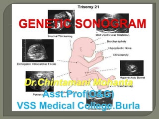
  • 42. MOST COMMON SONOGRAPHIC MARKERS NUCHAL FOLD THICKENING ECHOGENIC INTRACARDIAC FOCUS SHORTENED LONG BONES HYPERECHOIC BOWEL RENAL PYELECTASIS CHOROID PLEXUS CYST CLINODACTYLY HYPOPLASTIC OR ABSENT NASAL BONE
  • 43. EXCESS SOFT TISSUE IN THE POSTERIOR NECK AREA Measurement TS OF FETAL HEAD ANGLED POSTERIORLY TO INCLUDE THE CEREBELLUM & THE OCCIPITAL N T BONE OUTSIDE OF THE OCCIPITAL BONE OUTER SKIN EDGE
  • 44. THE NUCHAL SKIN FOLD MEASUREMENT THRESHOLD AUTHORS Gestational Threshold Age Gray & Crane 14-17 wks 5mm 18-20 wks 6mm Wilson < 17wks 5mm
  • 45. SENSITIVITY AUTHORS CUT OFF VALUE SENSITIVITY FALSE +VE Crane & >/= 5 mm 75% Gray Borrell & >/= 6 mm 33% 0.1% Colleagues Borrell & >/= 5mm 77.8% 2% Colleagues
  • 46. May persists throughout the 2nd trimester Or regression may occur
  • 47. ONCE AN ABNORMAL NUCHAL SKIN MEASUREMENT IS OBTAINED,THEREFORE ,AN AMNIOCENTESIS IS INDICATED, REGARDLESS OF WHETHER THE NUCHAL SKIN THICKNESS RESOLVES
  • 48. TRISOMY-21 oSHORT STATURED oSHORT FEMURS oSHORT HUMERI RATIOS OF THE MEASURED - TO - EXPECTED FL OF </= 0.91, BPD. EXPECTED FL = - 9.3105 + 0.9028 x BPD
  • 49. SENSITIVITY STUDY SENSITIVITY FALSE +VE RATE LOCKWOOD 50% 7% CALLEN 68% GRIST 50% 6.5%
  • 50. Study Parameter Sensitivity False +ve Anomalies rate Brumfield BPD: FL 40% 2.2% TRISOMY- et al; >/= 1.8 18 & 21 Ginsberg -do- 53% 7% - do- et al; Ginsberg + NT 81% 7% - do- et al; NOT A HELPFUL TOOL FOR SCREENING OF DS USEFUL IN COMBINATIONS WITH OTHER SONOGRAPHIC MARKER Eg: HUMERUS LENGTH & PYELECTASIS
  • 51. THE MEASURED- TO – EXPECTED HUMERUS LENGTH = - 7.9404 + 0.8492 x BPD < 0.90 as a cut - off STUDY METHODS SENSITIVITY FALSE +VE Callen HL 50% 6.25% Rodis et al; HL 54% FL 18% Biagiotti FL + HL ↑ ↓↓ Periti Cariati
  • 52. Nyberg et al; Short FL + HL 11 fold ↑ risk of DS Johnsons et al; FL+ HL 53% - sensitivity Foot Length 7% - false +ve rate
  • 53.
  • 54. An antero-posterior diameter of the renal pelvis >/=4 mm
  • 55. STUDY METHOD SENSITIVITY FALSE +VE Callen PYELECTAS 25% IS Crane & -do- 18.7% Gray Corteville , -do- 17% 2% Dicks & Crane ISOLATED PYELECTASIS- ↑risk NOT SUFFICIENT TO INDICATE AMNIOCENTESIS USED IN COMBINATION WITH OTHER
  • 56. THE BOWEL IS AS ECHOGENIC AS BONE 0.6% OF ALL 2ND - TRIMESTER FETUSES BE AWARE: High frequency transducer may tend to accentuate the echogenicity of the fetal bowel in NORMAL fetus
  • 57. ↑ RISK OF IUGR PREMATURITY FETAL DEMISE POOR PERINAL OUTCOME APH  CYSTIC FIBROSIS - Parental allele testing for CF carrier status is recommended  IN-UTERO CMV INFECTION
  • 58. MINERALIZATION IN THE PAPILLARY MUSCLE UNILATERAL BILATERAL
  • 59. 90% in the left ventricles When the Right ventricle or both ventricles are involved ↑ risk of Chromosomal anomalies FETAL STATUS EIF in Left EIF in Right / B/L Ventricle Normal 88% 12% Down Syndrome 78% 22%
  • 60. STUDY NORMAL TRISOMY- 21 TRISOMY- 13 Brown,Roberts 2% 16% 39% & Miller Callen 4.7% 18% Association of EIF & Chromosomal anomalies is low in low risk patient NO AMNIOCENTESIS Not associated with cardiac anomalies in low risk patient
  • 61.  NORMAL FETUSES - 0.3% TO 3.6%  1/3RD OF FETUSES WITH TRISOMY - 18 •16-21 WEEKS → TRANSIENT •BY 23RD WEEKS → USUALLY REGRESS •25-26 WEEKS → UNCOMMON
  • 62. U/L SINGLE SMALL B/L MULTIPLE LARGE SIZE = 0.5 cm – 2cm Very large CPC → Fill almost the entire lateral ventricle & expands its walls → FALSE VENTRICULOMEGALY
  • 63. + OTHER ISOLATED CPC SONOGRAPHIC FINDINGS CONSERVATIVE INVASIVE TESTING With detailed fetal sonographic anatomic survey by experts
  • 64. EXAMINING THE UA TRANSVERSE VIEW OF A FREE RUNNING ALONGSIDE & LOOP OF CORD AROUND THE BLADDER Transverse view of the pelvis
  • 65. 17% - CYTOGENETIC ABNORMALITY TRISOMY- 18 ( Most Common ) TRISOMY- 13 TURNERS SYNDROME (45X) TRIPLOIDY Commonly seen in normal fetuses It is non-specific The most common organ system involved – HEART , GI SYSTEM & CNS
  • 66. A targeted & detailed fetal ISOLATED SUA – anatomic survey should be No ↑ incidence for a done with detailed chromosome evaluation of the heart abnormality
  • 67. ILIAC WING ANGLE ILIAC LENGTH FRONTOTHALAMIC DISTANCE (BRACHYCEPHALY) SHORTENED FRONTAL LOBE ABNORMAL FHR ABNORMALLY SHORTENED EAR LENGTH FLAT FACIES CLINODACTYLY(with hypoplasia of the middle phalanx of the fifth digit) SANDAL GAP GREAT TOE SIMIAN CREASE OF THE PALM EAR LENGTH & WIDTH
  • 68. LOW RISK NO further Testing Normal USG No markers present HIGH RISK DS risk adjustment LOW RISK NO further Testing 1-ISOLATED MARKER (Except-Nuchal fold/ Absent Nasal Bone Genetic Amniocentesis HIGH RISK >/=2 MARKERS/Thick LOW RISK Genetic Amniocentesis Nuchal Fold/ Absent Nasal Bone/Structural Anomaly HIGH RISK Genetic Amniocentesis
  • 69. STRUCTURAL ABNORMALITIES ANEUPLOIDY MARKERS oCardiac defects Clinodactyly oCystic hygroma EIF oVentriculomegaly/hydrocephalus Hyperechoic bowel oEsophageal atresia Nuchal fold thickening /Tracheo-esophagial fistula Pyelectasis oDuodenal atresia Sandal gap oOmphalocele Short long bones Wide iliac angle OTHER FINDINGS SUA Brachycephaly Short ear length Flat facial profile Absent / hypoplastic Protruding tongue nasal bone Hydrops Hydrothorax Pericardial effusion Unfused amnion & chorion after 14 weeks
  • 70. STRUCTURAL ABNORMALITY Cardiac defect Cystic hygroma Diaphragmatic hernia Omphalocele Esophageal atresia+/- TOF CNS-agenesis of corpuscallosum,ventriculomegaly,hydrocephalus,large cisterna magna,Dandy-Walker Malformation,cerebellar dysgenesis,neural tube defect CRANIOFACIAL- Strawberry-shaped skull,prominent occiput,dolichocephaly,ocular anomalies,micrognathia,cleft lip/palata,small,low set ears GENITO-URINARY- Hydronephrosis,horseshoe kidney,BOO,duplication abnormality,genital abnormality
  • 71. EXTREMITY- Limb reduction abnormality,Radial aplasia,Clenched hands/Overlapping digits,Club feet,Rocker-bottom feet,Lower extremity/feet abnormality,Contracture/arthrogryposis/flexion deformity,movement disorders ANEUPLOIDY MARKERS- OTHER FINDINGS-  Choroid plexus cyst IUGR,Amniotic fluid Strawberry-shaped skull abnormality,Umbilical Nuchal thickening cord cyst,Non immune hydrops Single Umbilical artery Shortened limbs
  • 72. STRUCTURAL ABNORMALITIES- Cardiac defects,Cystic hygroma,Nuchal thickening,Omphalocele CNS- Holoprosencephaly,Agenesis of corpus callosum, Ventriculomegaly,Enlarged cisterna magna,Abnormal posterior fossa,Neural tube defects CRANIO-FACIAL- Microcephaly,Micrognathia,Cleft lip/palate,Facial defects,Ocular anomalies ,Sloping forehead,Small ears EXTREMITIES- Postaxial polydactyly,Overlapping digits,Camptodactyly,Radial aplasia,Rocker-bottom feet,Club feet UROGENITAL- Hydronephrosis,Cortical cysts,Horseshoe kidney,Echogenic kidney,Polycystic kidneys
  • 73. ANEUPLOIDY MARKERS Echogenic Intracardiac Focus Pyelectasis Single Umbilical Artery OTHER FINDINGS Microcephaly IUGR Polyhydramnios
  • 74.
  • 75.
  • 76.
  • 77.
  • 78.
  • 79.
  • 80.
  • 81.
  • 82.
  • 83.
  • 84.
  • 85.
  • 86.
  • 87.
  • 88.
  • 89.
  • 90.
  • 91.
  • 92.
  • 93.
  • 94.
  • 95.
  • 96.
  • 97.
  • 98.
  • 99. PAST Over the past decade & a half, AMNIOCENTESIS was reserved for woman of advanced maternal age PRESENT In the new millenium- major changes in the indications for INVASIVE GENETIC TESTING- such that advance maternal age alone will no longer be an indication FUTURE Whether a patient is at risk for fetal Aneuploidy will be based on the combination MATERNAL AGE,MULTIPLE BIOCHEMICAL SERUM MARKERS & perhaps a dozen SONOGRAPHIC MARKERS + a complete USG evaluation of the fetus.