First trimester screening Down's
•
62 likes•13,074 views

a powerpoint with the 11-13 week scan lecture of the FMF as its source of info

Recommended
More Related Content
What's hot
What's hot (20)
Viewers also liked
Viewers also liked (20)
Similar to First trimester screening Down's
Similar to First trimester screening Down's (20)
Recently uploaded
Recently uploaded (20)
First trimester screening Down's
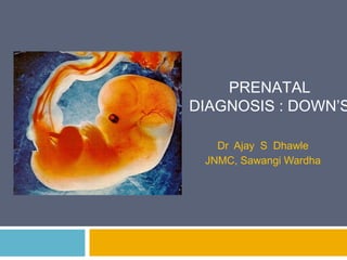
- 1. PRENATAL DIAGNOSIS : DOWN’S Dr Ajay S Dhawle JNMC, Sawangi Wardha
- 2. Screening I st Trimester II nd Trimester Diagnosis Chorionic Villus sampling Amniocentesis
- 3. Screening Every woman has a risk that her fetus/baby has a chromosomal defect The background or a priori risk depends on MA and GA The individual patient-specific risk : multiplying the a priori risk with a series of likelihood ratios, which depend on the results of a series of screening tests The likelihood ratio for a given sonographic or biochemical measurement is calculated by dividing the percentage of chromosomally abnormal fetuses by the percentage of normal fetuses with that measurement Every time a test is carried out the a priori risk is multiplied by the likelihood ratio of the test to calculate a new risk, which then becomes
- 4. The risk for trisomy 21: Increases with MA Decreases with GA (because about 30% of affected fetuses die between the 12th and 40th week of pregnancy)
- 5. In the 1970’s and 1980’s screening for trisomy 21 was based on maternal age and amniocentesis or CVS was offered to those aged 35 years or older. About 5% of pregnant women were ≥35 years and a policy of screening based on maternal age would result in: Invasive testing rate : 5% Detection rate of trisomy 21 :30%
- 6. In the last 30 years the maternal age of pregnant women has increased and now about 20% of pregnancies, including 50% of fetuses with trisomy 21, are in women aged ≥35 years
- 7. What is NT? Nuchal translucency is the sonographic appearance of subcutaneous accumulation of fluid behind the fetal neck in the first trimester of pregnancy.
- 8. When should NT be measured? GA : 11 – 13+6 weeks Fetal CRL : 45–84 mm.
- 9. Why 11 wks? CVS before this gestation is associated with transverse limb reduction defects. After 11 weeks, many major fetal defects can be diagnosed at the NT scan(eg. Anencephaly, abdominal wall defects, FB, 4 chamber cardiac view). Why 13+6 wks? Allow women the ease and safety of first trimester termination of pregnancy. incidence of abnormal accumulation of nuchal fluid in chromosomally abnormal fetuses is lower at 14–18 weeks than before 14 weeks. success rate for taking a measurement at 10–13 weeks is 98–100%, falling to 90% at 14 weeks because the fetus becomes vertical
- 10. Who should measure NT? A sonologist Accredited by the FMF (UK) Accredited by the NTQR programme (USA) Pretest and a Post test counseling MUST
- 11. How should NT be measured? Mid Sagittal Plane Neutral fetal position Proper magnification Proper placement of Calipers Adjustment of Gain Distinguishing fetal skin from the amnion Proper Reporting
- 12. Mid sagittal plane Para Sagittal view True sagittal view. Frontal process of the Maxilla in view
- 14. Fetal position Extended head Neutral position. Neutral position Hyper extended Hyperflexed Hyperextension can NT by upto 0.6mm Hyperflexion can NT by upto 0.4mm
- 15. Magnification Inadequate magnification Include only the fetal head and upper thorax in the image. The magnification should be as large as possible Adequate magnification (such that each slight movement of the callipers produces only a 0.1 mm change in the measurement).
- 16. Callipers The crossbar of the calliper should be hardly visible as it merges with the white line of the border and not in the nuchal fluid.
- 17. Amnion Both fetal skin and amnion appear as thin membranes at this gestation Wait for spontaneous fetal movement away from the amniotic membrane alternatively, Ask the mother to cough and/or by tapping the maternal abdomen the fetus is bounced off the amnion
- 18. Gain In magnifying the image, either pre or post freeze zoom, it is important to turn the gain down to avoid fuzzy edges & underestimation of NT
- 19. The NT thickness in euploid fetuses increases with fetal CRL In 75-80% of trisomy 21 fetuses the NT thickness is above the 95th centile of the normal range In trisomy 21 fetuses there is no relationship between NT thickness and maternal age Maternal age can be combined with fetal NT to provide effective first-trimester screening for chromosomal abnormalities
- 20. In a fetus with a given CRL, every NT measurement represents a likelihood ratio which is multiplied by the a priori maternal and gestational age-related risk to calculate a new risk NT : LR : Risk of Downs NT : LR : Risk of Downs The risk is greater in a woman of 20 years when the fetus has a high NT than in a woman of 40 years when the fetus has low NT
- 21. First Trimester Biochemical screening Trisomic pregnancies :altered maternal serum concentrations of various feto-placental products Screening in the second trimester by maternal age and various combinations of total or free ß-hCG, AFP, uE3 and Inhibin A can identify 56-71% of trisomy 21 pregnancies for a false positive rate of 5% Screening in the first trimester by a combination of maternal age, fetal NT, FHR and serum free ß-hCG and PAPP-A identifies about 90% of trisomy 21 pregnancies for a false positive rate of 3%
- 22. In trisomy 21 pregnancies maternal serum free ß-hCG is about twice as high and PAPP-A is reduced to about half compared to chromosomally normal pregnancies Performance of screening for trisomy 21 by maternal age and serum free ß-hCG and PAPP-A: Detection rate 65% False positive rate 5%
- 23. The measured concentration of free ß- hCG and PAPP-A is influenced by the machine and reagents used, gestational age, maternal weight, ethnicity, smoking status and method of conception In Black women the PAPP-A level is about 60% higher than in White women. Failure to take into account ethnic origin would result in substantial underestimate of the true risk of trisomy 21 in Black women In women who smoke and those conceiving by IVF serum PAPP-A is decreased and this could be misinterpreted for increased risk for
- 24. Free ß-hCG is higher than in euploid pregnancies and the difference between the two is higher at 13 than at 11 weeks Serum PAPP-A is lower than in euploid pregnancies and the difference between the two is higher at 11 than at 13 weeks The difference from euploid pregnancies in PAPP-A at 11 weeks is greater than the difference in ß-hCG at 13 weeks and therefore the overall performance of biochemical screening is better at 11 than at 13 weeks
- 25. Combined screening In trisomy 21 compared to euploid pregnancies: The difference in biochemical markers is greater at 11 than at 13 weeks The difference in fetal NT is greater at 11 than at 13 weeks Therefore the overall performance of screening is better at 11 than at 13 weeks
- 26. The overall performance of combined screening is better at 11 than at 13 weeks and may be best at 10 weeks Ultrasound scanning for fetal abnormalities is better at 12 than at 11 weeks and much better than at 10 weeks A good way of achieving a high performance of screening for trisomy 21 and diagnosing major fetal defects by ultrasound is to carry out the blood test at 10 or 11 weeks and the ultrasound scan at 12 weeks
- 27. Other defects In euploid pregnancies the average free ß-hCG is 1.0 MoM and PAPP-A is 1.0 MoM free ß-hCG PAPP-A Trisomy 21 2.0 0.5 Trisomy 18 0.2 0.2 Trisomy 13 0.3 0.4 Turner 1.2 0.5
- 28. Differences Fetal NT is higher in trisomies 18 and 13 than in trisomy 21 Serum PAPP-A is lower in trisomies 18 and 13 than in trisomy 21 Serum free ß-hCG in trisomy 21 is high whereas in trisomies 18 and 13 this is low Fetal heart rate in trisomy 13, unlike trisomies 21 and 18, is high
- 29. New Ultrasound Markers Assessment of the new Nasal bone markers improves the performance of combined screening by increasing the Facial angle detection rate and reducing the false Ductus venosus flow positive rate Chromosomal defects Examination of the new markers Major cardiac defects requires appropriate training of Fetal death sonographers and Certification of their competence in carrying out these Tricuspid flow scans Chromosomal defects The new markers can be assessed Major cardiac defects in all patients or only in the 15% of the total with an intermediate risk (1 in 51 to 1 in 1000) after combined screening
- 30. New Ultrasound Markers There are two strategies for assessment of the new markers in screening for trisomy 21 with similar detection and false positive rates: One, some, or all markers are examined in all cases The markers are examined only in the subgroup of pregnancies with an intermediate-risk (between 1 in 51 and 1 in 1000) after combined fetal NT, FHR, free ß-hCG and PAPP-A screening, which constitutes only one sixth (15%) of the total population
- 31. Reporting NEVER as an isolated measurement, NEVER without images Always mentions ‘a priory’ risk and ‘estimated ‘risk MOMs for the Free Sr B HCG & PAPP-A : MUST in the report
- 33. Increased NT+ Normal Karyotype: what next? Cardiac defects Neural tube defects Diaphragmatic hernia Skeletal dysplasias Genetic syndromes Higher risk of stillbirth Midtrimester ANOMALY scan + Fetal ECHO Does NOT warrant termination, still a good chance of having a healthy baby
- 34. Thank you
Editor's Notes
- Irrespective of whether it is septated and whether it is confined to the neck or envelopes the whole fetus. The incidence of chromosomal and other abnormalities is related to the size , rather than the appearance of NT.
- The maximum thickness of the subcutaneous translucency should be measured. More than one measurement must be taken and the maximum one should be recorded
- If Used in conjunction with FT biochemical screen, always check if the MOMs for the Free Sr B HCG & PAPP-A have been reported and appropriate software used.