Hi,Fi Call Girl In Mysore Road - 7001305949 | 24x7 Service Available Near Me
Connective.pptx
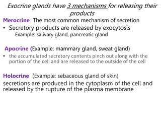
1. Exocrine glands have 3 mechanisms for releasing their
products
Merocrine The most common mechanism of secretion
• Secretory products are released by exocytosis
Example: salivary gland, pancreatic gland
Apocrine (Example: mammary gland, sweat gland)
• the accumulated secretory contents pinch out along with the
portion of the cell and are released to the outside of the cell
Holocrine (Example: sebaceous gland of skin)
secretions are produced in the cytoplasm of the cell and
released by the rupture of the plasma membrane
2.
3. Connective tissue
• Connective tissues are one of the most abundant and
widely distributed tissues in the body.
• They consist of two basic elements:
Cells
Extracellular matrix contain
Protein fiber and
Ground substance
4. • Consists of cells that are typically widely separated by lots of extracellular
material – referred to as the extracellular matrix
• Most cells are not in contact with each other but are distributed
throughout the extracellular matrix
Connective Tissue (CT)
5. Function
• Structural Support
• Protection
• Repair and Healing:
• Storage:
• Defense: WBC in blood and immune cells in
lymphoid tissue, play a role in defending the body
against infections.
• Transport
6. Common Characteristics
Cells are separated by a considerable amount
of extracellular matrix;
Do not occur on free body surface
Have blood and nerve supply except for
cartilage
Have common origin; arise from
mesenchyme
6
7. Epithelial Vs Connective Tissues
Epithelial
• Many cells are tightly
packed together
• Little or no ECM
• Has no blood vessels
• Form surface layers
• Rarely covered by another
tissue
Connective
• A few cells are usually widely
scattered.
• Large amount of ECM
• Has significant networks of
blood vessels.
• Forms underlying layers
• Usually covered by another
tissue
8. • The extra cellular matrix consists of two major components:
the ground substance and the fibers
Fibers
• Formed from polymerize proteins after secretion from
fibroblast
• Embedded in the extracellular matrix between the cells:
cells:
• Strengthen and support connective tissues.
• There are three types of fibers.
• Three types:
a) Collagen fibers
b) Elastic fibers
c) Reticular fibers
9. 1. Collagen fibers
• They are found in most types of connective tissues
• They are very strong and resist pulling forces (tension),
but they are not stiff, which allows tissue flexibility.
• Found in bone, cartilage, tendons and ligaments.
• Chemically, collagen fibers consist of the protein
collagen.
• There are 28 family of collagen protein exist in
vertebrates
11. Elastic Fibers
• Made primarily of a
protein called elastin,
whose coiled structure
allows it to stretch and
snap back like a rubber
band.
• Account for the ability of
the lungs, arteries, and
skin to spring back after
they are stretched.
In this slide, “A” is an elastic fiber
– “B” is collagenous Fiber
12. 3. Reticular fibers
• Are consisting of collagen
type III arranged in fine
bundles.
• They are produced by
fibroblasts, reticular fibers
are much thinner than
collagen fibers and form
branching networks.
• Found in spleen, lymph
nodes and basement
membrane
–Stained black in the
adjacent micrograph of the
liver.
13. Ground substance
• Amorphous gel-like substance in the extracellular
space
• Highly hydrated, transparent, complex mixture of
macro-molecules, principally of three classes
Glycosaminoglycans (GAGs)
Hyaluronic acid: largest & most ubiquitous
GAG
Proteoglycans
Multi-adhesive Glycoproteins.
• Fills the spaces between the cells and fibers that may
be fluid, semifluid, or calcified.
14. • Because it is viscous, acts as both a lubricant and a
barrier to penetration to invaders
• It supports cells, bind them together,
• Store water
• Provide a medium for exchange of substance between
blood & cells.
Ground substance…
15. Connective tissue cells
• The type of cells in connective tissue vary according to the
type of tissue and includes the following
1. Fibroblast
• Large and flat cells that present in all the general connective
tissues
• Are most numerous
• Secrete fibers (collagen, and elastin fibers) & certain components of
the ground substance of the ECM (glycosaminoglycans, proteoglycans)
• Involved in wound healing
• Seldom undergo mitosis unless in wound healing
17. 2. Macrophages
• They are developed from monocytes and type of white
white blood cell.
• They have an irregular shape with short branching
projections and are capable of engulfing bacteria and
cellular debris by phagocytosis.
• They have the ability to move throughout the tissue
and gather at sites of infection or inflammation.
• Have given different names in different organs
Kupffer cells in Liver, Microglial cells in CNS
Langerhans cells in Skin, Osteoclast in Bone
18. 3. Plasma cells
• They are type of white blood cell and developed from
B lymphocyte.
• They are small cells that secrete antibodies, proteins
that attack or neutralize foreign substances in the
body.
• An important part of the body’s immune response
19. 4. Adipocytes/Fat cells
• They are connective tissue cells that store triglycerides (fat)
• They are found deep to the skin and around organs such as the
heart and kidneys
• Serve to cushion & insulate the skin & other organs
20. 5. Mast cells
• Oval or irregularly shaped cells
• They are abundant alongside the blood vessels that
supply connective tissue.
• They produce histamine, Heparin, Serine protease,
cytokine, & phospholipid, a chemical that mediate
inflammatory response, immune and tissue repair.
• Release of certain chemical mediators stored in mast cells
also promotes the allergic reaction (immediate
hypersensitivity reaction)
21. 6. Leukocytes (white blood cells)
• Make up a population of wandering cells in
connective tissue
• They are not found in significant numbers in normal
connective tissues.
• However, in response to certain conditions they
migrate from blood into connective tissues
(diapedesis)
• This process increase greatly during inflammation
22. Types of Connective Tissue
22
• The difference in composition and amount of cells, fibers
and ground substance are responsible for the structural
and functional diversity of connective tissue.
• Connective tissue is classified broadly into
Embryonic connective tissue
Adult connective tissue
23. Embryonic CT
A. Mesenchymal CT
• Consists of irregularly shaped mesenchymal cells
embedded in a semifluid ground substance that
contains reticular fibers.
• Found under skin and developing bones of embryo,
in blood vessels of adult.
• Forms all other types of connective tissue.
24.
25. Mucous CT
• Widely scattered fibroblasts embedded in viscous,
jelly-like ground substance that contain fine collagen
fibers
• Found in umbilical cord or Wharton's jelly and fetal
tissues.
26. Classification of Adult Connective Tissues
A. Loose connective tissue
1. Areolar connective tissue
2. Adipose tissue
3. Reticular connective tissue
B. Dense connective tissue
1. Dense regular connective
tissue
2. Dense irregular connective
tissue
C. Cartilage
1. Hyaline cartilage
2. Fibro-cartilage
3. Elastic cartilage
D. Bone tissue
E. Liquid connective
tissue
1. Blood tissue
2. Lymph
Specialized connective tissue
Connective tissue Proper
27. Types of Adult Connective Tissue
• 2 types based on the relative abundance of
fibers
1. Loose Connective Tissue
• Lots of ground substance and cells. Fewer fibers.
• Leaves lots of empty space in tissue sections.
Eg,. Adipose, Areolar & Reticular
28. Types of loose CT
areolar connective tissue
• Consists of fibers (collagen, elastic, and reticular) and
several kinds of cells (fibroblasts, macrophages,
plasma cells, adipocytes, and mast cells) embedded in
a semifluid ground substance.
• Found in subcutaneous and dermis of skin; lamina
propria of mucous membranes; and around blood
vessels, nerves, and body organs.
• Provide Strength, elasticity, and support.
29. Adipose CT
• It contains mainly
adipocyte cells
Locations:
– Subcutaneous fat beneath skin
– Breast
– Heart surface
– Cushioning organs
• Kidneys and Eyes
Functions:
– Energy storage
– Thermal insulation
– Protective cushioning for some
organs
Loose Connective Tissue
32. 2. Dense Connective Tissue:
• Tissue has densely packed collagen fibers
• provides strength and resistance to stretching.
• Fibers occupy the most space.
• Much lower number of cells and less ground substance.
• Appears closely packed in tissue sections.
Types: Dense regular and Dense irregular
34. Dense Regular Connective Tissue
Locations:
– Tendons
– Ligaments
NOTE the waviness of the fibers.
Functions:
– Ligaments bind
bone tightly to
other bones
Resist stress
– Tendons attach
skeletal muscles to
bone
Transfer muscular
tension to bones
35. Dense Irregular CT
Microscopic Appearance:
– Densely packed, collagenous fibers running in random
directions.
– Scanty open space (ground substance)
and few visible cells
– Scarcity of blood vessels
Locations:
– Deeper portion of dermis of skin
– Capsules around visceral organs
such as the liver, spleen, and
kidneys
– Fibrous sheaths around cartilages
and bones
Functions:
– Provides a durable, hard to tear
structure that can withstand
stresses placed in unpredictable
36. Cartilage
• Cartilage is a specialized form of (Supportive) connective
tissue with
Cells chondrocytes
Extracellular matrix composed of
Fibers and ground substance
• Chondrocytes synthesize and secrete the ECM and the cells
themselves are located in matrix cavities called lacunae.
• Cartilage is avascular and is nourished by the diffusion of
nutrients
From capillaries in adjacent connective tissue
(perichondrium) or from synovial fluid in joint cavities
• There are three forms of cartilage,
• Each exhibiting variation in matrix composition.
37. cartilage…
1. hyaline cartilage
• Is most abundant type of cartilage.
• Consists of a bluish-white, shiny ground substance
with thin, fine collagen fibers and many chondrocytes;
• Type II collagen is the principal collagen type found in
the matrix.
Eg. at limb joints, ribs, nose, larynx & trachea
• Provides smooth surfaces for movement at joints, as
well as flexibility and support.
38. 2. Elasticcartilage
• Consists of chondrocytes located in a threadlike
network of elastic fibers within the extracellular
matrix.
• The more pliable and distensible possesses
• In addition to collagen type II, an abundance of elastic
fibers within its matrix.
Eg. external ear, epiglottis,
eustachian tube
• Gives support and maintains shape.
39. 2. Fibrocartilage
• Consists of chondrocytes scattered among thick bundles of
collagen fibers within the extracellular matrix.
• Present in regions of the body subjected to pulling forces,
• Characterized by a matrix containing a dense network of
coarse type I collagen fibers.
Eg. pubic symphysis, meniscus, annulus fibrosus of IVD
• Provide support and fusion.
40.
41. Specialized connective tissue
• Bone (Osseous Tissue)
• Blood Tissue
• Blood and Bone tissue will be cover in
circulatory and skeletal system
41