8. The Semicircular Canals
• posterior canal
shares plane with
contralateral
anterior canal.
• horizontal canals
share plane.
9. Stimulated by Angular Acceleration
• greatest when fulcrum
is within head
• induces relative motion
of endolymph
• crista is displaced by
fluid motion
10.
11. Responses of the Cristae
• All kinocilia are
oriented in the
same direction
• Crista in each pair
of canals respond
inversely to each
other
12. The
Otolithic
Organs
Saccule: roughly
vertical orientation,
responds to acceleration components within saggital
plane
Utricle: horizontal (+ 30 deg.) orientation
15. Vestibular Portion of C.N. VIII
superior division: utricle, anterior part of
saccule, and horiz & anterior canals
inferior division: posterior part of saccule,
and posterior canal
• to vestibular nuclei
• to cerebellum
16. Vestibulocochlear Nerve
• Exits temporal bone
near its medial edge.
• Enters lateral face of
brainstem at the level
of the lower pons.
• Synapsing in
(Cochlear and)
Vestibular Nuclei
17. Responses of Vestibular Neurons:
• To changes in acceleration, but onset
and fade slowly
• For most normal head movements
firing rates are in phase with head
VELOCITY.
20. From the Vestibular Nuclei:
• Vestibulo-Oculomotor Pathways:
– Direct: to oculomotor nuclei.
– Indirect: via reticular formation to
oculomotor nuclei (III IV and VI)
• Vestibulo-Spinal Pathways:
– Lateral V-S-throughout spinal cord
– Medial V-S-cervical & thoracic
– Reticulospinal tract-via brainstem reticular
formation
21. Median Longitudinal Fasciculus
• A tract linking Vest. Nuclei to
nuclei of CN III, IV, & VI;
• Supports conjugate eye
movement during movement of
the head.
• Continuous with the medial
vestibulospinal tract.
• The mlf runs near midline
ventral to ventricle IV and the
periaqueductal gray matter of
the midbrain
22.
23.
24. In the brainstem
• Vestibular inputs undergo integration
• Integrated signal is combined with
original (velocity driven) signal
• Processing to reset spatial map for eye
musculature
32. Eye Movements
• Saccades—rapid shift in gaze
• Pursuit—stabilize image of moving object
• Fixation—stabilize image of still object
• VOR—stabilize image during head motion
• OKN—backup for when VOR decays to
cont’d head rotation
• Vergent movements—change depth of focus
42. SENSORY ORGANIZATION
Determination of Body
Position
Compare, Select
& Combine Senses
Visual Vestibular Somatosensor
y
Processing of inputs from the periphery
Selection based on
Availability Accuracy
Value for the task at hand
The Balance System depends on a complex set of interactions between a variety of systems: Inner ears Brainstem and cerebellum Eyes Spinal cord Postural Muscles Cortices
Within the temporal bone lies the bony labyrinth a maze of tunnels lined with very dense and highly calcified bone. These we know as the cochlea, vestibule, and semicircular canals. Superior/Anterior Posterior Lateral/Horizontal These approximate (although not eactly) right angles to each other. This will allow for the analysis of curvilinear motion in terms of its components within each of these planes.
Within the bony labyrinth are spaces defined by soft tissue: the so-called membranous labyrinth. These divide the labyrinth into perilymphatic spaces and endolymphatic spaces. Just as in the cochlea, the endorgans for the balance half of the inner ear are contained within the endolymphatic spaces. Semicircular canals form partial circles whose diameter (if completed) would be on the order of 6.5 mm. The interior diameter of the endolymphatic tubes in the semicircular canals is up to 0.4 mm. Each of these canals has one swollen or ampulated end, which contains the endorgans. The posterior and superior canals share their non-ampulated ends in what is called the common crus.
Another view-- membranous labyrinth. These divide the labyrinth into perilymphatic spaces and endolymphatic spaces. Here we have labelled the utricle and saccule, the remaining endorgans for your sense of head motion. These endorgans are found in: the Ampullae--swollen end of each semicirc the Utricle the Saccule Also visible here is the endolymphatic sac, the subject of some surgical procedures.
Within each of the endorgans, the sensory transducers are hair cells just like we found in the cochlea. And they still come in two varieties, only here there is no “inner” or “outer” placement for reference. So they are labelled as Type and Type II. And they have a large cilium in addition to the sterecilia. This is known as the kinocilium. What breaks off to leave the basal body in the cochlea.
Here they are (in an example from the otolithic organ).
The sensory organ in both the saccule and utricle is the maccula. hair cells sit with their cilia embedded in a gelatinous membrane containing otoliths/otoconia/ stratoconia (calcium carbonate crystals). There is a curved stripe within each maccula that contains only type I (read "inner") hair cells, called the striola. The kinocilia on all of the hair cells are oriented toward the striola in the utricle and away from the striola in the saccule .
To Review: The vestibular system consists of the two otolithic organs, the utricle and the saccule (which are located in the vestibule, or central portion of the labyrinth), and the semicircular canals which run off of the vestibule. superior (anterior vertical) posterior (posterior vertical) horizontal (lateral) The vertical canals share a common opening to the vestibule, the common crus, at their non-ampullated ends. Their ampullar ends as well as both ends of the horizontal canal also open into the vestibule. The Saccule: roughly vertical orientation, responding to acceleration components within a saggital plane. The Utricle: horizontal (+ 30 degrees) orientation. The macculae are primarily sensitive to linear acceleration and gravity.
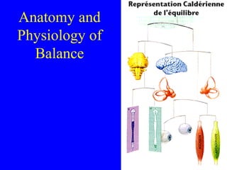
This chart represents the basic organization of the vestibular system. We see that the semicirc’s are primarily responding to head rotation or angular acceleration. The Saccule and Utricle are responsive to Linear acceleration and the pull of gravity. Together they form one of the three primary inputs to the central nervous system components dedicated to balance: visual// proprioception & tactile To foreshadow, the outputs of this system include: the occulomotor muscles, the spinal cord and cerebellum, and the forebrain.
This chart represents the basic organization of the vestibular system. We see that the semicirc’s are primarily responding to head rotation or angular acceleration. The Saccule and Utricle are responsive to Linear acceleration and the pull of gravity. Together they form one of the three primary inputs to the central nervous system components dedicated to balance: visual// proprioception & tactile To foreshadow, the outputs of this system include: the occulomotor muscles, the spinal cord and cerebellum, and the forebrain.
Smooth Pursuit: Slower, for keeping target on the fovea when there is relative movement. Controlled by the occipital cortex and are based on inputs from the retina via visual cortex. Saccadic : Fast, up to 70 degrees per .1 sec, ballistic movements which can come under voluntary control with input from the pre-frontal cortex. The pontine gaze center (within reticular formation) is involved with the initiation of these movements in the horizontal direction. VOR : to move eyes relative to head movements. Mediated via connections from the vestibular system. Accommodating Reflex : to keep target that is moving closer or farther away in focus. Mediated through the occipital cortex.
This chart represents the basic organization of the vestibular system. We see that the semicirc’s are primarily responding to head rotation or angular acceleration. The Saccule and Utricle are responsive to Linear acceleration and the pull of gravity. Together they form one of the three primary inputs to the central nervous system components dedicated to balance: visual// proprioception & tactile To foreshadow, the outputs of this system include: the occulomotor muscles, the spinal cord and cerebellum, and the forebrain.