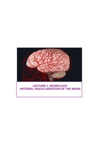
2. The vascularization of the brain is provided by 4 vertically stretched
arteries, directly or indirectly from the aortic arch, the internal carotid
arteries and the vertebral arteries.
These arteries anastomose to form an arterial circle at the base of the
brain, Willis polygon. The latter is at the origin of the cerebral arteries.
▪ The anterior circuit is supplied by the carotid system, the
internal carotid arteries supplied most of the brain;
▪ The posterior circuit supplied by the vertebro-basilar
system, which contributes to the posterior blood
supply (posterior circulation) of the brain and spinal cord.
3. The carotid system:
Anterior circulation
The anterior circulation involves all the arteries that originate from the internal
carotid arteries. It is responsible for the blood supply of the anterior and
middle aspect of the brain. The arteries of this anterior circuit are:
•The internal carotid arteries
•The anterior cerebral arteries
•The anterior communicating artery
•The middle cerebral arteries
Each of these arteries has:
- a basal segment that gives deep branches destined for the deep
territories of the brain;
- a cortical segment that gives superficial branches vascularizing the
cortical territories.
The internal carotid artery is one of two branches of the common carotid
artery. It is responsible for supplying a large portion of the anterior and middle
parts of the brain
4. The internal carotid artery
The internal carotid artery is one of two branches of the common carotid
artery. It is responsible for supplying a large portion of the anterior and
middle parts of the brain.
5. The anterior cerebral artery:
The anterior cerebral artery (ACA) is a much smaller branch of the internal
carotid artery (when compared to the middle cerebral artery).
It begins at the terminal portion of the internal carotid artery (after the
ophthalmic branch is given off) on the medial part of the Sylvian fissure.
It travels in an anteromedial course, superior to the optic nerve (CN II) towards
the longitudinal cerebral fissure. Here it anastomoses with the contralateral
counterpart via the short anterior communicating artery (AComm).
The paired arteries then travel through the longitudinal cerebral fissure along
the genu of the corpus callosum.
6. The anterior cerebral artery also gives off central and cortical branches. Central
branches arise from the anterior communicating artery (AComm). to perfuse the optic
chiasma, lamina terminalis, hypothalamus, para-olfactory areas, cingulate gyrus, and
anterior columns of the fornix.
Anterior cerebral artery
The cortical branches are named for the regions they supply.
They are responsible for the somatosensory and motor cortices of
the lower limbs.
7. The middle cerebral arteries or sylvian cerebral artery (MCA), is the
largest terminal branch of the internal carotid artery. It travels through the
Sylvian (lateral) fissure before coursing in a posterosuperior direction on
the island of Reil (insula). It subsequently divides to supply the lateral
cortical surfaces along with the insula.
• ascending cortical branches, for the lateral surface of the frontal and
parietal lobes; descending cortical branches for the temporal lobe and
deep branches, lateral striated arteries.
The cortical branches include the frontal, orbital, parietal, and temporal
branches:
•The frontal arteries perfuse the inferior frontal, middle, and precentral gyri.
•The lateral orbital parts of the frontal lobe, as well as the frontal gyrus, are
supplied by the orbital branches.
•The inferior parietal lobe, the inferior part of the superior parietal lobe, and the
postcentral gyrus receive blood from the parietal branch.
•Several temporal arteries then go on to perfuse the lateral aspect of
the temporal lobe.
8. Posterior circulation
The posterior circulation refers to all the blood vessels that arise from
the vertebrobasilar system. These blood vessels supply the hindbrain and
the occipital lobe of the cerebrum. The vessels of the posterior circuit include:
•The vertebral arteries
•The basilar artery and its branches
•The posterior cerebral arteries
•And the posterior communicating arteries
9. The vertebral arteries
The vertebral arteries gain access to the cranial vault via the foramen
magnum anterolateral to the brainstem. Concerning the branches, each vertebral
artery:
• Gives off a posterior inferior cerebellar artery
• Contributes to the formation of the anterior spinal artery via tributaries that
converge in the midline anterior to the medulla oblongata
• Contributes meningeal branches near the foramen magnum that supplies the falx
cerebelli and the surrounding bone
• May give off the posterior spinal artery; although this vessel usually arises from the
posterior inferior cerebellar artery
• Gives off medullary arteries that perfuse the medulla oblongata
The vertebral arteries unite in the midline at the pontomedullary junction to form
the basilar artery.
11. The basilar artery
The basilar artery is an important vessel found in the pontine cistern. It is posterior to
the clivus and anterior to the pons, as it ascends in the basilar groove. Its branches are
responsible for supplying the pons, cerebellum, internal ear, and other nearby
structures. There are three major branches of the basilar artery:
•Anterior inferior cerebellar
•Superior cerebellar
•Internal auditory (Labyrinthine)
There are also smaller pontine and posteromedial (paramedian) arteries that arise
from the lateral surface and distal bifurcation of the artery, respectively. The basilar
artery ends by dividing into two posterior cerebral arteries.
These vessels unite with the posterior communicating arteries to complete the circle
of Willis, posteriorly.
13. The posterior cerebral arteries (PCA)
The posterior cerebral arteries (PCA) are terminal branches arising from the
bifurcation of the basilar artery.
The division takes place behind the dorsum sellae. It is separated from the superior
cerebellar artery by the oculomotor nerve (CN III).
The artery continues in a course lateral to the midbrain (adjacent to the trochlear
nerve, CN IV). It gives off the posterior communicating artery, which completes the
circle of Willis.
The vessel then continues to course around the cerebral peduncles toward the
tentorial aspect of the cerebrum. Here, it supplies the occipital and temporal lobes.
14. The branches of the posterior cerebral artery
The branches of the posterior cerebral artery bring oxygenated
blood to the following areas:
• Anterior thalamus and subthalamus
• Lateral wall of the third ventricle and inferior horn of
the lateral ventricle
• Choroid plexus of third and lateral ventricles
• Globus pallidus
• Lateral and medial geniculate bodies
15. The posterior communicating artery (PComm)
The posterior communicating artery (PComm) is a long, slender vessel originating
from the posterior cerebral artery.
It is much longer than its anterior counterpart - the anterior communicating artery.
The vessel is medial to the uncus of the temporal lobe and lateral to the mammillary
bodies of the hypothalamus. The distal part of the vessel may overlap the proximal
part of the optic tract.
The posterior communicating artery completes the circle of Willis posteriorly.
Additionally, it gives tributaries to the optic tract, cerebral peduncles, internal
capsule, and thalamus.
16. Internal carotid arteries
Anterior cerebral arteries
Anterior communicating arteries
Middle cerebral arteries
Anterior circulation
Branch of the common carotid artery
Cincinnati classification and Newer 4 part classification
Mnemonic (excludes C1): Please Let Children Consume Our Candy
Carotid artery
Posterior cerebral arteries
Posterior communicating arteries
Vertebral arteries
Basilar artery
Posterior circuit
Branches: posterior inferior cerebellar artery (PICA), anterior and posterior
spinal, meningeal and medullary arteries
Vertebral arteries
Branches: Anterior inferior cerebellar, Superior cerebellar, Internal auditory
(Labyrinthine). Becomes the posterior cerebral artery
Basilar arteries
Union of anterior and posterior circulation
In the subarachnoid space, in the interpeduncular cistern
Surrounds optic chiasm and infundibulum
Circle of Willis