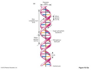
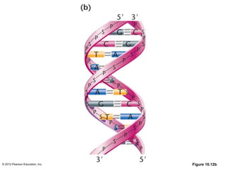
The document summarizes key evidence supporting DNA as the genetic material:
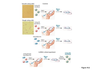
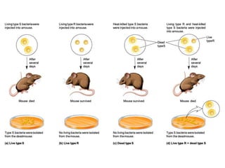
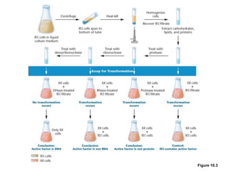
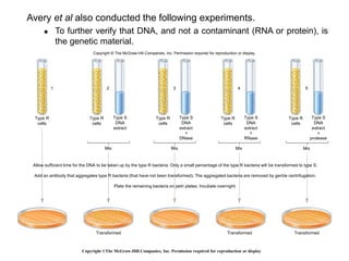
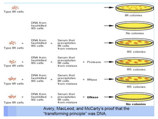
1. Avery, MacLeod and McCarty showed that purified DNA from one type of bacteria could transform another type of bacteria, but purified proteins and RNA could not.
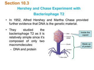
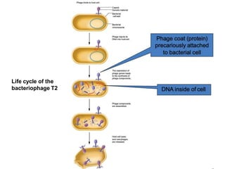
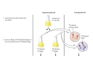
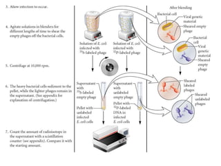
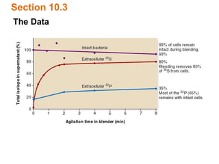
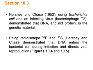
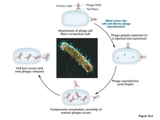
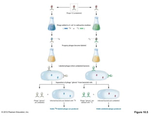
2. Hershey and Chase demonstrated through radioactive labeling that viral DNA, not proteins, enters host bacteria during viral infection.
3. Recombinant DNA technology provides direct evidence that isolated eukaryotic DNA sequences can function when inserted into bacteria.