The document describes the hERG assay, which is used to test for potential drug-induced prolongation of the QT interval. It discusses the hERG gene and potassium channel, how mutations can cause long QT syndrome. It then summarizes three methods for conducting the hERG assay: electrophysiological assay using whole-cell patch clamping, Fluorometric imaging plate reader-based thallium flux assay, and radioligand binding with 35S-MK-499. Details are provided on cell preparation and protocol for each type of hERG assay.

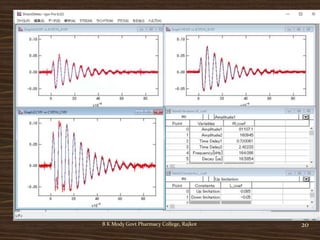
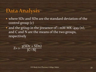
![ For determining compound 50% inhibition
concentration (IC50) values in the QPatch assay,
averaged percent control values were plotted as a
function of compound concentration and fitted using
a four parameter logistic equation with the minimum
fixed at zero and the maximum fixed to 100% of
control.
The inhibitor concentration against the percent
activity is plotted ([I]-Activity % graph). Using the
linear (y=mx+n) or parabolic (y=ax2+bx+c) equation
on this graph for y=50 value x point becomes IC50
value.
19B K Mody Govt Pharmacy College, Rajkot](https://image.slidesharecdn.com/hergassaynew-201215133919/85/hERG-Assay-19-320.jpg)
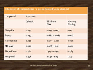
![B K Mody Govt Pharmacy College, Rajkot 34
The interaction of 35S-MK-499 with membranes prepared from
HEK293 cells expressing hERG
The 35S-MK-499 tracer was prepared at 50 pM in assay buffer
containing (in mM): 70 NaCl, 60 KCl, 1 CaCl2, 2 MgCl2 and 10
HEPES-NaOH (pH 7.4)
0.25 mL of the diluted tracer was dispensed into a 2 mL, 96-square-
well polypropylene deep-well block.
Nonspecific binding was defined in the presence of 1 mM unlabeled
MK-499
MK-499 [(+)-N-[1'-(6-cyano-1, 2, 3, 4-tetrahydro-2(R)-naphthalenyl)-3,
4-dihydro-4(R)-hydroxyspiro(2H-1-benzopyran-2, 4'-piperidin)-6-yl]
methanesulfonamide]](https://image.slidesharecdn.com/hergassaynew-201215133919/85/hERG-Assay-34-320.jpg)