Spines, plasticity, and cognition in alzheimer model mice.
poster Brussels final
1. HIGHLY DIFFERENTIATED IN VITRO PRIMARY MUSCLE FIBER:
A NOVEL MODEL RECAPITULATING PATHOLOGIC FEATURES OF G93A SOD1 MOUSE
Background
Amyotrophic lateral sclerosis (ALS) is a highly debilitating
fatal disease in humans with life expectancy ranging from
two to five years after diagnosis. This pathology is
characterized by weakness, muscle atrophy and spasticity
leading to death in patients mostly due to respiratory failure
caused by impaired diaphragm contraction. Muscle atrophy
is believed to be triggered by the loss of upper motor
neurons (UMNs) in the cortex and of lower motor neurons
(LMNs) in brainstem and spinal cord1. Genetic defects in the
zinc/copper superoxide dismutase (SOD1) gene, has been
linked to familial and sporadic ALS. While this model has
been extensively used to study loss of MNs and
neuromuscular junctions, less attention has been given to
the potential role of impaired muscle development.
Objective
Investigate the possible pathophysiological link between
muscle development and neuromuscular junction loss in the
SOD1G93A mice by :
i. setting up an in-vitro model that could recapitulate the
defects in muscle observed in ALS.
ii. investigating the dynamics of fiber differentiation and
neuromuscular features in G93ASOD1 myofiber
cultures.
Discussion & Conclusions
These preliminary data secure this protocol as a robust model to
study highly differentiated fibers recapitulating muscle development
in SOD1G93A mice.
Our findings from G93ASOD1 mice revealed that muscle
development and features of the post-synaptic apparatus are
impaired in fibers taken from young mice which are still
asymptomatic.
Our data supports the hypothesis that muscle development could
be an independent trigger of neuromuscular junction loss and muscle
atrophy.
This novel model could be used as a new tool to study time-based
development of neuromuscular structures in G93ASOD1 mice and to
test new therapeutic compounds for ALS.
VILMONT Valérie 1, CADOT Bruno 1, GOMES Rodrigues Edgar 1,2
1Myology Research Center, Sorbonne Universités, UPMC Université Paris, Paris, France, 2Instituto de Medicina Molecular, Lisbon, Portugal
Email address for correspondence: vilmont@myologygroup.net
Keywords: muscle, in-vitro differentiation, G93ASOD1
Model Set up
• Primary myoblasts were isolated from P7 transgenic pups
expressing the G93A mutant form of human SOD1 and from WT
littermates.
• Myoblasts proliferated to 70-80% confluence and were directed
to differentiate to advanced stages (Day 9-10) on Matrigel
support2.
• Features of high differentiation, such as:
triad formation (marked with DPHR) ,
movement of nuclei to the periphery ,
fiber thickness
were assessed at terminal differentiation (day 9).
• Three important post-synaptic features were also analyzed to
give an indication of the neuromuscular junction maintenance:
AchR (Acetylcholine receptor) clusters
MuSK/ phosphorylated MuSK
Rapsyn clusters
• Isolated fibers from symptomatic SOD1G93A mice as well as
from WT littermate were analyzed for the above post-synaptic
features to check for correlation between in-vitro differentiated
and in-vivo model.
In vitro DIFFERENTIATED FIBERS
Day 6 Day 9
Figure 1. A highly differentiated fiber showing peripheral nuclei and triads marked with DHPR
P7pups
Day 0
-Switch to
differentiation
medium + agrin
-Addition of
Matrigel
Myoblasts
Proliferation Differentiation
DHPR
References
1Wim Robberecht & Thomas Philips, Nat Rev Neuro, 2013
2Falcone S et al., EMBO Mol Med, 2014
3http://neuromuscular.wustl.edu/synmg.html
Acknowledgements
The authors are grateful to the following organizations for funding
Figure 2. AchR, MuSK and rapsyn localization at the
post-synaptic membrane upon agrin-induced AchR
clustering 3. Formation of this complex is important for the
maintenance of the post-synaptic structure and thereafter of
the neuromuscular junction.
Figure 7a. Comparison between SOD1 WT and SOD1G93A single fibers. In
SOD1G93A fibers, expression of Rapsyn, and MuSK is drastically reduced and the
pretzel structure of the AchR cluster is fragmented.
SYMPTOMATIC
Figure 3. Method for single fiber isolation. Symptomatic G93ASOD1 mouse showing
severe kyphosis and muscle atrophy. EDL muscles are sampled and flushed to obtain
single fibers. The latter are fixed and stained.
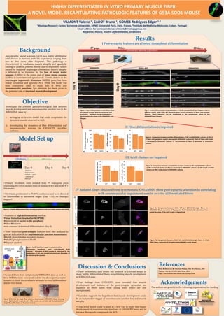
Results
I Post-synaptic features are affected throughout differentiation
G93ASOD1 MOUSE LIFESPAN
ASYMPTOMATIC
120daysP7 pups
SYMPTOMATIC
30days
Figure 4. Day 6 differentiated in-vitro fibers show
normal post-synaptic features expression and
localization. The fibers can be assimilated to
the asymptomatic phase of the G93ASOD1 mice
lifespan.
Day 9 in-vitro differentiated show expression of MuSK, phosphoMuSK and Rapsyn is lost to
different extent, while SOD1 WT littermate show normal expression of these post-synaptic
features. These alteration can be assimilated to the symptomatic phase of the
G93ASOD1mice lifespan.
G93ASOD1
Day6
G93ASOD1SOD1 WT
Day9
IV Isolated fibers obtained from symptomatic G93ASOD1 show post-synaptic alteration in correlating
with neuromuscular impairment seen in in-vitro differentiated fibers
a)
b)
Figure 7b. Comparison between SOD1 WT and G93ASOD1single fibers. In G93A
SOD1 fibers, expression of phosphorylayted MuSK is nearly absent.
III AchR clusters are impaired
Figure 6. Comparison between acetylcholine receptor clusters in WT and G93ASOD1 cultures.
a) The number of cluster per fiber is decreased in G93ASOD1 cultures. b) The length of AChR
clusters per fiber is decreased in G93ASOD1 cultures.
II Fiber differentiation is impaired
0
20
40
60
80
100
WT SOD1
%fiberswithperipheral
nuclei
0
10
20
30
40
50
WT SOD1
%fiberswithtriads
0
2
4
6
8
10
WT SOD1
Thickness(um)
Figure 5. Comparison between myofiber differentiation of WT and G93ASOD1 cultures. a) The %
of fibers with peripheral nuclei is decreased in G93ASOD1 cultures. b) The % of fibers with triads
is decreased in G93ASOD1 cultures. c) The thickness of fibers is decreased in G93ASOD1
cultures.
a) b) c)
0
0.2
0.4
0.6
0.8
1
1.2
1.4
WT SOD1
Numberofclusterperfiber
a)a)
0
5
10
15
20
25
30
WT SOD1
Lengthofclusterperfiber
b)