2. Get a modern PowerPoint Presentation that is beautifully designed. I
hope and I believe that this Template will your Time.
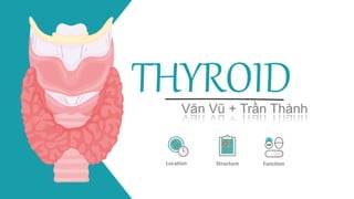
Location and Structure
01
Get a modern PowerPoint Presentation that is beautifully designed. I
hope and I believe that this Template will your Time.
Function
02
Table of Contents
4. The thyroid gland is located in the anterior neck and
spans the C5-T1 vertebrae.
Anatomical Location
It consists of two lobes (left and right), which are
connected by a central isthmus anteriorly – this produces
a butterfly-shape appearance.
The lobes of the thyroid gland are wrapped around the
cricoid cartilage and superior rings of the trachea.
The gland is located within the visceral compartment of
the neck (along with the
trachea, oesophagus and pharynx). This compartment is
bound by the pretracheal fascia.
Location and Structure
THYROID
5. Anteriorly – infrahyoid muscles, namely the
sternothyroid, superior belly of the omohyoid and
sternohyoid
Anatomical Relations
Laterally – carotid sheath, containing the common
carotid artey, internal jugular vein and vagus nerve
Medially –
Organs: larynx, pharynx, trachea and oesophagus
Nerves: external laryngeal and recurrent laryngeal
Location and Structure
THYROID
6. Superior thyroid artery – arises as the first branch of
the external carotid artery. It lies in close proximity to the
external branch of the superior laryngeal nerve
(innervates the larynx).
Arterial Supply
Inferior thyroid artery – arises from the thyrocervical
trunk (a branch of the subclavian artery). It lies in close
proximity to the recurrent laryngeal nerve (innervates the
larynx).
Thyroid ima artery : In a small proportion of people
(around 10%) there is an additional artery present. It
arises from the brachiocephalic trunk and supplies the
anterior surface and isthmus of the thyroid gland.
Location and Structure
THYROID
7. The superior and middle thyroid veins drain into the
internal jugular vein
Venous Drainage
The inferior thyroid vein empties into the
brachiocephalic vein
Location and Structure
THYROID
11. Basal Metabolic Rate
Glycogenolysis
Protein synthesis
Thermogenesis
THYROID
Metabolic processes increased
by thyroid hormones
This is achieved in a number of ways, such as:
1. Increasing the size and number of
mitochondria within cells
2. Increasing Na-K pump activity
3. Increasing the presence of β-adrenergic
receptors in tissues such as cardiac muscle.
Funtion of Thyroid
Gluconeogenesis
Lipogenesis
13. THANKS!
Does anyone have any questions?
Please keep this slide for
attribution
THYROID
BOYS GIRLS
s l i d e s p p t . n e t
Editor's Notes
Medially, the gland is related with larynx and trachea and is fixed to the cricoid cartilage, along with the first two tracheal rings, by the suspensory ligament of Berry. The cricothyroid muscles and the inferior constrictors of the pharynx are the medial muscular relations. The external laryngeal nerve passes by the gland along this border as well. Both the recurrent laryngeal nerve and the trachea are posteroinferiorly related to the medial border of the thyroid gland
Venous drainage is carried by the superior, middle, and inferior thyroid veins, which form a venous plexus around the thyroid gland.
The first of the three vessels arise from the upper pole of the thyroid gland and travels alongside the similarly named artery. It courses towards the carotid sheath and subsequently drains into the internal jugular vein. The middle thyroid vein exits from the lateral side of the gland, bringing deoxygenated blood from the inferior part of the gland and also drains into the internal jugular vein.
sách giáo khoa đoạn 1
Thyrotropin-releasing hormone (TRH) from the hypothalamus stimulates the anterior pituitary to secrete thyroid-stimulating hormone (TSH). TSH is involved in regulating each step in thyroid hormone synthesis. Thyroid follicular cells preferentially synthesize T4, which is then converted to T3 (a more physiologically active form) in the periphery. T3 causes feedback inhibition of TSH to maintain optimal thyroid hormone levels. (SIH, somatostatin)
T3 and T4 mainly circulate bound to thyroid-binding globulin (TBG). Thyroid hormones that are bound to plasma proteins are biologically inactive but provide a reservoir to buffer against changes in thyroid gland function or metabolic demand, which increases their half-life (decreased renal clearance).
T4 conversion to T3. T4 is converted to T3 in peripheral tissues by 5’– monodeiodination. This accounts for 80% of T3.
Reverse T3 (rT3) is an inactive metabolite of T4 that is formed during this deiodination.