Pavia 2013 dr. masciotra sonoelastography of the testis
UROP Poster
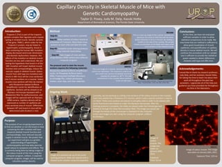
1. Method:
Step
#1:
Place
soleus
muscle
in
cryomold.
Step
#2:
Make
sec3ons
of
soleus
muscle
at
a
thickness
of
5
microns.
Place
three
cross-‐
sec3ons
on
each
slide
and
label
the
slide.
Step
#3:
Complete
Lec3n
staining
protocol
using
materials
listed
below.
Step
#4:
Take
images
of
cross-‐sec3ons
using
Olympus
DP80
camera
and
cellSense
computer
program.
The
protocol
used
to
stain
the
muscle
sec:ons
requires
the
following
materials:
Rhodamine-‐labeled
Griffonia
Simplificolia
I
Lec3n,
1X
Phosphate
Buffered
Saline
(PBS),
ProLong
Gold
An3fade
Mountant,
an
Immedge
Pen,
a
black
permanent
marker,
cover
slips,
and
clear
nailpolish.
Introduc:on:
Purpose:
The
purpose
of
our
on-‐going
experiment
is
to
find
out
whether
or
not
the
knock-‐in
mice
containing
the
A8V
muta3on
will
have
impaired
skeletal
muscle
func3on
and
correspondingly,
have
reduced
vascular
supply
in
skeletal
muscle.
We
predict
that
our
experiment
will
give
us
a
greater
understanding
of
hypertrophic
cardiomyopathy
and
how
daily
exercise
could
reduce
its
symptoms
and
effects.
This
is
currently
being
done
by
taking
images
of
the
soleus
muscle
of
the
mice
using
the
computer
program,
cellSens.
A
separate
computer
program,
ImageJ,
will
be
used
to
calculate
capillary
density.
Conclusions:
Acknowledgements:
Picture
Department
of
Biomedical
Sciences,
The
Florida
State
University
Capillary
Density
in
Skeletal
Muscle
of
Mice
with
Gene3c
Cardiomyopathy
Ongoing
Work:
Currently,
we
are
working
on
cuXng
cross-‐sec3ons
of
the
soleus
muscle
on
the
cryostat.
Each
cross-‐sec3on
has
a
thickness
of
5
microns,
which
is
why
a
special
machine
is
used
to
cut
them.
Three
cross-‐sec3ons
are
placed
on
one
glass
slide
in
order
to
be
stained
with
Lec3n
fluorescent
and
looked
at
under
the
Olympus
microscope.
A[er
the
staining
protocol
is
complete,
the
slides
are
placed
under
the
microscope
and
looked
at
through
the
Texas
Red
excita3on
filter.
The
image
you
would
see
through
the
lenses
of
the
microscope
is
displayed
on
the
computer
screen
and
images
are
seen
using
the
computer
program,
cellSens
By
Olympus.
Troponin
C
(TnC)
is
part
of
the
troponin
protein
complex
that
regulates
ac3n-‐myosin
cycling
in
striated
muscle.
Gene3c
variants
of
the
gene,
TNNC1,
which
codes
for
the
Troponin
C
protein,
may
be
linked
to
hypertrophic
cardiomyopathy.
Knock-‐in
mice
containing
the
human
A8V
muta3on
display
decreased
ventricular
dimensions
and
diastolic
dysfunc3on;
however,
the
effects
of
the
muta3on
on
skeletal
muscle
func3on
are
less
well
understood.
We
are
tes3ng
the
hypothesis
that
knock-‐in
of
the
A8V
muta3on
will
impair
skeletal
muscle
func3on
and
correspondingly,
reduce
the
vascular
supply
in
skeletal
muscle.
Soleus
muscle
from
wild
type
(no
muta3on)
and
knock-‐in
A8V
mice
will
be
cross-‐sec3oned
into
5
micron
sec3ons
on
a
cryostat.
Soleus
muscle
cross-‐sec3ons
will
then
be
stained
with
Rhodamine-‐labeled
Griffonia
Simplificolia
I
Lec3n
for
iden3fica3on
of
capillaries.
Sec3ons
will
be
viewed
on
an
Olympus
microscope
equipped
with
a
Rhodamine
filter
for
epifluorescence,
and
images
will
be
captured
with
an
Olympus
DP80
camera.
Capillary
density
will
be
expressed
as
number
of
capillaries
per
cross-‐sec3onal
area
of
muscle.
Differences
between
WT
and
knock-‐in
mice
will
be
determined
with
Student’s
t-‐test.
Taylor
D.
Posey,
Judy
M.
Delp,
Kazuki
Hoca
I
would
like
to
thank
my
research
professor,
Judy
Delp,
and
her
assistant,
Kazuki
Hoca,
for
taking
the
3me
to
teach
me
about
their
work
all
throughout
the
past
two
semesters.
I
appreciate
all
of
the
help
and
guidance
you
have
provided
me
throughout
my
3me
in
the
laboratory.
LEFT:
Image
of
soleus
muscle.
This
image
was
taken
on
January
26th,
2015.
Exposure
Time:
50
ms
ABOVE:
Image
of
soleus
muscle.
This
image
was
taken
on
January
26th,
2015.
Exposure
Time:
120
ms
ABOVE:
Image
of
a
standard
set-‐up
of
the
Lec3n
staining
protocol.
Slides
are
stained
with
a
diluted
Lec3n
solu3on
using
micropipeces.
rgwhite
and
PrometheusWiki
contributors.
"Cryostat
sec3oning
of
frozen
3ssues."
PrometheusWiki.
,
22
Jan.
2011
Web.
16
Mar.
2015.
ABOVE:
This
is
an
image
of
a
cryostat,
iden3cal
to
the
one
used
in
our
laboratory.
This
machine
is
used
to
cut
very
small
cross-‐sec3ons
of
skeletal
muscle
by
keeping
the
samples
at
-‐20
°C.
The
cold
environment
in
the
cryostat
allows
sec3ons
to
be
cut
with
ease.
BELOW:
This
is
a
close-‐up
image
of
the
cryostat.
Samples
are
placed
on
the
chuck
so
that
they
will
not
move.
Sec3on
thickness
is
set
to
5
microns.
By
rota3ng
a
handle
on
the
side
of
the
cryostat,
the
sample
moves
up
and
down
on
the
blade
and
makes
small
cross-‐sec3ons
of
the
skeletal
muscle.
At
this
3me,
we
have
not
evaluated
sufficient
samples
in
order
to
allow
sta3s3cal
comparisons
to
be
made.
We
now
know
that
our
method
is
sufficient
to
allow
good
visualiza3on
of
muscle
capillaries,
and
quan3fica3on
of
capillary
density
in
mouse
skeletal
muscle.
We
will
con3nue
this
procedure
un3l
our
sample
size
is
sufficient
to
provide
the
sta3s3cal
power
needed
to
detect
differences
between
wild
type
and
A8V
mice.

![Usability testingsp13 [compatibility mode]](data:image/gif;base64,R0lGODlhAQABAIAAAAAAAP///yH5BAEAAAAALAAAAAABAAEAAAIBRAA7)