1. Expression of KLF4, MKP-1 and Nrf2 at the presence of shear stress in vascular flow,
in vitro analysis
Tasnuva Humaira Molecular Medicine, Cardiovascular Pathway, The Medical School, Faculty of Medicine, Dentistry and Health
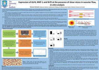
C e n te r P e rip h e ry
0
1
2
3
4
5
M C P 1
F lo w c o n d itio n
Foldchange
C
e
n
te
r
P
e
rip
h
e
ry
0 .0
0 .5
1 .0
1 .5
e N O S
F lo w c o n d itio n
Foldchange
C e n te r P e rip h e ry
0 .0
0 .5
1 .0
1 .5
K L F 4
F lo w c o n d itio n
Foldchange
**
C e n te r P e rip h e ry
0 .0
0 .5
1 .0
1 .5
M K P 1
F lo w c o n d itio n
Foldchange
*
C e n te r P e rip h e ry
0 .0
0 .5
1 .0
1 .5
N R F 2
F lo w c o n d itio n
Foldchange
n s
a b
c d
e
Vascular flow determines the characteristics of Endothelial Cells (EC) in numerous ways. The flow itself causes expression of different genes like KLF4, MKP-1, Nrf2. The genes
also have impacts on regulating the functions of EC as well the vascular damages, among which atherosclerosis is one of the majors.
Shear stress:
• It is the measure of the tendency of a material to slide on itself. Here the stress is parallel or tangential to a face of the material. This stress exerted on arterial walls
influence initiation of atherosclerotic lesion3.
• There are laminar flow and turbulent flow of blood in the vessels. Figure 1 shows, laminar flow and turbulent flow. When blood flows in a straight vessel it creates
Laminar flow and it causes high shear stress. Turbulent flow is the flow when the flow feels some obstruction causing at sites with complex vascular structures.
Atherosclerosis:
• Atherosclerosis is the build up of a waxy plaque on the inside of arteries. Artery wall has the layers tunica intima, media and adventitia (Figure 2)
• In atherosclerosis, the inner and middle layers harden. This phenomena is characterized by deposition of plaques. Thinning of the lumen impedes blood flow and
plaque forms due to collagen, elastin and necrotic debris deposition1.
• There are risk factors for atherosclerosis formation, however its distribution maybe governed by haemodynamic factors such as blood flow disturbance or even altered
genetic expressions. Endothelial dysfunction initiated by endothelial cell (EC) injury is considered to be one of the causative factors for atherosclerosis2. In such
scenario, shear stress can play a major role in the development of atherosclerosis. Figure 3 here shows the initiation of atherosclerotic plaque.
The experiment was done to check the expressions of KLF4, MKP-1 and Nrf2 in EC in context of their position, either central or peripheral, when they are exposed to shear
stress. Our hypothesis was to experiment their expression in the disrupted flow to see if the level of expression can alter vascular changes by the products of the said
genes. We used MCP1 and eNOS as our control genes during the experiment.
Figure 1: Types of blood flow inside vessels
Figure 2: Layers of
blood vessels
Figure 3: Adhesion cascaded initiating
atherosclerosis
Method and analysis
All data analysis was done in Microsoft Excel and the graphs are made using Graphpad Prism.
Figure 6: Orbital shaker. Left panel shows aligned EC from
periphery (non disturbed flow). Right panel shows polygonal EC
from center (disturbed flow) from the wells.
Discussion
• The functions and impacts of these genes suggest that their expressions in context of blood flow have major contribution in vascular
integrity. Lesser expressions of these genes show that the presence of shear stress in disrupted flow can lead to changes in vascular
pathology.
• Using the orbital shaker method here, the microplate fluid volume, well geometry, fluid surface tension, density and viscosity needed
to be considered. For our six well orbital shaker, 210 rpm speed was used at 37°C. The wells were kept in the incubator for 72 hours.
• The sample size in this experiment was rather low, because the primary cells were collected from only 3 umbilical cord donor. To get a
better and more accurate result, sample size should be larger. In this experiment, up to transcription level was observed. Protein
expressions can also be experimented using Western Blot method in other experiments.
• The genes are interlinked with each other by their functions and the signaling pathways in which they contribute. KLF4 expression
decreases in the presence of apyrase. ATP involvement can induce difference in KLF4 expression in EC. Disrupted shear stress
enhances chance of atherosclerosis formation5.
• Nrf2, on the other hand, has protective role from oxidative stress. Nrf2 pathway is highly sensitive to laminar fluid shear stress (FSS).
FSS has imperative influence on inflammatory and pro-inflammatory gene expression in EC and that highly affects vascular
dysfunction6.
• MKP1 induction is necessary for anti-inflammatory effects on EC, however its expression is elevated in increased shear stress4.
References
1.Ridger,V.,Krams,R.,Carpi,A.&Evans,P.C.Hemodynamicparametersregulatingvascularinflammationandatherosclerosis:Abriefupdate.Biomedicine&Pharmacotherapy62,536-540,doi:10.1016/j.biopha.2008.07.053(2008).
2.Kwak,B.R.etal.Biomechanicalfactorsinatherosclerosis:mechanismsandclinicalimplications.EuropeanHeartJournal35,3013-+,doi:10.1093/eurheartj/ehu353(2014).
3.Davies,P.F.,Civelek,M.,Fang,Y.&Fleming,I.Theatherosusceptibleendothelium:endothelialphenotypesincomplexhaemodynamicshearstressregionsinvivo.CardiovascularResearch99,315-327,doi:10.1093/cvr/cvt101(2013).
4.Zakkar,M.etal.Increasedendothelialmitogen-activatedproteinkinasephosphatase-1expressionsuppressesproinflammatoryactivationatsitesthatareresistanttoatherosclerosis.CirculationResearch103,726-732,doi:10.1161/circresaha.108.183913 (2008).
5.Sathanoori,R.etal.ShearstressmodulatesendothelialKLF2throughactivationofP2X4.PurinergicSignalling11,139-153,doi:10.1007/s11302-014-9442-3(2015).
Results
KLF4, MKP-1, Nrf2 genes paly major role in regulation of flow with the vascular changes that
occur regarding shear stress. Their expressions are less in the central area comparing to the
periphery. However their diverse effects, by themselves or by influencing other genes and
signalling pathways play important role in atherosclerosis. However only in vitro experiment
with only three donors do not conclude a hypothesis. In vivo or animal models should be
experimented with regarding the relationship of these genes with vascular pathologies.
Figure 7: Expression of MCP1, eNOS, KLF4, Nrf2 and MKP1 genes in response to shear stress. a
and b shows the control genes MCP1 and eNOS expressions. c, shows KLF4 expression is less in
center, where there is disturbed flow than in the periphery. d and e also show the same
expressions. However, the data for KLF4 gene expression is statistically highly significant. But it
has to be noted that the sample collected from only three donor cords. Also in vitro results need
to confirmed by doing in vivo experiments. c, d and e all are the expressions of genes in HUVEC.
Conclusion
KLF4, Nrf2 and MKP1 genes are expressed lesser in central or disrupted flow when compared to peripheral flow. All of them have some
protective property in the process of atherosclerosis formation. Any disruption of their expressions can lead to alteration of vascular
integrity. More experiments regarding this need to be conducted to conclude to a theory for this.