Cancer Precision Medicine Physiological Function of C MYC as Targeted Molecule
ACES-bulletpoint2
1. RESULTS
Demonstrate Quantification of Cell
Movement
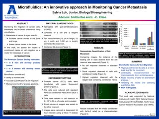
Comparing images, the distance of the
leading cell in each channel from the cell
reservoir was measured (Figure 3).
No cell response observed in standard
media.
Some migration observed in cells with
conditioned media (Figure 4).
Highest migration observed with the
reagent side containing conditioned media.
Results indicated that the media conditioned
with SaOs-2 acted as a chemoattractant
(Figure 5).
sylvia.loh@mavs.uta.edu
ABSTRACT
Monitoring the migration of cancer cells,
metastasis can be better understood using
microfluidics.
Metastasis of cancer is organ specific:
Prostate cancer moves to the bone
and lungs.
Breast cancer moves to the bone.
In this work, we assess the impact of
conditioned media on cell migration as a
model for metastasis of cancer.
INTRODUCTION
The American Cancer Society estimated:
1 in 6 men will develop prostate
cancer.
1 in 8 women will develop breast
cancer.
Microfluidics provide a(n):
Ability to monitor cells.
Accurate quantification of cell migration.
Microenvironment for precise gradients.
MATERIALS AND METHODS
Fabricated with poly-dimethylsiloxane
(PDMS).
Consisted of a cell and a reagent
reservoir.
Ten microchannels (10 μm in height, 25
μm in width and 1,000 μm in length)
connected the reservoirs.
Contained 32 devices (Figure 1).
EXPERIMENT SETTING
Prostate cancer (PC-3) cells were
transfected with green fluorescent
protein (Figure 2).
The cells were cultured with standard
RPMI or conditioned media from SaOs-2
cells.
Cells were seeded in cell reservoir at
3×10³ in 50 μL of media and incubated.
Equal volume of reagent was added to
the other reservoir.
Images were captured at designated 24
hour intervals using a Nikon Ti Eclipse
microscope.
Microfluidics: An innovative approach in Monitoring Cancer Metastasis
Sylvia Loh, Junior, Biology/Bioengineering
Advisors: Smitha Rao and J. –C. Chiao
SUMMARY
Microfluidic Devices for Cell Migration
Physical confinement to cells at a micro-
scale level for the movement.
Controllable microenvironment for
chemogradients, time, and temperature.
Allows recovery of cells for downstream
analyses.
Validating the results and collecting
quantitative data.
Work in Progress.
Figure 1: Microfluidic device mounted on a culture plate
A B C D
ACKNOWLEDGEMENTS
This work was supported by National
Institutes of Health (NIH) National Cancer
Institute grant R15CA133623, North Texas
Cancer Research Foundation and TxMRC.
Figure 2: PC3 cells transfected with GFP (green
fluorescent protein) in a cell reservoir under Brightfield
microscope and ultraviolet light
Brightfield UV
Figure 4: Microchannels containing cells with
conditioned media, showing migration
Figure 5: Measurement of migration of PC-3 cells with
and without SaOs-2 conditioned media against
standard media or SaOs-2 conditioned media
0
2
4
6
8
10
12
14
16
18
Media vs.
Media
Media vs.
Conditioned
Conditioned
vs. Media
Conditioned
vs.
Conditioned
Migrationdistance(μm)
Day 1
Day 2
Day 3
Day 4
Day 5
Figure 3: Migration of a PC-3 cell in RPMI in a single
channel over a 5 day period with SaOs2 conditioned
media as attraction
Day 1 - 31.39 μm
Day 2 - 203.16 μm
Day 3 - 283.51 μm
Day 4 - 285.37 μm
Day 5 - 290.05 μm