International Journal of Biometrics and Bioinformatics(IJBB) Volume (3) Issue...
CCRC Clonality Poster
1. Methylation Profiling as a Tool for
Investigating Clonality in Prostate Cancer
Birbal Prasad1, Atsunari Kawashima2, Nathan How2, John B.A. Okello2, David M Berman2, and Robert J Gooding1
Department of Physics, Engineering Physicis, and Astronomy, Department of Pathology and Molecular Medicine,
Queen’s Cancer Research Institute, Queen’s University, Kingston, Ontario
BACKGROUND
RESULTS
METHODS
AKNOWLEDGEMENTS
Prostate cancer is known to be multi-focal, but little is definitively known about the clonal relationship of discrete cancer foci. Questions remain concerning the origin of
tumours, whether low grade cancers have the potential to progress to high grade ones, and the proper evaluation of extent of disease when faced with small, discontinuous
cancer foci. Our project sheds light on these issues with the aid of epigenetic profiling. The prevalence and heritability of methylation events make them suitable for
characterizing clonality in cancer, especially early prostate cancer in which genomic aberrations are otherwise rare. An abundance of well established, cancer-specific
methylation events in prostate cancer provide guidance for target selection.
Using Methylation-Specific PCR (MSP), we built a panel of several CpG islands that
are heterogeneously methylated across cases. We tested this panel on archival radical
prostatectomy tissues that were reviewed and graded by expert pathologists to
identify spatially separated cancers. MSP values for each gene in the panel were
obtained and classified into either ‘high’ or ‘low’ categories based on whether they
exceeded the median methylation value for that assay across all samples.
Figure 3. Analysis of the MSP results showed that the methylation data was
distributed exponentially
This project would not have been possible without the close supervision, advice, and mentoring of the Berman
Laboratory as well as the financial and structural support of Queen’s University, Kingston General Hospital, Ride
for Dad, Movember, and Prostate Cancer Canada.
FUTURE DIRECTIONS
Now that it has been developed, this tool can be used in future studies
analyzing the ability of the tool to differentiate histopathologically between
tumour discontinuities that represent a true multiplicity of clones and those
that are simply due to sampling methods.
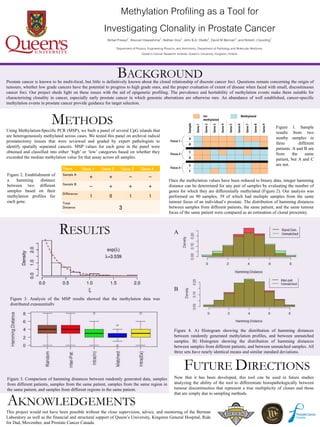
Tissue Gene 1 Gene 2 Gene 3 Gene 4
Sample A
+ + - -
Sample B
- + + +
Difference
1 0 1 1
Total
Distance 3
Figure 4. A) Histogram showing the distribution of hamming distances
between randomly generated methylation profiles, and between unmatched
samples. B) Histogram showing the distribution of hamming distances
between samples from different patients, and between unmatched samples. All
three sets have nearly identical means and similar standard deviations.
Figure 3. Comparison of hamming distances between randomly generated data, samples
from different patients, samples from the same patient, samples from the same region in
the same patient, and samples from different regions in the same patient.
Figure 1. Sample
results from two
nearby samples in
three different
patients. A and B are
from the same
patient, but A and C
are not.
Figure 2. Establishment of
a hamming distance
between two different
samples based on their
methylation profiles for
each gene.
Once the methylation values have been reduced to binary data, integer hamming
distance can be determined for any pair of samples by evaluating the number of
genes for which they are differentially methylated (Figure 2). Our analysis was
performed on 90 samples, 39 of which had multiple samples from the same
tumour focus of an individual’s prostate. The distribution of hamming distances
between samples from different patients, the same patient, and the same tumour
focus of the same patient were compared as an estimation of clonal proximity.
A
B