
Recommended
More Related Content
What's hot
What's hot (20)
Similar to GINGIVAL SURGICAL TECHNIQUES IN PERIODONTOLOGY
Similar to GINGIVAL SURGICAL TECHNIQUES IN PERIODONTOLOGY (20)
Recently uploaded
Recently uploaded (20)
GINGIVAL SURGICAL TECHNIQUES IN PERIODONTOLOGY
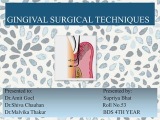
- 1. GINGIVAL SURGICAL TECHNIQUES Presented to: Presented by: Dr.Amit Goel Supriya Bhat Dr.Shiva Chauhan Roll No.53 Dr.Malvika Thakur BDS 4TH YEAR
- 2. CONTENTS Introduction History Gingival curettage Rationale for curettage Indications & contraindications Procedure Other techniques Healing after curettage Clinical appearance after curettage Gingivectomy Indication & contraindications Various techniques Healing after gingivectomy Gingivoplasty Conclusion References
- 3. INTRODUCTION Bacterial Plaque causes the formation of Periodontal Pockets and resorption of alveolar bone due to apical migration of Junctional Epithelium. Limited to gingiva and not involving underlying osseous structures Gingival curettage Gingivectomy Gingivoplasty Current understanding of disease etiology and therapy limits the use of both techniques, but their place in surgical therapy is essential.
- 4. TREATMENT OF PERIODONTAL POCKETS Pocket reduction Pocket elimination
- 5. PERIODONTAL POCKET MANAGEMENT SUPRA/SUBGINGIVAL DEBRIDEMENT ORAL HYGIENE INSTRUCTIONS RE-EVALUATION POCKET ELIMINATION POCKET REDUCTION SUPPORTIVE PERIODONTAL CARE •GINGIVECTOM Y •APICALLY REPOSITIONE D FLAP •ACCESS FLAP •MODIFIED WIDMAN •ENAP
- 6. HISTORY John W Riggs (1811-1885)- In an 1876 paper,he developed the concept of oral prophylaxis and prevention, advocated for the cleanliness of the mouth, and opposed surgery, which at the time consisted of gingival resection. In 1935, Kronfeld proved that the bone in the periodontal pockets was neither necrotic nor infected but rather destroyed by inflammatory process, and the era of tissue curettage began as the attention was shifted to the soft tissue surrounding the tooth as the source of infection. Salomon Robiscek (1845-1928) - He developed a surgical technique consisting of a scalloped continuous gingivectomy excision , exposing the marginal bone for subsequent curettage & remodeling. The rationale for the radical treatment was supported by the authors such as Neuman, Widman , Robicsek ,Zemsky , Ceszynki and Nodine who popularized the surgical procedures for the elimination of periodontal pocket.
- 7. WHAT IS CURETTAGE? The word curettage is used in periodontics to mean the scraping of the gingival wall of a periodontal pocket to separate diseased soft tissue. ( Carranza 10th edition) Curettage is a closed, definitive surgical procedure performed under local anesthesia and aimed at pocket reduction, elimination, reattachment, or new attachment. It is performed with sharp curettes in an attempt to remove: (1) The sulcular epithelium and the epithelial attachment (2) The inflammed connective issue of the pocket wall
- 8. TERMINOLOGIES Gingival curettage- Removal of the inflammed soft tissue lateral to the pocket wall. Subgingival curettage -Procedure that is performed apical to the epithelial attachment, severing the connective tissue attachment down to the osseous crest. Inadvertent curettage- Some degree of curettage is done unintentionally when scaling and root planing is performed. Extent of gingival curettage Subgingival curettage
- 9. RATIONALE Accomplishes the removal of chronically inflamed granulation tissue that forms lateral wall of periodontal pocket. Inflamed Granulation Tissue Barrier to the attachment of new fibers Root Planing Pocket pathologic changes resolve
- 10. According to Academy report in 2002, short and long term clinical trails have confirmed that gingival curettage provides no additional benefits when compared to SRP alone. After comparing SRP alone to curettage plus SRP, it was concluded that curettage “did not serve any additional useful purposes”. Additionally, the American Academy of Periodontology did not include gingival curettage as a method of treatment in its guidelines for periodontal therapy. J periodontol,2002
- 11. INDICATIONS CONTRAINDICATIONS 1) Edematous and inflammed tissues 2) Shallow pockets 3) Suprabony pockets 4) As part of initial preparation prior to open surgical procedures in an attempt to achieve tissue quality that can be handled easily 5) Progressive attachment or alveolar bone loss 6) Increased levels of pathogenic microorganism 1) Fibrotic tissue 2) Deep pockets ≥ 5mm 3) Furcation involvements 4) Medically compromised patient
- 12. Basic Technique with a Curette Instruments: Gracey curettes Universal curettes Isolation & Anesthetize : Local infiltration is given to anesthetize the isolated selected site. Insertion of Curettage Sharp Gracey(Gracey #13-14 for mesial surfaces,Gracey #11-12 for distal surfaces) or Universal curette(4R-4L) is inserted with the cutting edge against the tissue so as to engage the inner lining of pocket wall & junctional epithelium.
- 13. Curette the soft tissue wall : Curette is carried along the soft tissue in a horizontal stroke The pocket wall is supported by gentle finger pressure on the external surface. Several overlapping strokes are used to completely remove the epithelium & underlying granulation tissue. Gingival curettage performed with a horizontal stroke. In subgingival curettage, the tissue attached to the bottom of the pocket & alveolar crest are removed with a scooping motion of the curette to the tooth surface. A-elimination of pocket lining B-elimination of junctional epithelium C-procedure completed
- 14. Other Techniques Other techniques for gingival curettage include 1. The excisional new attachment procedure(ENAP) 2. Ultrasonic curettage, 3. The use of caustic drugs. ENAP is the surgical procedure in which an internal bevel incision is made to remove the epithelial lining of the crevice and the junctional epithelium, allowing root visibility. Definitive subgingival curettage performed with knife. ENAP Gain new attachment Decrease probing depth Access root surface
- 15. After anesthesia, an internal bevel incision is given from the margin of the free gingiva apically to a point below the bottom of the pocket. Remove the excised tissue with a curette and perform root planing on all exposed cementum to achieve a smooth hard surface. Approximate the wound edges, if they do not meet passively, recontour the bone until good adaptation of the wound edges is achieved. Place sutures and a periodontal dressing
- 16. ENAP Modification In 1977, Fredi and Rosenfeld modified the technique by advocating a partial-thickness inverse beveled incision down to the crest of bone to completely remove tissue about the periodontal ligament. Initial incision made to crest of pocket Inner wall remove to crest of bone&pdl Healed tissue
- 17. ULTRASONIC CURETTAGE Ultrasound is effective for debriding the epithelial lining of periodontal pockets. It results in a narrow band of necrotic tissue (microcauterization), which strips off the inner lining of the pocket. The Morse scaler-shaped and rod-shaped ultrasonic instruments are used for this purpose. Nadler H-1962 found ultrasonic instruments to be as effective as manual instruments for curettage but resulted in less inflammation and less removal of underlying connective tissue.
- 18. CAUSATIVE AGENTS The use of caustic drugs has been recommended to induce a chemical curettage of the lateral wall of the pocket or even the selective elimination of the epithelium. Drugs such as sodium sulfide, alkaline sodium hypochlorite solution (Antiformin)and phenol, have been proposed and then discarded after studies indicated their ineffectiveness. The extent of tissue destruction with these drugs cannot be controlled, and they may increase rather than reduce the amount of tissue to be removed by enzymes and phagocytes.
- 19. Healing after curettage Immediately after curettage - a blood clot Hemorrhage-polymorphonuclear leukocytes appear shortly Rapid proliferation of granulation tissue Restoration and epithelialization of the sulcus require 2 to 7 days Restoration of the JE - 5 days after treatment Immature collagen fibers - 21 days.
- 20. Immediately after curettage- gingiva appears hemorrhagic and bright red. After 1 week- the gingiva appears reduced in height- apical shift in the position of the gingival margin. The gingiva is also slightly redder than normal After 2 weeks - normal color, consistency, surface texture, and contour of the gingiva are attained and gingiva margin is well adapted to the tooth. Clinical Appearance After Scaling And Curettage
- 21. DURATI- ON CONNECTIVE TISSUE CHANGES EPITHELIAL CHANGES CLINICAL CHANGES Immediat ely • Hemorrhage • a/c inflammatory reaction • Removal of ep. lining • Few cells may remain • Blood & exudate 1st day • Marked inflammation • Epithelial migration begins (0.5-1mm/day) • Edematous • Discoloration persists 2nd day • Inflammation • Vascularity • Epithelium begins to cover the gingival corium •Discoloration • Edema still present 4th-6th day • Chr inflammation • Collagenation • Matrix formation • Restoration of junctional & sulcular epithelium - 7th-10th day • Collagen formation& organisation • Epithelium formation is complete • Edema • Rigid & well adapted gingival wall 10th-14th day • Repair of C.T • vascularity • Surface keratinization • Normal color • Stippling appears • Gingival shrinkage After 2 weeks • Mature collagen • New sub sulcular & marginal vessels - • Color, contour, consistency, texture. • Well adapted marginal gingiva
- 22. GINGIVECTOMY GINGIVECTOMY means EXCISION OF THE GINGIVA. Removal of pocket wall Visibility & accessibil ity for complete calculus removal Proper smoothen ing of roots Favourabl e environm ent for gingival healing Restorati on of physiologi c gingival contour
- 23. INDICATIONS CONTRAINDICATIONS 1) Elimination of suprabony pockets. 2) Elimination of gingival enlargement . 3) Elimination of suprabony periodontal abscess. 4) The presence of furcation involvement (without associated bone defects) where there is wide zone of attached gingiva. 5) Pericoronal flap 1) The need for bone surgery or examination of the bone shape and morphology. 2) Situations in which the bottom of the pocket is apical to the mucogingival junction. 3) Esthetic considerations, particularly in the anterior maxilla
- 24. Various techniques of gingivectomy Surgical gingivectomy Gingivectomy by electrosurgery Laser gingivectomy Gingivectomy with chemosurgery
- 25. SURGICAL TECHNIQUES INSTRUMENTS Mouth mirror Probe Pocket markers Kirkland and orban interdental knives Surgical blade,bard parker handle Surgical curette, gracey curette Tissue forceps Scissors Periodontal dressing
- 26. Procedure Step 1: The pocket on each surface are explored with a periodontal probe and marked with a pocket marker. Each pocket is marked in several areas to outline its course on each surface. Step 2: Periodontal knives are used for incisions on the facial and lingual surfaces and those distal to terminal tooth in the arch. Bard-parker knives #11 and #12 and scissors are used as auxiliary instruments.
- 27. The incision is started apical to the points marking the course of the pocket (Orban 1952) and is directed coronally to a point between the base of the pocket and crest of the bone. It should be as close as possible to the bone. Discontinuous or continuous incisions may be used. Incision should be beveled at 450 to the tooth surface
- 28. Step 3: Remove excised pocket wall Clean the area Examine the root surface Step 4 : Curette granulation tissue Remove any remaining necrotic cementum or calculus Step 5: Cover the area with surgical pack.
- 30. HEALING AFTER SURGICAL GINGIVECTOMY Initial response 1. Blood clot formation 2. Underlying tissue is acutely inflamed and necrotic and soon replaced by the granulation tissue. 24 hours later 1. Increased connective tissue cells ( mainly angioblasts) 2. Epithelial cells at the margins of the wound start migrating over the granulation tissue. Epithelial activity reaches a peak in 24 to 36 hours.
- 31. 3 days later 1. Young fibroblasts are seen 2. Highly vascular granulation tissue grows coronally creating free gingival margin and sulcus 2 weeks 1. Capillaries from vessels of periodontal ligament migrate into connective tissue and connect with gingival margins 2. After 5 to 14 days, surface epithelialization is generally complete 3. Complete repair takes about 1 month. – Connective tissue repair in 7 weeks and the pigmentation is diminished .
- 32. By electrosurgery(surgical diathermy) Uses high frequency current of 1.5 to 7.5 million cycles per second. ADVANTAGES Control of hemorrhage Adequate contouring of the tissue DISADVANTAGES Cannot be used in patients who have poorly shield cardiac pacemakers. Unpleasant odour If the electro surgery point touches the bone irreplacable damage can be done. When electrode touches the root,cementum burn are produced.
- 33. INSTRUMENTS: Needle electrode 0.0075inch – 0.015inch PROCEDURE: Area must be slightly moist Blended cutting and coagulating current is used Electrode must be in constant motion to prevent heat build- up . Debris should be cleaned with isopropyl alcohol from the electrode for each motion.
- 34. INDICATIONS Removal of gingival enlargement. Gingivoplasty Relocation of frenum and muscle attachments. Incision of periodontal and pericoronal abscess. TECHNIQUE For gingivoplasty: needle electrodes and diamond shaped electrodes are used for festooning. In all reshaping procedures electrodes are activated and moved in a concise “shaving” motion. For abscess drainage: incision can be made with the needle electrode. For hemostasis: ball electrode is used. For relocation and of frenum muscle attachment: loop electrode is used.
- 36. LASER GINGIVECTOMY The laser used in dentistry are the carbon dioxide and the Nd:YAG which have wave lengths of 10,600nm and 1064nm. Advantages : Completely dry, bloodless surgery. Surgical time is reduced Instant sterlization of the area, decreasing the chances of bacteremia. Minimal postoperative pain, swelling and scarring. Disadvantages Protective eyewear should be used High cost of the equipment.
- 38. CHEMOSURGICAL GINGIVECTOMY 5% paraformaldehyde –Orban B 1942 Pottasium hydroxide –Loe 1961 Disadvantages: Depth of action cannot be controlled Gingival remodelling cannot be accomplished. Epithelialization & reformation of JE , re-establishment of the alveolar crest fiber system areoccursslowly – TonnaE1967 Not recommended
- 40. GINGIVOPLASTY Recontouring of the gingiva in the absence of pockets Used to correct deformities like: Gingival clefts & craters Shelf like interdental papilla - ANUG Gingival enlargements Instruments: Periodontal knife & scalpel Rotary coarse diamond stones Electrodes
- 41. Procedure Tapering the Gingival Margin Creating scalloped marginal outline Thinning of attached gingiva Creating vertical interdental grooves Shaping interdental papilla
- 42. Current understanding of disease etiology & therapy limits the use of these techniques, but their place in surgical therapy is essential. Current periodontal surgery must consider the conservation of keratinized gingiva, minimal gingival tissue loss to maintain esthetic, adequate access to osseous defects for definitive defect correction, minimal post operative discomfort and bleeding by attempting surgical procedures that will allow primary closure. CONCLUSION
- 43. REFERENCES Carranza’s clinical periodontology: 10th edition Essentials of clinical periodontology:3rd edition Shantipriya Reddy Textbook of periodontics, Shalu Bathla Lindhe, Karring, Lang: Clinical Periodontology & Implant Dentistry. Blackwell Munksgaard; 5th Edititon The American Academy of Periodontology statement regarding curettage.J periodontol 2002