WEBINAR Characterisation of human pluripotent stem cells (ESCs and IPSC) and ...
antigen presentation machinery in ctcs (1)
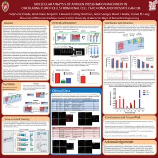
1. MOLECULAR ANALYSIS OF ANTIGEN PRESENTATION MACHINERY IN
CIRCULATING TUMOR CELLS FROM RENAL CELL CARCINOMA AND PROSTATE CANCER.
Stephanie Thiede, Jacob Tokar, Benjamin Casavant, Lindsay Strotman, Jamie Sperger, David J. Beebe, Joshua M. Lang
University of Wisconsin Carbone Cancer Center, University of Wisconsin Dept. of Biomedical Engineering
Clinical Data
Blood from patients with metastatic prostate cancer or RCC were were drawn under a UW-IRB approved
protocol and their CTCs were stained and analyzed for intact nuclei (hoechst), cytokeratins, CD45 and
HLA expression. Below is the total number of CTCs identified and those expressing HLA.
Conclusions and Future Work
The VERSA:
Isolation:
bead-bound
cells of interest
Non-target cells
Magnet
2) Magnet pull
1) Add PMPs
Vertical Exclusion Based
Rare Sample Analysis
Principle:
Two aqueous solutions can be placed in adjacent
wells, and due to the relative dominance of surface
tension on the microscale, stay pinned, creating a
‘virtual wall’at the interface.
virtual
wall
Aqueous phases
(cell suspensions, stains, washes)
Oil phases
INTRODUCTION AND OBJECTIVES: Tumor cells develop many mechanisms by
which to avoid detection and destruction by the immune system, namely down-
regulation or silencing of genes critical for antigen expression, processing and
presentation. These mechanisms of evasion have been identified in nearly all
tumor types, including prostate cancer and renal cell carcinoma (RCC). However,
there is a lack of available biomarkers to identify patients with advanced cancer.
A relatively new area of interest is the use of circulating tumor cells (CTCs) as an
accessible source of tumor cells for molecular analysis. These cells could provide
us with more information on HLA expression across disease types and even
across patients. However, interrogation of these cells with current techologies is
limited. We have designed a novel platform to permit the use of any antibody of
interest bound to paramagnetic particles (PMPs) to isolate and purify PMP-bound
cells via immiscible oil barriers. After isolation, using this same device, we are
able to stain and image proteins of interest and extract nucleic acids for gene ex-
pression analysis.
METHODS: The VERSA platform was designed using the relative dominance of
surface tension in the microscale to create virtual walls between oil and aqueous
phases filtering contaminants in a single step, while maintaining cell viability for
further analysis. In previous experiments, the isolation of CTCs in patients with
prostate cancer was optimized using a known prostate cancer cell line (LnCAP)
spiked into whole blood and captured with EpCAM. To optimize the isolation of
CTCs in patients with RCC, samples of whole blood were spiked with known RCC
cell lines (786-0 and 769-P) and captured with Carbonic Anhydrase IX (CAIX). Pa-
tient CTCs were then isolated to interrogate for HLA Expression and tumor asso-
ciated antigen expression.
RESULTS: Initial results investigating isolation of CTCs in patients with RCC show a
capture efficiency of about 50-70% when spiking known cell lines into blood.
RCC and prostate CTCs were successfully isolated from 7.5mL of blood and ca-
pable of being stained both extracellularly with HLA-ABC (W6/32 antibody) and
intracellularly with cytokeratin. Nucleic acids extracted from these patient
samples were sufficient enough to detect tumor specific antigens of interest in-
cluding PAP, AR and SSX2.
Abstract
Sieve-Assisted Staining
A microporous membrane is fabricated into one of the VERSA wells such that
fluid can be added and replaced from an adjacent well without touching the
sample, critical for rare or delicate cell samples and enabling sophisticated flu-
idic procedures in-device.
Membrane
Aspirate
Add Fluid (Wash, Fixative, etc.)
Incubate
Repeatasnecessary
Top View Side View
Fluid Exchanges
Rear
Well
Front
Well
Magnet
PMP Removal
0.5
0.6
0.7
0.8
0.9
1
1.1
1.2
1.3
1 2 3 4 5
Wash Number
Normalized
NumberofCells
Loss Due to Washing
Vertical Cell Isolation
Vertical device orientation, featuring the long axis on the vertical instead of
horizontal axis, allows non-target cells to passively settle out of the opera-
tional path of the PMP-bound target cells
FG FM
Input
Magnet
Oil
Traverses
Wash Output
0
100
200
300
400
500
600
700
800
900
NumberofCells
PBMC
LNCaP
Purity
Traverse Number
1 2 3
77% 82%
86%
1 2 3
VERSA
Device Side-View
Force
Vectors Cell Data
This allows us to capture a few cells from a large background:
0
20
40
60
80
100
5M 20M 100M
PercentRecovery
Background PBMCs
(M = million)
One cell in 20M PBMCs
target well
Variable PBMCs
PMPs were bound to an antibody specific for CAIX or EpCAM. The immortalized
RCC lines, 786-O and 769-P, were incubated with CAIX-PMPs with and without
EpCAM-PMPs in the VERSA chip, then captured as above. The best capture effi-
ciency in both cell lines occured when using CAIX-PMPs alone.
Total Nucleic Acid Extraction
Patient blood samples were processed using the VERSA and mRNA extracted for gene expres-
sion analysis of a housekeeping gene, P0, and tumor specific antigens, AR, PAP and SSX2.
Acknowledgements
This work was supported by a Movember-Prostate Cancer Foundation
Challenge award and a PCF Young Ivestigator Award to Dr. Lang, grants
from the DOD PCRP Physician Research Training Award W81XWH-09-1-
0192, Wisconsin Partnership Program, UWCCC Investigator Initiated Pilot.
Tumor Associated Antigen Expression in Prostatic CTCs
Patient Sample
RelativeExpression
0.00
0.01
0.02
0.03
0.04
0.1
0.2
0.3
0.4
0.5
0.6
0.7
0.8
AR 1/2
PAP
SSX2
43 71 98 99 100 159 165124
After imaging, cells are lysed and PMPs bound to an oligo-dT are added to capture mRNA. The
PMP-mRNA conjugates is magnetically are purified into the final rear well of the chip. A high
salt buffer is then added to lyse nuclei. Silica beads are then added to bind DNA and the PMP-
DNA conjugates are purified into the final front well. Nucleic acids are eluted from PMPs in a
20uL volume for qPCR or sequencing.
Input
Live Cell
Staining
Intracellular
Staining
DNA
Purification
mRNA
Purification
1
10
100
1,000
10,000
100,000
1000 100 10 1
RelativeDNASignal
Number of Cells
Qiagen AllPrep
VERSA
1
10
100
1,000
10,000
100,000
1000 100 10 1
RelativeRNASignal
Number of Cells
Qiagen AllPrep
VERSA
Direct DNA Sequencing of 10 LNCaP cells Direct RNA Sequencing of 10 LNCaP cellsC) D)
A) B)qPCR for DNA on Low Cell Numbers qRT PCR for mRNA on Low Cell Numbers
Circulating tumor cells can be assayed for expression of molecular machinery critical to
immune recognition and antigen expression.
Significant heterogeneity exists in CTC populations with regards to MHC expression that
may have potential as a predictive and pharmacodynamic biomarker for immunotherapies.
Tumor associated antigen expression can be identified in CTCs as a potential predictive
marker for vaccine based immunotherapies.
Prospective clinical trials will be needed to validate the utility of these assays.
Prostate
Patient #
Age Pathology Sites of Disease Treatment History Total CTC Number
(EpCAM+/CK+/CD45-)
HLA+
CTCs
103 74 Gleason
4+5
Bone Mets Docetaxel, Enzalutamide 30 4
79 64 Gleason
4+4
Lymph Node and
Bone Mets
PAP Vaccine, Provenge,
Docetaxel, Abiraterone
10 7
84 58 Gleason
4+4
Bone Mets Docetaxel, TAK700,
Enzalutamide
58 24
71 64 Gleason
3+3
Lymph Node and
Bone Mets
Docetaxel, TAK700,
Axitinib, Enzalutamide
469 171
Renal
Patient #
Age Pathology Sites of Disease Treatment History Total CTC Number
(CAIX+/CK+/CD45-)
HLA+
CTCs
143 73 Clear Cell
Carcinoma
Pancreatic Mass Tivozanib 4 4
111 69 Clear Cell
Carcinoma
Nephrectomy Bed Sunitinib, Bevacizumab 14 14
142 74 Clear Cell
Carcinoma
Bone Mets Tivozanib, Everolimus 7 7
145 76 Clear Cell
Carcinoma
Lymph Node
Mets
Axitinib, Everolimus,
Pazopanib
46 40
Hoescht Cytokeratin
CD45
Hoescht Cytokeratin
CD45HLA-ABC
Merged
HLA-ABC
Merged
Pt 84 CTCs
Hoescht Cytokeratin
CD45HLA-ABC
Merged
Pt 145 CTCs Hoescht
HLA-ABC
Merged
CD45
Cytokeratin
Merged
Hoescht
Hoescht Cytokeratin
HLA-ABC CD45
Merged