Drug induced liver disorders for Pharm.D
•Download as PPTX, PDF•
112 likes•19,449 views

Definition, Patterns/types and mechanisms of drug induced liver disorders, assessment of drug induced liver disorders and its treatment (pharmacotherapeutics-3)

Recommended
Recommended
More Related Content
What's hot
What's hot (20)
Similar to Drug induced liver disorders for Pharm.D
Similar to Drug induced liver disorders for Pharm.D (20)
More from Soujanya Pharm.D
More from Soujanya Pharm.D (20)
Recently uploaded
Recently uploaded (20)
Drug induced liver disorders for Pharm.D
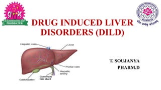
- 1. DRUG INDUCED LIVER DISORDERS (DILD) T. SOUJANYA PHARM.D
- 2. CONTENTS: • Definition • Patterns/types of drug induced liver disorders • Mechanisms of DILD • Assessment • Treatment • Reference/bibliography
- 3. DEFINITION: The liver disease/disorder resulting from the inhalation, ingestion or parenteral administration of any pharmacological or chemical agent is called as drug induced liver disease or drug induced liver disorder (DILD).
- 4. PATTERNS/TYPES OF DILD The most common and serious drug induced liver disorders are as follows: 1. Idiosyncratic reactions 2. Allergic hepatitis 3. Toxic hepatitis 4. Chronic active hepatitis 5. Toxic cirrhosis 6. Liver vascular disorders
- 5. 1. IDIOSYNCRATIC REACTIONS: For some drugs, a genetic or acquired abnormality must exist in a particular metabolic pathway for a toxic reaction to take place. In other cases, the reactions are typically associated with a drug concentration and often respond to simply lowering the dose of the drug. Idiosyncratic reactions tend to occur without association to particular blood concentrations or specifically identified metabolic abnormalities. e.g. sulfonylureas like glipizide and antibiotics like ciprofloxacin. Idiosyncratic reactions are rare and are sometimes described as liver hypersensitivity to a drug.
- 6. 2. ALLERGIC HEPATITIS: Allergic reactions in the liver can be caused by many drugs and result in many different kinds of hepatic damage. It is marked by fever, pruritus, rash, eosinophilia, arthritis, and haemolytic anaemia. The formation of granulomas within the liver is often seen on biopsy. The reaction reverses with discontinued therapy and reappears upon rechallenge. e.g. Most antibiotics have been associated with this type of reaction, including the fluoroquinolones, macrolides, β-lactams, trimethoprim- sulfamethoxazole, penicillinase-resistant Penicillins such as dicloxacillin and allopurinol.
- 7. CONTD… SIGNS & SYMPTOMS: i) The onset of symptoms is 1 to 6 weeks after initiation of therapy. ii) The incidence, like all the allergic liver reactions, is low, estimated at less than 1%. iii) The clinical presentation includes eosinophilia, fever, rash, and arthritis. The biopsy may show a pattern of fibrin-ring granulomas similar to those seen in Q fever.
- 8. 3. TOXIC HEPATITIS: Toxic reactions are predictable, often dose-related effects in the liver due to specific agents. e.g. Paracetamol (acetaminophen). When taken in overdose, acetaminophen becomes bioactivated to a toxic intermediate known as N-acetyl-p-benzoquinone imine (NAPQI). NAPQI is very reactive, with a high affinity for sulfhydryl groups and requires glutathione for further metabolism to non-toxic metabolites. After glutathione supplies are exhausted, the toxic metabolite binds to sulfhydryl-containing proteins in the liver cell and causes lipid peroxidation disrupting the cell membrane. These events eventually lead to cell death.
- 9. CONTD… Acetaminophen P450 (2E1) N-acetyl-P- benzoquinone imine (NAPQI) (toxic) Glutathione Cysteine & mercapturic acid (non-toxic) e.g. Reye’s syndrome is an aggressive form of toxic hepatitis often associated with aspirin use in children. Valproate toxicity can also present in this pattern.
- 10. CONTD… SIGNS & SYMPTOMS: i) Early in the process of Reye’s syndrome, mitochondrial dysfunction leads to the depletion of acyl coenzyme A and carnitine. ii) Fatty acids accumulate and gluconeogenesis is impaired, resulting in hypoglycaemia. iii) A concurrent disruption of the urea cycle occurs, leading to a decrease in the removal of ammonia and a slowing of protein use. iv) A threefold rise in the blood ammonia level and an increase in the prothrombin time are common findings. v) In advanced stages of Reye’s syndrome, many patients develop intracranial hypertension that can be life threatening and refractory to therapy.
- 11. 4. CHRONIC ACTIVE TOXIC HEPATITIS: Patients experience periods of symptomatic hepatitis followed by periods of convalescence, only to repeat the experience months later. It is a progressive disease with a high mortality rate and is more common in females than males. Antinuclear antibodies appear in most patients. These drugs appear to form anti-organelle antibodies. The exact identification of a causative agent is sometimes difficult since diagnosis requires multiple episodes occurring long after exposure to the offending drug. e.g. Dantrolene, isoniazid, phenytoin, nitrofurantoin, and trazodone have been reported in association with a type of autoimmune mediated disease in the liver.
- 12. 5. TOXIC CIRRHOSIS: The scarring effect of hepatitis in the liver leads to the development of cirrhosis. Some drugs tend to cause such a mild case of hepatitis that it may not be detected. Mild hepatitis can be easily mistaken for a more routine generalized viral infection. If the offending drug or agent is not discontinued, this damage will continue to progress. The patient eventually presents not with hepatitis, but with cirrhosis. e.g. Methotrexate and vitamin A toxicity.
- 13. CONTD… i) Methotrexate causes periportal fibrosis in most patients who experience hepatotoxicity. The lesion results from the action of a bioactivated metabolite produced by cytochrome P450. This process has most commonly been noted in patients treated for psoriasis and arthritis. The extent of damage can be reduced or controlled by increasing the dosage interval to once weekly or by routine use of folic acid supplements. ii) Vitamin A is normally stored in liver cells, and causes significant hypertrophy and fibrosis when taken for long periods in high doses. Hepatomegaly is a common finding, along with ascites and portal hypertension. In patients with vitamin A toxicity, gingivitis and dry skin are also very common. This is accelerated by ethanol, which competes with retinol for aldehyde dehydrogenase.
- 14. 6. LIVER VASCULAR DISORDERS: Focal lesions in hepatic venules, sinusoids, and portal veins occur with various drugs. e.g. Cytotoxic agents used to treat cancer, the pyrrolizidine alkaloids, and the sex hormones. A centralized necrosis often follows and can result in cirrhosis. Azathioprine and herbal teas that contain comfrey (a source of pyrrolizidine alkaloids) are associated with the development of veno- occlusive disease. The exact incidence is rare and may be dose related.
- 15. CONTD… “Peliosis hepatitis” is a rare type of hepatic vascular lesion that can be seen as both an acute and a chronic disease. The liver develops large, blood-filled lacunae within the parenchyma. Rupture of the lacunae can lead to severe peritoneal haemorrhage. e.g. Peliosis hepatitis has been associated with exposure of the liver to androgens, oestrogens, tamoxifen, azathioprine, and danazol.
- 16. MECHANISMS OF DILD: The common mechanisms involved in drug induced liver disorders are: 1. Centro lobular necrosis 2. Steatohepatitis 3. Phospholipidosis 4. Generalized hepatocellular necrosis 5. Cholestatic jaundice 6. Mixed hepatocellular necrosis and cholestatic disease 7. Neoplastic jaundice
- 17. 1. CENTRO TUBULAR NECROSIS: Centro lobular necrosis is often a dose-related, predictable reaction secondary to drugs such as acetaminophen. However, it also can be associated with idiosyncratic reactions, such as those caused by halothane. It is also called as “direct or metabolite-related hepatotoxicity”. Centro lobular necrosis is usually the result of the production of a toxic metabolite. The damage spreads outward from the middle of a lobe of the liver. More severe forms of Centro lobular necrosis are accompanied by nausea, vomiting, upper abdominal pain, and jaundice.
- 18. 2. STEATOHEPATITIS: Steatohepatitis (also known as steatonecrosis) is a specialized type of acute necrosis resulting from the accumulation of fatty acids in the hepatocyte. Drugs or their metabolites that cause steatonecrosis do so by affecting fatty-acid oxidation within the mitochondria of the hepatocyte. Hepatic vesicles become engorged with fatty acids, eventually disrupting the homeostasis of the hepatocyte. The liver biopsy is marked by a massive infiltration by polymorphonuclear leukocytes, degeneration of the hepatocytes, and the presence of Mallory bodies.
- 19. CONTD… Drugs/metabolites Affect fatty acid oxidation Accumulation of fatty acids in hepatocytes Necrosis of hepatocyte Steatohepatitis
- 20. CONTD… Alcohol is the drug that most commonly produces steatonecrotic changes in the liver. When alcohol is converted into acetaldehyde, the synthesis of fatty acids is increased. When the hepatocyte has become completely engorged with micro vesicular fat, it often breaks open, spilling into the blood. If enough hepatocytes break open, an inflammatory response begins. If the offending agent is withdrawn before significant numbers of hepatocytes become necrotic, the process is completely reversible without long-term sequelae. In non-alcoholic steatohepatitis the same endpoint is often achieved through oxidation of lipid peroxidases. e.g. Tetracycline, sodium valproate produces non-alcoholic steatohepatitis.
- 21. 3. PHOSPHOLIPIDOSIS: Phospholipidosis is the accumulation of phospholipids instead of fatty acids. The phospholipids usually engorge the lysosomal bodies of the hepatocyte. e.g. Amiodarone Patients treated with amiodarone who develop overt hepatic disease tend to have received higher doses of the drug. These patients also have higher amiodarone to N-desethyl-amiodarone ratios, indicating a greater accumulation of the parent compound. Amiodarone and its major metabolite N-desethyl-amiodarone remain in the liver of all patients for several months after therapy is stopped. Usually the phospholipidosis develops in patients treated for more than 1 year. The patient can present with either elevated transaminases or hepatomegaly; jaundice is rare.
- 22. 4. GENERALISED HEPATOCELLULAR NECROSIS: Generalized hepatocellular necrosis mimics the changes associated with the more common viral hepatitis. The onset of symptoms is usually delayed as much as a week or more after exposure to toxin. Bioactivation is often important for toxic hepatitis to develop, but may not be the immediate cause of damage. Many drugs that are associated with toxic hepatitis produce metabolites that are not inherently toxic to the liver. Instead, they act as haptens, binding to specific cell proteins and inducing an autoimmune reaction. The rate of bioactivation can vary between males and females and between individuals of the same sex. The cytochrome P450 system (CYP) tends to metabolize lipophilic substrates which are actively pumped into the hepatocyte by an organic anion (or cation) transporting protein. The CYP subspecies 1A, 2B, 3A, and 4A are regulated by the highly inducible xenobiotic receptor on complementary DNA.
- 23. CONTD… The receptor is found in the liver, and to a lesser extent in the cells lining the intestinal tract, and is responsible for cholesterol catabolism and bile acid homeostasis. The activity of this receptor is subject to genetic polymorphism as well. This results in a wide variation in the sensitivity of the population to “generalized hepatocellular necrosis” and other forms of hepatic damage. e.g. i) The long-term administration of isoniazid can lead to hepatic dysfunction in 10% to 20% of those receiving the drug. ii) Ketoconazole produces generalized hepatocellular necrosis or milder forms of hepatic dysfunction in 1% to 2% of patients treated for fungal infections.
- 24. 5. CHOLESTATIC JAUNDICE: Cholestatic jaundice, or cholestasis, can be classified by the area of the bile canalicular or ductal system that is impaired. Canalicular cholestasis is very often associated with long-term high-dose oestrogen therapy. Clinically, these patients are often asymptomatic and present with mild to moderate elevations of serum bilirubin. e.g. i) An intravenous form of vitamin E, α-tocopherol acetate, causes cholestatic jaundice primarily involving the canalicular duct in premature infants. ii) The administration of total parenteral nutrition for periods greater than 1 week induces cholestatic changes and nonspecific enzyme elevations in some patients. iii) This reaction also has been reported to occur rarely with sulfonamides, sulfonylureas, erythromycin estolate and ethyl succinate, captopril, lisinopril, and other phenothiazines.
- 25. 6. MIXED HEPATOCELLULAR NECROSIS & CHOLESTATIC JAUNDICE: Patients infrequently present with a purely hepatocellular necrosis or cholestatic damage, but rather with a mixed picture of damage. e.g. i) Flutamide causes a mix of lesions that appear at or about the 48th week of treatment. ii) Niacin in doses greater than 3 g/day, or in doses greater than 1 g/day of sustained-release formulations, causes the same mixed pattern of damage. iii) These patients often present with only a few signs or symptoms at first, but can progress rapidly to fulminant hepatic failure. Additionally, niacin- induced and other drug-induced mixed hepatocellular disease can be misinterpreted as hepatobiliary cancers.
- 26. 7. NEOPLASTIC JAUNDICE: A large body of the current literature on adverse reactions and the liver addresses the development of neoplasms following drug therapy. Both carcinoma- and sarcoma-like lesions have been identified. Fortunately, hepatic tumours associated with drug therapy are usually benign and remit when drug therapy is discontinued. Except in rare instances, these lesions are associated with long-term exposure to the offending agent. e.g. i) Androgens, oestrogens, and other hormonal-related agents are the most frequently associated causes of neoplastic disease. ii) The model for drug-induced hepatic cancer is polyvinyl chloride exposure. Used in the production of many types of plastic products, polyvinyl chloride induces angiosarcoma in exposed workers after as few as 3 years of exposure.
- 27. ASSESSMENT: Assessment and monitoring of DILD is important. DILD may cause death if they are not treated. Assessment done based on various factors like patient’s clinical presentation and patient history. 1. Patient history: Social habits like alcohol consumption Previous medications or reactions Occupational and environment factors Alternative/non drug therapy 2. Liver enzyme levels: Serum proteins: increased albumin indicates impaired liver function Prothrombin time 3. Liver biopsy 4. Nutritional status
- 28. TREATMENT: • Current therapy should be stopped. • Rechallenge – if rechallenge is negative then continue the treatment. • Management with drugs include antidote (if available), corticosteroids, supportive treatment.
- 29. REFERENCE/BIBLIOGRAPHY: Textbook of Pharmacotherapy: A Pathophysiologic approach by Joseph T. Dipiro.