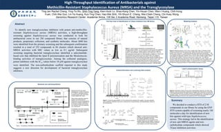
1. High-Throughput Identification of Antibacterials against
Methicillin-Resistant Staphylococcus Aureus (MRSA) and the Transglycosylase
Ting-Jen Rachel Cheng, Ying-Ta Wu, Shih-Ting Yang, Kien-Hock Lo, Shao-Kang Chen, Yin-Hsuan Chen, Wen-I Huang, Chih-Hung
Yuan, Chih-Wei Guo, Lin-Ya Huang, Kuo-Ting Chen, Hao-Wei Shih, Yih-Shyun E. Cheng, Wei-Chieh Cheng, Chi-Huey Wong
Genomics Research Center, Academia Sinica, 128 Sec 2 Academia Road, Nankang, Taipei 115, Taiwan
Abstract
To identify new transglycosylase inhibitors with potent anti-methicillin-
resistant Staphylococcus aureus (MRSA) activities, a high-throughput
screening against Staphylococcus aureus was conducted to look for
antibacterial cores in our 2M compound library that consists of natural
products, proprietary collection, and synthetic molecules. About 3600 hits
were identified from the primary screening and the subsequent confirmation
resulted in a total of 252 compounds in 84 clusters which showed anti-
MRSA activities with MIC values as low as 0.1 µg/ml. Subsequent
screening targeting bacterial transglycosylase identified a salicylanilide-
based core that inhibited the lipid II polymerization and the Moenomycin-
binding activities of transglycosylase. Among the collected analogues,
potent inhibitors with the IC50
values below 10 µM against transglycosylase
were identified. The non-carbonhydrate scaffold reported in this study
suggests a new direction for development of bacterial transglycosylase
inhibitors.
Figure 2. High-throughput identification of antibacterials and
transglycosylase inhibitors. (A) The fluorescence intensity and the coefficient
of variation (CV) values vs. bacteria culture in various dilutions. Bacterial
growth in 1536-well plates. Various dilutions of SA culture at 0.1 OD600/ml
were dispensed into the wells of a 1536-well plate, incubated for 17 hours and
the cell growth was monitored using 10% Alamar blue. The image and
fluorescence intensities were taken with Viewlux. (B) HTS campaign of
antibacterial screens. The remaining activity after inhibition of each individual
compound was counted and accumulated for the decision of hits. In this
activity histogram, a remaining activity less than 0.6 was used as the
threshold to obtain the primary screening hits. (C) High-throughput assay
development and validation of antibacterial screening in 1536-well plates.
Figure 1. The pathway of bacterial cell-wall biosynthesis, highlighting the glycosyltransfer steps and their inhibitors
Figure 3. Active hits against MRSA. (A) The confirmed hits consisted of 84 clusters. Gray bars represent
known antibiotics while black bars are for molecules from compound library. (B) Representative clusters of
the confirmed hits.
Figure 4. Compounds that showed potent
inhibition activities against bacterial TGase.
(A) Inhibitors identified in this study.
(B) Salicylanilides can be grouped into two
scaffolds. Molecules with scaffold I showed no
TGase inhibition activities whereas molecules
with scaffold II can inhibit TGase activities.
Figure 5. Modeling analysis of 42-10 (2) in complex with TGase. (A) Results of blind
docking of compound 2 to the surface of the TGase (PDB code 3hzs). Compound 2
mostly (~32%; avg score = -9 kcal/mole) fell within the moenomycin binding site.19, 20
(B)
The modeled complex of compound 2 with E. coli PBP (3fwl, blue stick presentation). The
main residues for interaction, Asn275, Tyr310, Tyr315, Ile314, Glu323, Arg325, Val354,
and Ala357 were labeled. Compound 2 was shown in green stick. The reference
structure (PDB coded 3hzs) was shown in grey wire. Nitrogen is in blue, oxygen in red,
bromine in dark red, and chlorine in green. (C) The structures of the compounds 6–11
prepared to identify the essential moieties in compounds 42-10 (2). Except compound 10,
the other five compounds showed no TGase inhibition activities at 100 µM.
Figure 6. Preparation of lipid II (A) and lipid II polymerization assay (B). (A) Lipid II was
prepared using chemoenzymatic method and conjugated with a fluorophore as described
in the Experimental sections. (B) NBD (marked in red)-labeled lipid II was polymerized by
functional TGase. The polymerized lipid II was further digested with muramidase and the
digested peptidoglycan monomer was analyzed by HPLC chromatography.
Summary
We decided to conduct a HTS of 2 M
compounds in our library by using the GNF
HTS system capable of screening nearly 1M
molecules a day for identification of new
hits against wild-type Staphylococcus
aureus. This strategy led to the identification
of new anti-MRSA agents and non-
carbohydrate compounds which showed
TGase inhibition activities.
Modeling analysisThe flow chart of identification of TGase inhibitors from 2M library.
Bacterial cell wall biosynthesis
Lipid II polymerization assay
Chemoinformatics and screening data analysis
Bacterial cell wall, also called
peptidoglycan, consists of repeating
β-1,4-linked N-acetylglucosaminyl-
N-acetylmuramyl units cross-linked
through peptide side chains.
Most of these antibiotics target the
peptide cross-linking step by
inhibiting the enzyme transpeptidase
directly (e.g. β-lactam antibiotics), or
by forming a complex with the last
two residues D-ala-D-ala dipeptides
(e.g. vancomycin).
No antibiotics have been developed
to target TGase except moenomycin
A , which has only been used in
animals.