Continuous Exposure to Chrysotile Asbestos Can Cause
1. See related Commentary on page 1378.
TUMORIGENESIS AND NEOPLASTIC PROGRESSION
Continuous Exposure to Chrysotile Asbestos Can Cause
Transformation of Human Mesothelial Cells via HMGB1 and
TNF-a Signaling
Fang Qi,*y
Gordon Okimoto,* Sandro Jube,* Andrea Napolitano,*y
Harvey I. Pass,z
Rozalia Laczko,* Richard M. DeMay,x
Ghazal Khan,x
Maarit Tiirikainen,* Caterina Rinaudo,{
Alessandro Croce,{
Haining Yang,*k
Giovanni Gaudino,* and
Michele Carbone*k
From the University of Hawai’i Cancer Center,* and the Department of Molecular Biosciences and Bioengineering,y
University of Hawai’i, Honolulu,
Hawaii; the Department of Pathology,k
John A. Burns School of Medicine, University of Hawai’i, Honolulu, Hawaii; the Division of Thoracic Surgery,z
Department of Cardiothoracic Surgery, Langone Medical Center, New York University, New York, New York; the Section of Cytopathology,x
Department of
Pathology, University of Chicago Medical Center, University of Chicago, Chicago, Illinois; and the Department of Science and Technological Innovation,{
University of Piemonte Orientale “Amedeo Avogadro,” Alessandria, Italy
Accepted for publication
July 17, 2013.
Address correspondence to
Michele Carbone, M.D., Ph.D.,
University of Hawai’i Cancer
Center, University of Hawai’i,
701 Ilalo St., Honolulu,
HI 96813. E-mail: mcarbone@
cc.hawaii.edu.
Malignant mesothelioma is strongly associated with asbestos exposure. Among asbestos fibers,
crocidolite is considered the most and chrysotile the least oncogenic. Chrysotile accounts for more than
90% of the asbestos used worldwide, but its capacity to induce malignant mesothelioma is still debated.
We found that chrysotile and crocidolite exposures have similar effects on human mesothelial cells.
Morphological and molecular alterations suggestive of epithelialemesenchymal transition, such as
E-cadherin down-regulation and b-catenin phosphorylation followed by nuclear translocation, were
induced by both chrysotile and crocidolite. Gene expression profiling revealed high-mobility group box-1
protein (HMGB1) as a key regulator of the transcriptional alterations induced by both types of asbestos.
Crocidolite and chrysotile induced differential expression of 438 out of 28,869 genes interrogated by
oligonucleotide microarrays. Out of these 438 genes, 57 were associated with inflammatory and immune
response and cancer, and 14 were HMGB1 targeted genes. Crocidolite-induced gene alterations were
sustained, whereas chrysotile-induced gene alterations returned to background levels within 5 weeks.
Similarly, HMGB1 release in vivo progressively increased for 10 or more weeks after crocidolite exposure,
but returned to background levels within 8 weeks after chrysotile exposure. Continuous administration
of chrysotile was required for sustained high serum levels of HMGB1. These data support the hypothesis
that differences in biopersistence influence the biological activities of these two asbestos fibers.
(Am J Pathol 2013, 183: 1654e1666; http://dx.doi.org/10.1016/j.ajpath.2013.07.029)
Malignant mesothelioma (MM) is an aggressive cancer of the
pleura and peritoneum, and less frequently of other meso-
thelial linings; it is strongly associated with asbestos expo-
sure and affects approximately 3200 individuals annually in
the United States.1
The median survival of MM patients is
approximately 1 year from diagnosis, despite surgical re-
section, chemotherapy, and radiotherapy.2,3
Asbestos is a nonspecific term commonly used to describe
any of six types of naturally occurring fibrous silicate minerals
that were widely used commercially during the 20th century.4
Asbestos fibers are divided into two major groups, serpentine
and amphibole, and are further distinguished based on their
chemical composition and crystalline structure.5
Serpentine
asbestos is chrysotile (white asbestos); amphibole asbestos
Supported by NIH grants NCI R01 CA106567 (M.C.), NCI R01
CA160715-0A (H.Y.), P01 CA114047 (M.C.), and the P30 CA071789
(UHCC Genomics Shared Resource); the Mesothelioma Applied Research
Foundation (H.Y.), the United-4 A Cure (H.Y.), the Hawai’i Community
Foundation (H.Y. and G.G.), the V foundation (H.Y.), and the University of
Hawai’i Foundation (M.C.).
Current address of R.L., Cardiovascular Research Center, University of
Hawai’i, Honolulu, HI.
Copyright ª 2013 American Society for Investigative Pathology.
Published by Elsevier Inc. All rights reserved.
http://dx.doi.org/10.1016/j.ajpath.2013.07.029
ajp.amjpathol.org
The American Journal of Pathology, Vol. 183, No. 5, November 2013
2. includes crocidolite (blue asbestos), amosite (brown asbestos),
anthophyllite, actinolite, and tremolite. It has been esti-
mated that chrysotile accounts for approximately 95% of
all asbestos used in the United States6
and 90% of asbestos
used worldwide.7,8
In the human body, amphibole fibers tend
to persist at sites of deposition, with fiber concentration
increasing with prolonged exposure, whereas chrysotile
fibers are usually rapidly cleared from the lungs.6
It is well
accepted that amphibole asbestos cause MM.9
Although
chrysotile can induce MM in animal experiments,10e16
its
carcinogenic role in humans is still debated, because epide-
miological studies have not proven a definitive causal asso-
ciation between chrysotile and MM.6,17,18
It has been
proposed that the mechanisms of asbestos carcinogenesis
may vary among different species19
; however, few studies
have investigated the molecular pathways induced by
chrysotile that may eventually lead to MM.5,20
Exposure to crocidolite induces necrosis of primary human
mesothelial (HM) cells, which is accompanied by passive
release of the damage-associated molecular pattern high-
mobility group box-1 protein (HMGB1).21
In the extra-
cellular space, HMGB1 leads to chronic inflammation
through the recruitment and accumulation of macro-
phages, which in turn actively secrete HMGB1 along
with several other cytokines, including tumor necrosis
factor (TNF-a), which plays a critical role in crocidolite-
mediated carcinogenesis.22
Epithelialemesenchymal transition (EMT) is a physio-
pathological process by which epithelial cells acquire mes-
enchymal shape and properties associated with cell migration
and cancer progression.23
EMT contributes to the histo-
morphological features of MM (ie, epithelioid versus biphasic
and sarcomatoid subtypes).23e25
TNF-a has been shown to
induce EMT in epithelial cells26,27
and in mesothelial cells,28
and HMGB1 has been also associated with EMT in alveolar
epithelial cells.29,30
EMT is characterized by increased ex-
pression of mesenchymal markers such as the cytoskeletal
proteins, vimentin, and a-smooth muscle actin31
and by
decreased expression of the epithelial cell adhesion molecule
E-cadherin,either at the transcriptional level26,32,33
or through
ubiquitin-mediated degradation.34,35
E-cadherin forms ad-
herent junctions that maintain cell adhesion in a multiprotein
complex that includes b-catenin.36
During EMT, phosphor-
ylation of b-catenin on tyrosine 142 (Y142) by receptor
tyrosine kinases (eg, c-Met) eventually results in disassem-
bling of the adhesion junction complex, degradation of
E-cadherin, and release of b-catenin.37
Depending on the
upstream messages, b-catenin can be either degraded or
translocated to the nucleus, where it is transcriptionally
active.34,38
The majority of established MM cell lines exhibit
nuclearaccumulation of b-catenin,39e41
suggesting a possible
contributory role of b-catenin in MM development.
In the present study, we compared the biological,
morphological, and transcriptional effects of crocidolite
and chrysotile on primary HM cells in tissue culture and in
mice.
Materials and Methods
Cell Lines and Culturing Conditions
HM cells are routinely cultured in our laboratory (University
of Hawai'i Cancer Center), isolated from pleural fluids from
patients with congestive heart failure or other benign
conditions.42
HM cells are established in cell culture in
Dulbecco’s modified Eagle’s medium containing 20% fetal
bovine serum (FBS) and are characterized morphologically
and by positive immunostaining for cytokeratin, HBME-1,
and calretinin and negative staining for LeuM1, Ber-Ep4,
B72.3, and carcinoembryonic antigen.42
THP-1 human
monocytes (ATCC TIB202; ATCC, Manassas, VA) are
cultured in RPMI 1640 medium supplemented with 10%
FBS and 0.05 mmol/L 2-mercaptoethanol. Experiments were
performed using Dulbecco’s modified Eagle’s medium
containing 10% FBS, unless otherwise specified.
Fiber Preparation
Chrysotile and crocidolite fibers were obtained from the
International Union against Cancer (Union Internationale
Contre le Cancer; UICC Lyon, France). Fibers were pro-
cessed as described previously.22,43
In brief, fibers were
baked at 150
C for 18 hours, suspended in PBS at 4 mg/mL,
triturated 10 times through a 22-gauge needle, and auto-
claved. Reference chrysotile B, Canadian, consisted of
a mixture of fibers from the following companies or mines:
Bell, Carey, Cassiar, Flintkote, Johns-Manville, Lake,
Normandie, and National. Reference chrysotile A, histori-
cally called Rhodesian, consisted of asbestos fibers mined in
Zvishavane (also spelled Shabani or Shavani), Matabeleland
South Province, Zimbabwe. Both chrysotile A and B are
predominantly made up of hydrous silicates of magnesia.
Reference crocidolite consisted of fibers from the Koegas
mine, a large asbestos mine in the Northern Cape province of
South Africa. This mineral is predominantly made up of
hydroxyl silicates of sodium, magnesium, and iron. The cell-
culture experiments were performed with 5 mg/cm2
for each
of the asbestos fiber types, unless specified otherwise. Cells
were cultured in the presence of fibers for different durations,
depending on the type of assay performed.
Asbestos Fiber Electron Microscopy
A drop of fiber suspension (40 mg/mL) was deposited onto
carbon conductive tape and placed on pin stubs for analysis
under a scanning electron microscope. Finally, the liquid
phase was evaporated by placing the sample in a drying
glass cloche for 24 hours. The chrysotile dimensional study
was performed under an Quanta 200 environmental scan-
ning electron microscope (FEI, Hillsboro, OR), equipped
with an energy-dispersive spectroscopy (EDS) microprobe
system (EDAX, Mahwah, NJ) in low-vacuum operational
mode, allowing sample characterization without coating by
Transforming Potential of Chrysotile
The American Journal of Pathology - ajp.amjpathol.org 1655
3. carbon or gold. The pressure used was 90 Pa, with accel-
erating voltage of 20 kV and a working distance of 10 mm.
The fibers were identified at Â2000 magnification and
counted. Length and diameter of each fiber were determined
at Â500, Â2000, or Â4000, depending on the dimensions.
In each sample, 200 fibers were measured, for a total of 600
crystals for each mineral phase. Samples were analyzed in
triplicate. Average length and diameter of chrysotile B fibers
were 19.9 Æ 19.6 mm and 0.47 Æ 0.27 mm, respectively
(Supplemental Figure S1). Average length and diameter of
reference crocidolite fibers were 13.60 Æ 20.24 mm and
0.60 Æ 0.45 mm, respectively (Supplemental Figure S2).
TNF-a Signaling Studies
To stimulate TNF-a signaling, HM cells were incubated with
10 ng/mL recombinant TNF-a (Sigma-Aldrich, St. Louis,
MO). To inhibit endogenous TNF-a activity, a neutralizing
antibody (antieTNF-a; RD Systems, Minneapolis, MN)
was used at a concentration of 1 mg/mL.
Immunofluorescence Staining
Asbestos Staining
Asbestos fibers were stained as described.44
In brief,
asbestos fibers were preincubated with normal rabbit serum
(Santa Cruz Biotechnology, Santa Cruz, CA) and added to
HM cells for 24 hours. The HM cells were then fixed in 4%
paraformaldehyde, washed in PBS, permeabilized using 0.2%
Triton X-100, and saturated with bovine serum albumin.
Incubation was performed with Alexa Fluor 594econjugated
anti-rabbit secondary antibody and Alexa Fluor 488e
conjugated phalloidin (Life Technologies, Carlsbad, CA).
Slides were washed in PBS and mounted with aqueous
mounting medium premixed with the nuclear marker DAPI
(Vector Laboratories, Burlingame, CA) for fluorescence
microscopy.
b-Catenin Staining
Cells were cultured and fixed as described above. Bovine
serum albumin (1% solution) was used to block nonspecific
immunoreactivity. Incubation with antieb-catenin or control
rabbit IgG (Santa Cruz Biotechnology) was followed by
labeling with Alexa Fluor 594econjugated secondary anti-
body and Alexa Fluor 488econjugated phalloidin (Life
Technologies). Slides were mounted as described above for
fluorescence microscopy.
Cell Proliferation and Viability Assay
To evaluate and compare effects on cell proliferation and
viability, HM cells were seeded in 96-well plates with six
replicates per treatment condition. Experiments were per-
formed in the presence or absence of crocidolite or chrysotile
asbestos fibers. Cells were preincubated with 10 ng/mL
TNF-a or PBS for 24 hours and then were exposed to asbestos
for an additional 24 or 48 hours before viability was assessed
using an MTS assay according to the manufacturer’s protocol
(Promega, Madison, WI). MTS assays were quantified by
using a colorimetric reaction mix and microplate reader at
490 nm (Bio-Rad Laboratories, Hercules, CA). To avoid the
intrinsic variability of this method, we performed the MTS
assay for all time points in one measurement on the same day.
Cell Cytotoxicity Assay
Cytotoxicity was assessed by measuring the amount of
lactate dehydrogenase (LDH) released from the cytosol of
damaged cells. A LDH cytotoxicity detection kit (Roche
Diagnostics, Indianapolis, IN) was used according to the
manufacturer’s protocol. In brief, HM cells were seeded in
a 96-well tissue culture plate at a density of 5 Â 103
cells per
well. The next day, the medium was changed, and 100 mL of
asbestos fiber suspensions at different concentrations was
added. After 24 hours, 100 mL of supernatant per well was
harvested and transferred into a new 96-well, flat-bottom
plate. LDH substrate was added to each well and incubated
at room temperature protected from light. The absorbance of
the samples was measured at 490 nm with an enzyme-linked
immunosorbent assay (ELISA) reader. Cytotoxicity was
calculated as % cytotoxicity Z [(experimental value À low
control) Â100]/(high control À low control), where the low
control is assay medium plus cells and the high control is
assay medium with 2% Triton X-100 plus cells.
Western Blotting
For total cell protein extraction, cells were washed with cold
PBS, scraped in radioimmunoprecipitation assay lysis buffer
(100 mmol/L NaCl, 10 mmol/L Tris, 0.1% SDS, 1% Triton
X-100, and 5 mmol/L EDTA at pH 7.2) containing a
protease inhibitor cocktail (Roche Diagnostics), and incu-
bated for 30 minutes on ice.
A nuclear extraction kit (Active Motif, Carlsbad, CA)
was used according to the manufacturer’s protocol to per-
form cell fractionation. In brief, cultured cells were rinsed in
cold PBS, scraped, and centrifuged in PBS supplemented
with phosphatase inhibitors. The cell pellets were lysed in
hypotonic buffer supplemented with detergents for 15
minutes on ice to access cytoplasmic proteins. The nuclear
pellets were lysed separately in nuclear lysis buffer for 30
minutes on ice to access the nuclear proteins.
Equal amounts (20e50 mg) of cell lysate per lane were
applied on 4% to 12% gradient gels (NuPAGE; Life Tech-
nologies). Proteins separated on bis-tris gels were transferred
to polyvinylidene difluoride membranes (Immobilon-P;
EMD Millipore, Billerica, MA) and blocked with 5% bovine
serum albumin before incubation with specific primary and
secondary antibodies. The antibodies used were used histone
1, E-cadherin, b-catenin, vimentin (Santa Cruz Biotech-
nology); GAPDH (Chemicon; EMD Millipore); Y142-
phosphorylated b-catenin (ECM Biosciences, Versailles,
Qi et al
1656 ajp.amjpathol.org - The American Journal of Pathology
4. KY); and HMGB1 and a-smooth muscle actin (Abcam,
Cambridge, MA). The relative density of Western blot bands
was evaluated by the ImageJ software version 1.45s (NIH,
Bethesda, MD). Briefly, the density of target bands were
normalized versus the corresponding loading control band
density and compared to vehicle exposed controls and
expressed as relative density.
Quantitative RT-PCR
HM cells were exposed to asbestos fibers at 5 mg/cm2
for
24 hours. Total RNAs were isolated using an RNeasy kit
(Qiagen, Valencia, CA) according to the manufacturer’s
protocol and were treated with RNase-free DNase (Qiagen).
For each sample, 1 mg RNA was used. GAPDH was used as
the internal control. Quantitative RT-PCR (RT-qPCR) for
TNF-a and GAPDH mRNA was performed in triplicate in
25-mL total reaction volumes using a SYBR Green master
mix (Bio-Rad Laboratories). A LightCycler 480 RT-qPCR
system (Roche Diagnostics) was used according to standard
procedures. The primers were as described previously.22,45
Cell Transformation Assay
HM cells were cultured and seeded in six-well plates at
a density of 3 Â 105
cells per well. The next day, the medium
was switched to Dulbecco’s modified Eagle’s medium with
10% FBS. In the TNF-aedependent transformation assays,
HM cells were pretreated with 10 ng/mL TNF-a or with PBS
(vehicle) control for 24 hours before exposure to 5 mg/cm2
asbestos fibers or PBS control for another 48 hours. Culture
medium containing 10 ng/mL TNF-a was replaced twice
a week.
For coculture cell transformation assays, the HMe
macrophage coculture assay was performed as described
previously.46
THP-1 peripheral blood monocytes were dif-
ferentiated into macrophages as described previously.46
In
brief, tissue-culture inserts containing differentiated macro-
phages were placed above HM cell cultures. Membranes of
the inserts allowed the cytokines and growth factors produced
by macrophages to reach the lower chamber, where HM cells
were cultured. HM cells were cocultured with macrophages
and then were exposed to crocidolite fibers, chrysotile fibers,
or vehicle alone for 48 hours. Fibers not engulfed by cells and
floating in the medium were washed off. Freshly differenti-
ated macrophages and culture medium were replaced twice
a week. After 6 to 8 weeks, foci were identified by crystal
violet staining and were counted under a light microscope.
Microarray Profiling
Whole-genome expression arrays were used to characterize
the transcriptional response of HM cells exposed to crocidolite
or chrysotile fibers. HM cells were cultured in six-well plates
with 2 Â 105
cells/well. Experimentalconditionswere HM cells
with PBS (vehicle control), HMemacrophage cocultures46
prepared as described above, and HM cells cultured in the
presence of TNF-a exposed to crocidolite, chrysotile, or PBS
for 48 hours and 5 weeks. The experiments were performed in
duplicate to verify reproducibility of the findings. RNA was
extracted using a miRNeasy micro kit (Qiagen), and RNA
quality was determined using an Agilent Bioanalyzer system
with RNA 6000 Nano or Pico chips (Agilent Technologies,
Santa Clara, CA). The average RNA integrity number (RIN)
was 9.2. GeneChip human gene 1.0 ST arrays (Affymetrix,
Santa Clara, CA) were used to interrogate expression of 28,869
genes, with two replicate samples for each condition, by
applying the Affymetrix GeneChip whole-transcript sense
target labeling protocol with 100 ng of total RNA.
Microarray Data Analysis
The raw .CEL file data for each array were quantified,
quantile-normalized, and log2etransformed, to facilitate
comparisons between different samples. The array data were
modeled using two-factor analysis of variance with experi-
mental condition (seven levels) and time (two levels) as the
two factors. The experimental design also included two
biological replicates per level for each factor, as required for
such an analysis. The two-factor analysis of variance model
was applied to all 28 arrays. A set of 57 genes were even-
tually identified that were both highly variable between two
or more of the seven conditions [false discovery rate (FDR)
0.05] and highly enriched for biological processes associated
with inflammatory and immune response and cancer as
determined by Ingenuity Pathway Analysis software version
6 (IPA; Ingenuity Systems, Redwood City, CA). The false
discovery rate based on the BenjaminieHochberg method
was used to control for multiple comparisons. Subsequent
pairwise analyses that compared fiber-treated HM cells with
vehicle and HMemacrophage cocultures using a noise-
adaptive fold change to identify differentially expressed
genes (DEGs) for each comparison.47
IPA was then used to
predict upstream regulators and downstream effects based on
the observed patterns of pairwise differential expression.
Microarray Data Validation
For the technical and biological validation and replication of
gene expression levels generated in the microarray expres-
sion profiling assay, RT-qPCR was used to measure gene
expression levels for six genes. These genes were chosen
because they were either differentially expressed between the
different treatment conditions (ATL3, CCL20, IL24, and
PTGS2 from 438 DEGs) or represented a stable gene across
our various experimental conditions (TERF2IP). In addition,
a reference gene (GAPDH ) was used to normalize mRNA
expression levels. In brief, validation RT-qPCR was per-
formed on the same samples that were used in the microarray
assays and on the samples from repeated experiments.
The cDNA was prepared from 1 mg of untreated total RNA
using an Applied Biosystems high-capacity cDNA reverse
Transforming Potential of Chrysotile
The American Journal of Pathology - ajp.amjpathol.org 1657
5. transcription kit (Life Technologies). qPCR was performed
for the seven genes using TaqMan gene expression assays
and TaqMan universal PCR master mix with the recom-
mended thermal profiles on a 7900HT fast real-time PCR
system (Life Technologies). Relative normalized expression
levels between samples were calculated using the 2(ÀDDCT)
method.
Prediction of Upstream Transcriptional Factors
IPA was used to predict the likely upstream transcription
factors (TFs) for a given set of DEGs (for here, 438 DEGs) in
terms of activation z-score and number of downstream targets
that are also DEGs. For a given TF, the direction of change of
each target in our set of DEGs was compared with the pre-
dicted effect based on the current literature to arrive at a z-
score that implies activation or inactivation of the TF of
interest. If the change in expression across all target genes is
positively correlated with prediction, then the TF is viewed as
relatively more activated in the treated samples, compared
with the controls. If the target genes are negatively correlated
with prediction, then the TF is viewed as less activated,
compared with the controls. The intersection P value
measures the probability that an observed number of down-
stream targets in the data are due to chance. A given TF is
significant if it has a z-score of 2 in absolute value and has an
intersection P value of 0.01.
Animal Experiments and Cytokine Detection in Serum
Human HMGB1 ELISA was used to measure the levels of
HMGB1 in asbestos-exposed mice sera. BALB/c (BALB/
cAnNCrl) female mice aged 6 to 8 weeks (Charles River
Laboratories International, Wilmington, MA) were housed
and handled under aseptic conditions, in accordance with
University of Hawai’i Institutional Animal Care and Use
Committee (IACUC) guidelines. Mice were weighed and
randomly assigned to three different groups: i) negative
control (vehicle PBS); ii) high-dose, short-term injection
with crocidolite or chrysotile; and iii) low-dose, long-term
injection with crocidolite or chrysotile (n Z 10 animals per
group). Mice in the high-dose, short-term groups received
two intraperitoneal injections, 1 week apart, of 2.5 mg of the
respective asbestos fiber; mice in the low-dose, long-term
groups received 10 intraperitoneal injections, 1 week apart,
of 0.5 mg of the respective asbestos fiber. Thus, animals in
all of the asbestos-exposed groups received a total of 5 mg
of fiber. Each fiber injection had a volume of 500 mL. The
control group received intraperitoneal injection of 500 mL
PBS vehicle on the same schedule as the corresponding
high-dose or low-dose treatment protocol. In all groups,
blood was drawn every 2 weeks. The sera were collected
and used for the detection of HMGB1 levels with an ELISA
kit (IBL International, Toronto, ON, Canada). After
completion of the experiment, mice were euthanized and
necropsied according to IACUC regulations.
An ELISA kit (RD Systems) was used to measure
TNF-a levels in the conditioned medium of HM cells ex-
posed to either crocidolite or chrysotile asbestos fibers. For
the detection of extracellular TNF-a released by HM cells,
2 Â 105
cells were cultured in Dulbecco’s modified Eagle’s
medium with 1% FBS for 24 hours. The culture medium
was then collected and concentrated by ultrafiltration using
Amicon Ultra centrifugal filters (EMD Millipore), and
50-mL aliquots were assayed in duplicate by ELISA. All
culture media were collected under identical conditions.
Statistical Analysis
All experiments were performed at least three times. One-
way analysis of variance was used for MTS and LDH data
analysis. Unpaired U-test was used for microarray validation.
For other data analysis, unpaired Student’s t-test was per-
formed using GraphPad Prism software version 5 (GraphPad
Software, La Jolla, CA). P 0.05 was considered significant.
Results
Chrysotile Induces Cell Death and Morphological
Changes in Primary HM Cells
To compare the biological effects of crocidolite and chrys-
otile on HM cells, the cells were first exposed to 5 mg/cm2
of
each of the asbestos fibers for 48 hours and analyzed for the
viability and morphology. Fiber analyses showed that the
majority of fibers were short and measured within the ranges
found in human MM48
(Supplemental Figures S1 and S2).
Fewer chrysotile-exposed HM cells than crocidolite-exposed
HM cells were adherent on tissue culture dishes (46 Æ 9%
versus 73 Æ 12%; P 0.0001). Moreover, in chrysotile-
exposed HM cells, a greater number of attached (ie,
surviving) cells acquired a spindle-shaped morphology,
compared with crocidolite-exposed cells (48 Æ 6% versus 26
Æ 8%; P Z 0.0173), a possible indication of EMT (discussed
below) (Figure 1A and Supplemental Figure S3). The greater
numbers of dead and spindle-shaped surviving cells
suggest that chrysotile induces higher cellular stress than
crocidolite, eventually resulting in cell death. Viability and
cytotoxicity assays revealed that both fibers are cytotoxic in
a density-dependent manner, with chrysotile being signif-
icantly more cytotoxic than crocidolite (Figure 1, B and C).
TNF-a Significantly Reduces Chrysotile Cytotoxicity
We previously showed that crocidolite-exposed HM cells
are able to engulf asbestos fibers and that some HM cells
survive, despite asbestos-induced damage. Cell survival is
favored by a proinflammatory microenvironment, mostly
mediated by HMGB1 and TNF-a,21,22
which are released
by both inflammatory cells (ie, macrophages) and HM cells.
In the present study, chrysotile caused the release of high
levels of HMGB1 into the conditioned medium in a fiber
Qi et al
1658 ajp.amjpathol.org - The American Journal of Pathology
6. densityedependent manner (Figure 2). In fact, chrysotile
caused HM cells to express and release higher levels of
TNF-a, compared with crocidolite (Figure 3, A and B).
To investigate the role of TNF-a in cell survival, HM cells
were pretreated for 24 hours with 10 ng/mL TNF-a before
exposure to asbestos for 48 hours. In TNF-aepretreated HM
cells, cytotoxicity of both crocidolite and chrysotile fibers
was significantly reduced (Figure 3C). These results were
confirmed by cell counting (data not shown).
These results reveal that chrysotile induces HMGB1 and
TNF-a secretion, an effect that has been linked to crocid-
olite carcinogenicity.
Chrysotile Has in Vitro Transforming Potential
To study whether chrysotile has in vitro transforming
potential, we cultured HM cells in the presence of 10 ng/mL
TNF-a and either chrysotile or crocidolite and then
compared the number of three-dimensional foci induced by
these fibers. Foci formation is a measure of carcinogenicity
in tissue culture.5,49
After 4 weeks in culture, chrysotile-
exposed HM cells formed fewer foci, compared with
crocidolite-exposed HM cells (21 Æ 1 versus 53 Æ 5; P Z
0.021), whereas HM cells exposed to crocidolite or chrys-
otile alone (ie, without TNF-a) died within 2 weeks, which
supports the critical role of TNF-a in maintaining HM cell
viability after asbestos exposure (Figure 4). To validate
these results, HM cells were exposed to asbestos fibers in
the presence of macrophages in a coculture system that
mimics the process of macrophage recruitment and activa-
tion to sites of fiber deposition leading to the secretion of
HMGB1, TNF-a, and other cytokines, as described previ-
ously.21,46
After 8 weeks in culture, greater numbers of
three-dimensional foci developed in crocidolite-exposed
HM cells than in chrysotile-exposed HM cells (12 Æ 2
versus 3 Æ 1 foci; P Z 0.008). No foci developed in
HMemacrophage cocultures without asbestos exposure
(Supplemental Figure S4).
These data revealed that, although both types of asbestos
induce HMGB1 and TNF-a release, crocidolite induced
greater numbers oftransformed foci, comparedwithchrysotile.
Genome-Wide Transcriptional Response of HM Cells to
Chrysotile and Crocidolite Fibers over Time
To determine whether the differing ability of crocidolite and
chrysotile to induce three-dimensional foci is linked to
different gene pathways induced by these fibers, we studied
the expression levels of more than 28,000 genes at two
different time points (48 hours and 5 weeks) under the
following experimental conditions: i) HM cells with PBS
(vehicle); ii) HMemacrophage cocultures (MF) or the
coculture with either crocidolite or chrysotile fibers
(MFþCROC, MFþCHRY); and iii) HM cells treated with
TNF-a only (TNF-a) or with TNF-a and either crocidolite
or chrysotile (TNF-aþCROC, TNF-aþCHRY).
Gene expression profiling at the 48-hour time point
characterizes the early events related to short-term cell
exposure to fibers, such as cell survival or change of
morphology, whereas gene expression at the 5-week time
point is expected to correspond to events that occur after
long-term exposure, such as foci formation.
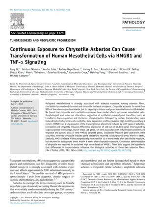
Figure 2 Chrysotile induces HMGB1 secretion. Western blotting
revealed HMGB1 released into the conditioned medium on exposure of HM
cells to either crocidolite or chrysotile fibers. GAPDH was used as loading
control. CM, conditioned medium; IC, intracellular; M, untreated HM cells.
Figure 1 Morphological changes and cell death induced by asbestos
fibers. A: Representative light microscopy images showing the morpho-
logical change of HM cells from predominantly rounded and epithelial to
spindle and fibroblast-like after 48-hour exposure to crocidolite (CROC) or
chrysotile (CHRY), compared with PBS (vehicle). Adherent cells were
counted in five different fields, and percentages were calculated for control
versus asbestos-exposed HM cells. B and C: HM cells were exposed to
different densities of asbestos (1 to 10 mg/cm2
) for 24 hours and subjected
to MTS viability assay (B) or LDH cytotoxicity assay (C). Data are repre-
sentative of one experiment out of three performed and expressed as means
Æ SD. *P 0.05. Original magnification, Â100.
Transforming Potential of Chrysotile
The American Journal of Pathology - ajp.amjpathol.org 1659
7. Total RNA was extracted from cells for each condition
of interest at each time point, and global gene expression
was assayed using Affymetrix GeneChip human gene 1.0
ST arrays. After data normalization and log2 transformation,
two-way analysis of variance, hierarchical cluster analysis,
438 DEGs were identified, and among these 438 DEGs, IPA
were used to identify a subset of 57 genes that were highly
variable over the seven experimental conditions of interest and
that followed a coordinate expression pattern of gene
expression. These 57 genes were statistically enriched for
biological processes associated with carcinogenesis, inflam-
matory response, and immune response (Figure 5A and
Table 1). Heat mapping of the 57 genes (Figure 5B) showed
persistent gene activation over time (ie, from the 48-hour to
the 5-week time point) in TNF-aetreated HM cells, inde-
pendent of asbestos fiber exposure (TNF-a, TNF-aþCROC,
TNF-aþCHRY), as well as in crocidolite-exposed HMe
macrophage coculture (MFþCROC). In chrysotile-exposed
HMemacrophage coculture (MFþCHRY), however, acti-
vation of those genes was observed at 48 hours but had
declined to baseline levels at 5 weeks (Figure 5, A and B). To
validate the microarray experiment, we performed RT-qPCR
for 4 of the 438 differentially expressed genes (DEGs) and 1
Figure 3 TNF-a is induced in HM cells after asbestos exposure and signifi-
cantly reduces asbestos cytotoxicity. A: HM cells were exposed to 5 mg/cm2
of
crocidoliteorchrysotilefibersortoPBS(vehiclecontrol)for24hoursandqPCRfor
TNF-a mRNA was performed, using GAPDH as reference. B: TNF-a protein levels in
conditioned medium from vehicle control HM cells and HM cells exposed to
crocidolite or chrysotile at 5 mg/cm2
for 24 hours. C: HM cells were preincubated
for 24 hours either with 10 ng/mL TNF-a (solid lines) or with PBS (dashed lines),
andthen5mg/cm2
crocidoliteorchrysotilefiberswereaddedandcellviabilitywas
measured by MTS assay after an additional 24 and 48 hours. As control, HM cells
were pretreated with PBS for 24 hours. Data are representative of one experiment
out of three performed and expressed as means Æ SD. *P 0.05.
Figure 4 Transforming potential of crocidolite and chrysotile fibers in
presence of TNF-a. HM cells were pretreated with 10 ng/mL TNF-a and
exposed to crocidolite, chrysotile, or PBS (vehicle). A: HM cells were
pretreated with PBS and exposed to PBS (top left image) or were pre-
treated with TNF-a and exposed to PBS (top right image), to crocidolite
(bottom left image), or to chrysotile (bottom right image). After 4
weeks, three-dimensional foci were observed in both crocidolite- and
chrysotile-exposed HM cells, but not in PBS-exposed cells. B: Foci were
identified by crystal violet staining and were counted under light
microscopy. Data are expressed as means Æ SD. Experiments were per-
formed three times, and data are representative of one experiment out of
three performed. *P 0.05. Original magnification: Â100.
Qi et al
1660 ajp.amjpathol.org - The American Journal of Pathology
8. non-DEG. The results confirmed the reliability of the micro-
array experiment (Supplemental Figure S5).
To isolate the DEGs associated with exposure to specific
fibers, pairwise analyses of whole-genome expression data
were performed from HMemacrophage samples exposed to
either chrysotile (MFþCHRY) or crocidolite (MFþCROC)
at 48 hours and 5 weeks. Upstream regulatory factors and
downstream effects associated with DEGs at 48 hours and 5
weeks were identified by IPA.
Notably, at 48 hours, HMGB1 was a top predicted
upstream regulator of DEGs observed for both experimental
conditions. Statistically significant downstream effects were
observed on canonical pathways, biological processes, and
pathways related to cancer, inflammatory response, immu-
nological disease, and IL-17 signaling. Venn diagramming
(Figure 5C) and IPA network analysis (Supplemental
Figure S6) showed that 10 DEGs downstream of HMGB1
were shared between the two experimental conditions
(MFþCROC and MFþCHRY) at 48 hours. These results
indicate that similar inflammatory and immune response
pathways were activated early after exposure to either
crocidolite or chrysotile fibers in the coculture system. At
the 5-week time point, however, only crocidolite still in-
duced a similar pattern of transcriptional activation among
the HMGB1 downstream target genes (Figure 5C and
Supplemental Figure S6).
The IPA analysis showed that nuclear factor NF-kB was
also among the top predicted upstream regulators of DEGs.
Unlike HMGB1, NF-kB was activated in all of the experi-
mental conditions, including MFþCHRY at 5 weeks
(Supplemental Tables S1 to S6). To identify the subset of
genes that are uniquely regulated by HMGB1 or synergisti-
cally regulated by both HMGB1 and NF-kB, the HMGB1 and
NF-kB downstream DEGs were compared (Supplemental
Figure S7). Because TNF-a is heavily involved in asbestos-
induced chronic inflammation and is a known major acti-
vator of NF-kB, the HMGB1 and TNF-a downstream DEGs
were also compared (Supplemental Figure S8). The results
showed that the downstream targets of HMGB1 are also
downstream targets of NF-kB and TNF-a, but not vice versa.
Moreover, IFIT1 was identified as a downstream target
that is specifically related to HMGB1 but not to NF-kB
(Supplemental Figures S7 and S8).
Altogether, these results suggest that crocidolite and
chrysotile induce similar transcriptional response, that
Figure 5 Gene expression analysis of HM cells exposed to crocidolite and
chrysotile. A: Steps leading to the identification of 57 DEGs. Out of 28,869
genes tested via microarray profiling, 438 genes were identified as highly
variable between two or more of the 14 conditions (FDR 0.05). Among these,
57 DEGs were highly enriched for biological processes associated with
inflammatory and immune response and cancer and were selected for further
analysis. B: Clustered heat mapping shows the expression pattern for the 57
genes identified by analysis of variance, with the rows hierarchically clustered
into groups of genes with similar expression profiles at 48 hours and at 5 weeks.
Activation is shown in red and inhibition in green. The HM cells were cocultured
with macrophages and exposed to no fiber (MF), crocidolite (MFþCROC), or
chrysotile (MFþCHRY) or were treated with TNF-a alone (TNF-a) or TNF-a plus
crocidolite (TNF-aþCROC) or chrysotile (TNF-aþCHRY). C: The expression of
a total of 14 HMGB1 downstream target genes was altered in cells exposed to
chrysotile and crocidolite. Overall, genes alterations were sustained in cells
exposed to crocidolite, while were short lasting in cells exposed to chrysotile.
Areas in pink indicate genes whose expression was altered by crocidolite; areas
in blue, genes altered by chrysotile; overlapping areas, genes expressions
altered by both fibers. At 5 weeks, IFIT1 gene expression was upregulated in
chrysotile exposed HM-macrophage cocultures and down-regulated in crocid-
olite exposed cultures, thus IFIT1 is reported twice in the respective areas.
Table 1 The Set of 57 Genes Coordinately Expressed under
Crocidolite and Chrysotile Exposure
Symbol Symbol Symbol Symbol
AMIGO2 HIVEP2 NFKBIA SLC25A37
ARFGAP3 IFNGR2 NFKBIZ SLC7A2
C5orf62* IL7R NKX3-1 SMOX
CCL2 IL8 NR4A2 SNX9
CCL20 IRAK2 NRG1 SOD2
CSF3 ITPRIP PANX1 TMPRSS15
CXCL1 KIAA0247 PID1 TNFAIP2
CXCL2 KRTAP21-2 PIM3 TNFAIP3
CXCL3 KYNU PPAP2B TNFAIP6
CXCL5 LUZP1 PRRX1 TNFAIP8
CXCL6 MAP2K1 PTGS2 TNIP1
DENND5A MRGPRX3 RIPK2 TRAF1
EHD1 NFKB1 RNF19B UBTD2
G0S2 NFKB2 SLC25A37 UGP2
GFPT2
Crocidolite and chrysotile induced coordinate expression of 57 out
of 28,869 genes interrogated by oligonucleotide microarrays (Figure 5,
A and B).
*C5orf62 is now reclassified as SMIM3.
Transforming Potential of Chrysotile
The American Journal of Pathology - ajp.amjpathol.org 1661
9. crocidolite induces a more persistent transcriptional response
over time compared with chrysotile, and that HMGB1 acti-
vates a unique pattern of downstream target genes (compared
with NF-kB and TNF-a) that appear to play an important role
in both crocidolite-mediated and chrysotile-mediated cell
transformation.
In Vivo, Prolonged Chrysotile Exposure Induces
Sustained HMGB1 Serum Levels
Our microarray data showed that crocidolite induces a
more persistent transcriptional response over time, and that
HMGB1 is one of the top upstream mediators. To validate
these results in vivo, HMGB1 levels were assayed in the sera
of BALB/c mice injected intraperitoneally with crocidolite or
chrysotile fibers in a high-dose, short-term protocol (two
injections of 2.5 mg separated by 1 week, for a total 5.0 mg) or
in a low-dose, long-term protocol (10 weekly injections of 0.5
mg each, for a total 5.0 mg). Blood was collected every 2
weeks, and sera were assayed for HMGB1 by ELISA. For
mice injected according to the high-dose, short-term protocol,
HMGB1 serum levels increased constantly over time in
crocidolite-injected mice, whereas in chrysotile-injected mice
the levels of HMGB1 in the sera dropped to levels similar to
those of noninjected mice within 6 to 10 weeks from the last
chrysotile injection (Figure 6A). In contrast, in mice injected
according to the low-dose, long-term protocol, HMGB1
levels were higher than the control group up to 10 weeks,
regardless of the type of fiber (Figure 6B).
In summary, under a high-dose, short-term injection
protocol, crocidolite induced increasing levels of HMGB1
over the course of the experiment, whereas the HMGB1
increase caused by chrysotile was transient, declining to
background levels within 6 to 10 weeks from exposure. In
contrast, prolonged chrysotile exposure (low-dose, long-
term protocol) induced the same sustained HMGB1 sera
levels as were observed for crocidolite.
Asbestos Fibers Suppress E-Cadherin Expression and
Modulate b-Catenin Signaling
The microarray data showed that E-cadherin, a key mediator
of b-catenin signaling and EMT, was among the most down-
regulated genes in our experimental conditions (data not
shown). This evidence matches the initial observation that
asbestos fibers caused morphological changes, possibly
consistent with EMT induction (Figure 1A and Supplemental
Figure S3). Based on these data, we performed further
investigations of the association between asbestos fibers,
expression of E-cadherin and other EMT markers, and b-
catenin signaling.
A significant reduction in the levels of the epithelial
marker E-cadherin was detected when HM cells were
exposed to either crocidolite or chrysotile (Figure 7A). In
contrast, two mesenchymal markers (ie, vimentin and
a-smooth muscle actin) were up-regulated (Supplemental
Figure S9). Taken together, these findings are supportive
of EMT induced by both chrysotile and crocidolite. Having
already shown that TNF-a is secreted by HM cells exposed
to both chrysotile and crocidolite and that TNF-a secretion
is related to cell survival and transformation (Figures 3 and
4), we tested whether TNF-a, either secreted on asbestos
fiber exposure or introduced exogenously, modulates E-
cadherin expression and EMT in HM cells. The cells were
pretreated with TNF-a or with TNF-a neutralizing antibody
for 24 hours, then exposed to crocidolite or chrysotile fibers or
to PBS (vehicle) for an additional 48 hours. Both fibers
induced E-cadherin down-regulation; however, chrysotile
induced a significantly sharper reduction, compared with
crocidolite. Moreover, the treatment with exogenous TNF-a
further induced E-cadherin down-regulation. TNF-a neu-
tralizing antibody impaired E-cadherin down-regulation
(Figure 7A). Taken together, these results suggest that TNF-a
signaling is directly related to the changes in E-cadherin and
to EMT in HM cells exposed to asbestos.
Phosphorylation of b-catenin at Y142 is known to be
a key event in the disassembling of the multiprotein com-
plex including E-cadherin that results in loss of adherent
junctions.37
We therefore investigated b-catenin phosphor-
ylation at Y142 in HM cells exposed to either crocidolite or
Figure 6 HMGB1 serum levels are long-term persistent after exposure
to crocidolite but not to chrysotile. HMGB1 serum levels were measured in
mice injected with a total of 5 mg of crocidolite or chrysotile in either
a high-dose, short-term protocol with two weekly injections of 2.5 mg each
(A) or a low-dose, long-term protocol with 10 weekly injections of 0.5 mg
each (B). Data are expressed as means Æ SD. n Z 10 animals per group.
Qi et al
1662 ajp.amjpathol.org - The American Journal of Pathology
10. chrysotile fibers for 16 and 24 hours. Y142-phosphorylated
b-catenin substantially increased in cells exposed for 24
hours to either type of fiber, compared with controls
(Figure 7B). Subsequently, we investigated the distribution
of b-catenin in HM cells exposed to either crocidolite or
chrysotile fibers. In vehicle-exposed HM cells, b-catenin
localized in the cytoplasm. HM cells exposed to either
crocidolite or chrysotile exhibited a marked increase of
b-catenin in the nuclear fraction, which was more evident in
chrysotile-exposed cells (Figure 7C). These results were
validated by immunofluorescence. Both cytoplasmic and
nuclear staining for b-catenin were observed in HM cells
exposed to either crocidolite or chrysotile fibers, whereas
only cytoplasmic staining was evident in vehicle-exposed
cells (Figure 7D). A further increase in the amount of
nuclear b-catenin, paralleled by a decrease in cytoplasmic
b-catenin, was observed in crocidolite- or chrysotile-exposed
HM cells treated with TNF-a. A marked decrease in nuclear
b-catenin was observed in crocidolite- or chrysotile-exposed
HM cells in the presence of antieTNF-a (Figure 7E). The
downstream targets of b-catenin COX2 and MMP7 were
also transcriptionally induced (data not shown).
Taken together, these data indicate that asbestos induces
both down-regulation of E-cadherin and up-regulation of
mesenchymal markers (ie, vimentin and a-smooth muscle
actin) associated with EMT and also induces phosphoryla-
tion of b-catenin at Y142, with release of b-catenin from
the adherent junctions and its accumulation, followed by
nuclear translocation.
To further confirm these findings, all of the in vitro
experiments except the microarray analysis were repeated
with chrysotile fibers of a different origin (ie, chrysotile A,
from Zimbabwe). Chrysotile A exerted effects largely
overlapping with those obtained with chrysotile B (from
Canada) (Supplemental Figure S10).
In summary, the present results show that chrysotile has
the capacity to induce in vitro and in vivo molecular changes
that are similar to those observed with crocidolite. We
Figure 7 Chrysotile induces more E-cadherin down-regulation and b-catenin nuclear translocation, compared with crocidolite. A: Representative Western blots
show E-cadherin expression in HM cells exposed to crocidolite (C), chrysotile (Y), or mock treatment (M) with no asbestos fiber in the presence of PBS (vehicle),
TNF-a, or TNF-a neutralizing antibodies (antieTNF-a) (top panel). Asbestos fibers induce endogenous TNF-a secretion into cultured medium (Figure 3), and
antieTNF-a neutralizes the secreted endogenous TNF-a. A: The relative expression of E-cadherin versus GAPDH was evaluated by bands relative density using
ImageJ software (NIH, Bethesda, MD) (bottom panel). B: Representative Western blots of HM whole-cell lysates show expression of Y142-phosphorylated
b-catenin in different conditions. C: Representative Western blots of b-catenin in nuclear and cytoplasmic extracts of HM cells in control mock treatment or
increasing densities of crocidolite or chrysotile. D: Immunofluorescence staining shows b-catenin (Texas Red) distribution in HM cells exposed to crocidolite or
chrysotile as indicated. DAPI (blue)and phalloidin (green) were usedas reference markers labelingHM cell nuclei and F-actin, respectively. E: Western blot ofnuclear
and cytoplasmic b-catenin in HM cells exposed to crocidolite, chrysotile, or no fiber in the presence of PBS (vehicle), TNF-a, or TNF-a neutralizing antibodies. In all
experiments, GAPDH was used as loading marker for whole cell lysate or for cytoplasmic fraction. Histone 1 was used as loading marker for the nuclear fraction. Data
are representative of one experiment out of three performed and expressed as means Æ SD. Original magnification, Â400.
Transforming Potential of Chrysotile
The American Journal of Pathology - ajp.amjpathol.org 1663
11. characterized HMGB1 and TNF-a as key regulators of the-
se processes, and our data suggest that b-catenin and E-cad-
herin signaling are early cellular events that follow asbestos
exposure and contribute to oncogenic transformation.
However, crocidolite-induced effects last over the course of
several weeks or longer, whereas chrysotile signaling is of
only short duration, unless exposure is continued over time.
Discussion
Chrysotile accounts for 90% or more of commercially used
asbestos fibers worldwide. Although its use is banned in the
European Union and in many other countries, chrysotile is
still widely mined and is still exported to developing
countries,8
under the assumption that its carcinogenicity is
not conclusively proven. Some argue that because the great
majority of asbestos exposure comes from chrysotile, even
if chrysotile were to be significantly less carcinogenic than
crocidolite, it would still account for a large percentage of
MM cases.50
Others argue that epidemiological and bio-
logical data do not support a causative role for chrysotile in
MM.6
The issue is complicated by many billions of dollars
in litigation and chrysotile exports that might be influenced
by research linking or not linking chrysotile to MM.5,7,51
Our present results show that, compared with crocidolite,
chrysotile causes more mesothelial cell death and increased
release of HMGB1 and TNF-a (at picogram levels). TNF-a
reduced asbestos cytotoxicity. On long-term HM cell culture
with exogenous TNF-a or macrophages, crocidolite ex-
hibited greater transforming potential than chrysotile, as
measured by the number of HM three-dimensional foci that
were induced by these fibers. A possible explanation is that,
because chrysotile is more cytotoxic than crocidolite,
chrysotile exposure results in fewer surviving HM cells,
which in turn may account for the fewer foci observed in
chrysotile-exposed cells.
When HM cells were exposed to asbestos fibers in the
HMemacrophage coculture system (in which the macro-
phages are also exposed to asbestos and thus release
HMGB1), or when HM cells were cultured in the presence
of exogenous TNF-a, we observed the activation of up-
stream regulatory elements associated with MM, including
HMGB1. A number of genes differentially expressed in
HMemacrophage coculture exposed to asbestos were pre-
dicted to be downstream targets of HMGB1. There was no
bias toward HMGB1-inducible targets, because each pair-
wise analysis was based on an unbiased, genome-wide scan
of more than 28,000 genes over seven experimental condi-
tions, with two time points per condition and two replicates
per time point. IPA of the top 200 DEGs identified in each
analysis predicted HMGB1 as a significant regulator of the
observed DEGs (Supplemental Tables S1 to S6) for both
fibers at 48 hours, and only crocidolite at 5 weeks.
Similarly, 57 DEGs were identified based on an unbiased,
genome-wide scan of gene expression. The row-clustered
heat map of these 57 genes with samples grouped by time
revealed that, although both fibers showed similar activation
patterns at 48 hours, only crocidolite showed persistent
activation at the 5-week time point. The IPA knowledge
base to predict which transcriptional factors are involved in
regulation of the DEGs showed that HMGB1 was among
the seven top predicted regulators in all but two of the
conditions, the exceptions being MF only and MFþchry-
sotile at 5 weeks (Supplemental Tables S1 to S6).
As expected, NF-kB was also among the top predicted
regulators of DEGs, but with a different temporal pattern of
activation than was observed for HMGB1. Long-term
coculture of HM cells and MF with crocidolite resulted in
persistent transcriptional activity of both NF-kB and
HMGB1 and a large number of transforming foci. In the
case of chrysotile, NF-kB transcriptional activity was
maintained, but fewer foci were observed, correlating with
the reduced activation of HMGB1. This finding suggests
that NF-kB signaling alone is not sufficient to fully account
for asbestos carcinogenesis, and that sustained HMGB1
signaling seems to be required. HMGB1 selection as a target
in our investigation was therefore unbiased (Figure 5A). The
critical role of HMGB1 is also reinforced by the evidence
that HMGB1 specifically regulates only a subset of NF-kBe
regulated genes, and that not all of the HMGB1 targets are
shared with other transcriptional regulators.
The microarray data also showed that persistent transcrip-
tional activation of HMGB1 and downstream genes 5 weeks
from exposure was present in HM cells exposed to high dose
of exogenous TNF-a, regardless of fiber presence. The
observation that TNF-a is able to induce persistent tran-
scriptional activation validates the crucial role of TNF-a in
promoting mesothelial cell survival and transformation.22
A possible explanation for the absence of persistent
HMGB1 signaling in HM cells cocultured with macro-
phages and exposed to chrysotile is that chrysotile forms
silky fibril bundles (in contrast to the needle-shaped fibers
of crocidolite), which are more easily washed out during
cell culture procedures, resulting in a loss of physical
persistence and biological signaling over time. Also,
crocidolite fibers persist at sites of deposition in vivo, with
the fiber concentration increasing with prolonged expo-
sure,6
whereas chrysotile fibers are rapidly cleared from the
lung.6
We therefore, further tested in vivo the hypothesis
that the biopersistence of chrysotile fibers is required for
sustained HMGB1 signaling, comparing HMGB1 serum
levels in short-term and long-term injection protocols.
Increasing the biopersistence of chrysotile fibers through
repeated injections resulted in HMGB1 secretion to the
same extent as that of crocidolite.
Finally, we observed that cells surviving exposure to either
type of asbestos fibers developed a spindle-like morphology
suggestive of EMT. Chrysotile appeared to have a stronger
effect than crocidolite in decreasing E-cadherin expression.
We concluded that the observed morphological changes were
associated with E-cadherin down-regulation, a result that was
Qi et al
1664 ajp.amjpathol.org - The American Journal of Pathology
12. further promoted by exogenous TNF-a. One of the hallmarks
of EMT is E-cadherin down-regulation also at the gene
expression level,33
a marker we found in the microarray gene
expression profiling, supporting the occurrence of EMT in
HM cells exposed to asbestos. Chrysotile- and crocidolite-
induced E-cadherin down-regulation (possibly both at tran-
scriptional and post-translational levels) was also associated
with b-catenin phosphorylation, nuclear translocation, and
transcriptional activity.
Potential limitations to the present study involve both
in vitro and in vivo aspects. First, the concentration of as-
bestos used in our experiments, although suitable for in vitro
studies to allow measurement of a biological response within
a limited time span, likely exceeds concentrations achieved in
the pleura of individuals exposed to asbestos. A second
limitation is the absence of a long-term experiment assessing
MM incidence in vivo with the two different injection
protocols. (Our research group is currently conducting such
an experiment, with results expected within approximately
2 years.) The chrysotile samples used in this study closely
match the average dimensions of chrysotile fibers found in
occupation-exposed MM patients to asbestos, strengthening
the validity of our results (Supplemental Figure S1 and
Supplemental Figure S2). Moreover, samples from
a different chrysotile source (chrysotile A; Zvishavane,
Matabeleland South, Zimbabwe) produced similar results
(see Supplemental Figure S10 for a representative experi-
ment), supporting the reliability of our findings.
In summary, our results show that, in HM cells, chrysotile
has the capacity to induce molecular changes similar to the
molecular changes induced by crocidolite, but that these
changes are transitory. HMGB1 and TNF-a proved to be
key mediators of these processes, for both chrysotile and
crocidolite. Moreover, E-cadherin down-regulation and b-
catenin signaling pathways were induced by both chrysotile
and crocidolite, and were enhanced by TNF-a.
Although our results do not address the overall issue of
chrysotile carcinogenesis in humans, they highlight for the
first time similarities and differences between crocidolite
and chrysotile in inducing biological changes that may lead
to MM. Sporadic exposure to crocidolite was sufficient to
induce some of the molecular changes associated with HM
transformation, such as sustained gene alterations and
HMGB1 secretion. Sporadic exposure to chrysotile did not
induce these effects. Instead, repeated exposure to chrysotile
and crocidolite led to similar molecular changes and similar
amount of HMGB1 secretion in vitro and in vivo.
Our data suggest that fiber biopersistence is one of the
main differences between the carcinogenicities of crocido-
lite and chrysotile. The different fiber physical characteris-
tics are associated with different biopersistence,52
but it
cannot be excluded that different morphometric parameters
may result in different biological activities per se.
The present results underscore the importance of asbestos
fiber biopersistence in inducing sustained HMGB1 levels and
HM malignant transformation, and support the notion that
only continuous exposure to chrysotile is able to maintain the
processes that may lead to MM over a prolonged time span.
Acknowledgments
We thank Dr. Zeyana Rivera for help in conducting animal
experiments, Brian Kendrick for help in characterizing
primary human mesothelial cells, and Dr. Yurii Shvetsov,
Dr. Thomas Wenska, and Michael Loomis for fruitful
discussions and advice on microarray data management and
bioinformatics analysis.
Supplemental Data
Supplemental material for this article can be found at
http://dx.doi.org/10.1016/j.ajpath.2013.07.029.
References
1. Henley SJ, Larson TC, Wu M, Antao VC, Lewis M, Pinheiro GA,
Eheman C: Mesothelioma incidence in 50 states and the District of
Columbia, United States, 2003-2008. Int J Occup Environ Health
2013, 19:1e10
2. Flores RM, Pass HI, Seshan VE, Dycoco J, Zakowski M, Carbone M,
Bains MS, Rusch VW: Extrapleural pneumonectomy versus pleur-
ectomy/decortication in the surgical management of malignant pleural
mesothelioma: results in 663 patients. J Thorac Cardiovasc Surg
2008, 135. 620e626, 626.e1e3
3. Pass HI, Vogelzang NJ, Hahn SM, Carbone M: Benign and malignant
mesothelioma. DeVita, Hellman, and Rosenberg’s Cancer: Principles
and Practice of Oncology. ed 9. Edited by De Vita VT Jr,
Lawrence TS, Rosenberg SA, DePinho RA, Weinberg RA. Phila-
delphia, Lippincott Williams Wilkins, 2011, pp 2052e2080
4. Baumann F, Ambrosi JP, Carbone M: Asbestos is not just asbestos:
an unrecognised health hazard. Lancet Oncol 2013, 14:576e578
5. Carbone M, Ly BH, Dodson RF, Pagano I, Morris PT, Dogan UA,
Gazdar AF, Pass HI, Yang H: Malignant mesothelioma: facts, myths,
and hypotheses. J Cell Physiol 2012, 227:44e58
6. Britton M: The epidemiology of mesothelioma. Semin Oncol 2002,
29:18e25
7. Tweedale G: Asbestos and its lethal legacy. Nat Rev Cancer 2002,
2(4):311e315
8. Burki T: Health experts concerned over India’s asbestos industry.
Lancet 2010, 375:626e627
9. McDonald JC: Epidemiology of malignant mesotheliomaean outline.
Ann Occup Hyg 2010, 54:851e857
10. Smith WE, Miller L, Elsasser RE, Hubert DD: Tests for carcinoge-
nicity of asbestos. Ann N Y Acad Sci 1965, 132:456e488
11. Wagner JC, Berry G, Skidmore JW, Timbrell V: The effects of the
inhalation of asbestos in rats. Br J Cancer 1974, 29:252e269
12. Davis JM: Histogenesis and fine structure of peritoneal tumors
produced in animals by injections of asbestos. J Natl Cancer Inst
1974, 52:1823e1837
13. Glickman LT, Domanski LM, Maguire TG, Dubielzig RR, Churg A:
Mesothelioma in pet dogs associated with exposure of their owners to
asbestos. Environ Res 1983, 32:305e313
14. Friemann J, Brinkmann O, Pott F, Müller KM: Peritoneale Differ-
enzierungsstörungen als Reaktion auf Asbest- und Asbestersatzstoffe.
Tierexperimentelle Untersuchungen [Disturbances in peritoneal
differentiation as a reaction to asbestos and asbestos substitutes.
Experimental animal studies]. German. Verh Dtsch Ges Pathol 1988,
72:312e316
Transforming Potential of Chrysotile
The American Journal of Pathology - ajp.amjpathol.org 1665
13. 15. Minardi F, Maltoni C: Results of recent experimental research on the
carcinogenicity of natural and modified asbestos. Ann N Y Acad Sci
1988, 534:754e761
16. Hasanoglu HC, Bayram E, Hasanoglu A, Demirag F: Orally ingested
chrysotile asbestos affects rat lungs and pleura. Arch Environ Occup
Health 2008, 63:71e75
17. Pott F: Asbestos use and carcinogenicity in Germany and a compar-
ison with animal studies. Ann Occup Hyg 1994, 38:589e600
18. Yano E, Wang ZM, Wang XR, Wang MZ, Lan YJ: Cancer mortality
among workers exposed to amphibole-free chrysotile asbestos. Am J
Epidemiol 2001, 154:538e543
19. Mossman BT, Churg A: Mechanisms in the pathogenesis of asbes-
tosis and silicosis. Am J Respir Crit Care Med 1998, 157:1666e1680
20. Nagai H, Ishihara T, Lee WH, Ohara H, Okazaki Y, Okawa K,
Toyokuni S: Asbestos surface provides a niche for oxidative modi-
fication. Cancer Sci 2011, 102:2118e2125
21. Yang H, Rivera Z, Jube S, Nasu M, Bertino P, Goparaju C,
Franzoso G, Lotze MT, Krausz T, Pass HI, Bianchi ME, Carbone M:
Programmed necrosis induced by asbestos in human mesothelial cells
causes high-mobility group box 1 protein release and resultant
inflammation. Proc Natl Acad Sci USA 2010, 107:12611e12616
22. Yang H, Bocchetta M, Kroczynska B, Elmishad AG, Chen Y, Liu Z,
Bubici C, Mossman BT, Pass HI, Testa JR, Franzoso G, Carbone M:
TNF-alpha inhibits asbestos-induced cytotoxicity via a NF-kappaB-
dependent pathway, a possible mechanism for asbestos-induced
oncogenesis. Proc Natl Acad Sci USA 2006, 103:10397e10402
23. Fassina A, Cappellesso R, Guzzardo V, Dalla Via L, Piccolo S,
Ventura L, Fassan M: Epithelial-mesenchymal transition in malignant
mesothelioma. Mod Pathol 2012, 25:86e99
24. Merikallio H, Pääkkö P, Salmenkivi K, Kinnula V, Harju T, Soini Y:
Expression of snail, twist, and Zeb1 in malignant mesothelioma.
APMIS 2013, 121:1e10
25. Casarsa C, Bassani N, Ambrogi F, Zabucchi G, Boracchi P,
Biganzoli E, Coradini D: Epithelial-to-mesenchymal transition, cell
polarity and stemness-associated features in malignant pleural
mesothelioma. Cancer Lett 2011, 302:136e143
26. Baran B, Bechyne I, Siedlar M, Szpak K, Mytar B, Sroka J, Laczna E,
Madeja Z, Zembala M, Czyz J: Blood monocytes stimulate migration
of human pancreatic carcinoma cells in vitro: the role of tumour
necrosis factor-alpha. Eur J Cell Biol 2009, 88:743e752
27. Wu Y, Deng J, Rychahou PG, Qiu S, Evers BM, Zhou BP: Stabili-
zation of snail by NF-kappaB is required for inflammation-induced
cell migration and invasion. Cancer Cell 2009, 15:416e428
28. Demir AY, Groothuis PG, Dunselman GA, Schurgers L, Evers JL, de
Goeij AF: Molecular characterization of soluble factors from human
menstrual effluent that induce epithelial to mesenchymal transitions in
mesothelial cells. Cell Tissue Res 2005, 322:299e311
29. Lynch J, Nolan S, Slattery C, Feighery R, Ryan MP, McMorrow T:
High-mobility group box protein 1: a novel mediator of
inflammatory-induced renal epithelial-mesenchymal transition. Am J
Nephrol 2010, 32:590e602
30. He M, Kubo H, Ishizawa K, Hegab AE, Yamamoto Y, Yamamoto H,
Yamaya M: The role of the receptor for advanced glycation end-
products in lung fibrosis. Am J Physiol Lung Cell Mol Physiol
2007, 293:L1427eL1436
31. Kalluri R, Weinberg RA: The basics of epithelial-mesenchymal
transition [Erratum appeared in J Clin Invest 2010, 120:1786]. J
Clin Invest 2009, 119:1420e1428
32. Bellovin DI, Bates RC, Muzikansky A, Rimm DL, Mercurio AM:
Altered localization of p120 catenin during epithelial to mesenchymal
transition of colon carcinoma is prognostic for aggressive disease.
Cancer Res 2005, 65:10938e10945
33. Cano A, Pérez-Moreno MA, Rodrigo I, Locascio A, Blanco MJ, del
Barrio MG, Portillo F, Nieto MA: The transcription factor snail
controls epithelial-mesenchymal transitions by repressing E-cadherin
expression. Nat Cell Biol 2000, 2:76e83
34. Cavallaro U, Christofori G: Cell adhesion and signalling by cadherins
and Ig-CAMs in cancer. Nat Rev Cancer 2004, 4:118e132
35. Pece S, Gutkind JS: E-cadherin and Hakai: signalling, remodeling or
destruction? Nat Cell Biol 2002, 4:E72eE74
36. Reynolds AB, Daniel JM, Mo YY, Wu J, Zhang Z: The novel catenin
p120cas binds classical cadherins and induces an unusual morpho-
logical phenotype in NIH3T3 fibroblasts. Exp Cell Res 1996, 225:
328e337
37. Lilien J, Balsamo J: The regulation of cadherin-mediated adhesion by
tyrosine phosphorylation/dephosphorylation of beta-catenin. Curr
Opin Cell Biol 2005, 17:459e465
38. Clevers H, Nusse R: Wnt/beta-catenin signaling and disease. Cell
2012, 149:1192e1205
39. Uematsu K, Kanazawa S, You L, He B, Xu Z, Li K, Peterlin BM,
McCormick F, Jablons DM: Wnt pathway activation in mesothe-
lioma: evidence of Dishevelled overexpression and transcriptional
activity of beta-catenin. Cancer Res 2003, 63:4547e4551
40. Abutaily AS, Collins JE, Roche WR: Cadherins, catenins and APC in
pleural malignant mesothelioma. J Pathol 2003, 201:355e362
41. Dai Y, Bedrossian CW, Michael CW: The expression pattern of beta-
catenin in mesothelial proliferative lesions and its diagnostic utilities.
Diagn Cytopathol 2005, 33:320e324
42. Bocchetta M, Di Resta I, Powers A, Fresco R, Tosolini A, Testa JR,
Pass HI, Rizzo P, Carbone M: Human mesothelial cells are unusu-
ally susceptible to simian virus 40-mediated transformation and
asbestos cocarcinogenicity. Proc Natl Acad Sci USA 2000, 97:
10214e10219
43. Xu A, Zhou H, Yu DZ, Hei TK: Mechanisms of the genotoxicity of
crocidolite asbestos in mammalian cells: implication from mutation
patterns induced by reactive oxygen species. Environ Health Perspect
2002, 110:1003e1008
44. MacCorkle RA, Slattery SD, Nash DR, Brinkley BR: Intracellular
protein binding to asbestos induces aneuploidy in human lung
fibroblasts. Cell Motil Cytoskeleton 2006, 63:646e657
45. Zhang L, Qi F, Gaudino G, Strianese O, Yang H, Morris P, Pass HI,
Nerurkar VR, Bocchetta M, Carbone M: Tissue tropism of SV40
transformation of human cells: role of the viral regulatory region and
of cellular oncogenes. Genes Cancer 2010, 1:1008e1020
46. Carbone M, Baris YI, Bertino P, Brass B, Comertpay S, Dogan AU,
Gaudino G, Jube S, Kanodia S, Partridge CR, Pass HI, Rivera ZS,
Steele I, Tuncer M, Way S, Yang H, Miller A: Erionite exposure in
North Dakota and Turkish villages with mesothelioma. Proc Natl
Acad Sci USA 2011, 108:13618e13623
47. Szauter KM, Jansen MK, Okimoto G, Loomis M, Kimura JH,
Heller M, Ku T, Tiirikainen M, Boyd CD, Csiszar K, Girton RA:
Persistent inflammatory pathways associated with early onset
myocardial infarction in a medicated multiethnic Hawaiian cohort.
Biochem Insights 2011, 2011(4):13e27
48. Suzuki Y, Yuen SR, Ashley R: Short, thin asbestos fibers contribute
to the development of human malignant mesothelioma: pathological
evidence [Erratum appeared in Int J Hyg Environ Health 2005, 208:
439e444]. Int J Hyg Environ Health 2005, 208:201e210
49. Jube S, Rivera Z, Bianchi ME, Powers A, Wang E, Pagano IS,
Pass HI, Gaudino G, Carbone M, Yang H: Cancer cell secretion of the
DAMP protein HMGB1 supports progression in malignant meso-
thelioma. Cancer Res 2012, 72:3290e3301
50. Kanarek MS: Mesothelioma from chrysotile asbestos: update
[Erratum appeared in Ann Epidemiol 2012, 22:377]. Ann Epidemiol
2011, 21:688e697
51. Lagnese JA: Economic aspects of mesothelioma. Malignant Meso-
thelioma: Advances in Pathogenesis, Diagnosis, and Translational
Therapies. Edited by Pass HI, Vogelzang NJ, Carbone M. New York,
Springer, 2005, pp 821e832
52. Bernstein DM, Rogers R, Smith P: The biopersistence of Canadian
chrysotile asbestos following inhalation [Erratum appeared in Inhal
Toxicol 2004, 16:67]. Inhal Toxicol 2003, 15:1247e1274
Qi et al
1666 ajp.amjpathol.org - The American Journal of Pathology