
Recommended
Recommended
More Related Content
Similar to mandibular recnstruction.ppt
Similar to mandibular recnstruction.ppt (20)
Recently uploaded
Recently uploaded (20)
mandibular recnstruction.ppt
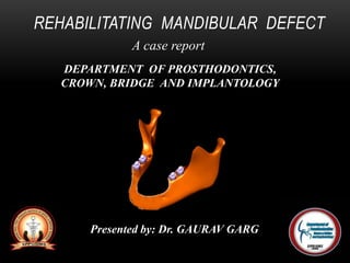
- 1. REHABILITATING MANDIBULAR DEFECT A case report Presented by: Dr. GAURAV GARG DEPARTMENT OF PROSTHODONTICS, CROWN, BRIDGE AND IMPLANTOLOGY
- 2. INTRODUCTION • Mandible reconstruction has been a challenge for the clinicians for more than a century. • Mandible defects resulting in face deformity of various stages are mostly the consequence for ablative surgery for malignancies, huge jaw cysts, infections as osteomyelitis and trauma, that may compromise orofacial functions and cause subsequent psychological disorders.
- 3. • Even new surgical reconstructive techniques may not sufficiently restore sensory-motor functions, and in most instances they fail to provide adequate support for dental prostheses.Loss of the proprioceptive sense of occlusion leads to the uncoordinated, less precise movement of the mandible. • Poor tissue support after mandibular reconstruction along with the addition of impaired tongue function, may totally compromise mastication and has hindered prosthodontists in constructing stable and functional dental prostheses for these patients.
- 4. CASE REPORT • A 32 Years old male patient reported to our Out Patient Department with the chief complaint of missing teeth in the lower arch. • The clinical examination and past history revealed that that the patient had an infected Radicular Cyst in the mandibular region.The enucleation of the cyst was done followed by chemical cauterization.The procedure also accompanied extraction of tooth #31, # 32, # 33, # 34, # 35, # 41, # 42, # 43, # 44.
- 10. CANTOR AND CURTIS CLASSIFICATION OF PARTIAL MANDIBULECTOMY •CANTOR AND CURTIS in 1971 classified mandibular resection patients by amount of mandible that remains after resection and surgical reconstruction.
- 11. TYPE I - Mandibular resection involving alveolar defect with preservation of mandibular continuity
- 12. • TYPE II - Resection defects involve loss of mandibular continuity distal to the canine area
- 13. • TYPE III - Resection defect involves loss up to the mandibular midline region.
- 14. • TYPE IV - Resection defect involves the lateral aspect of the mandible, but are augmented to maintain pseudo articulation of bone and soft tissues in the region of the ascending ramus.
- 15. • TYPE V - Resection defect involves the symphysis and parasymphysis region only, augmented to preserve bilateral temporomandibular articulations.
- 16. •The criteria of this classification suggested that this case will come under Type I resection of the mandible as the inferior border was intact. •For Type I resection the denture bearing area is compromised by closure of defect with the use of adjacent lining mucosa which reduces the bucco-lingual width.
- 17. TREATMENT OPTIONS 1) Mandibular reconstruction by autogeneous Bone Graft 2) Implant supported fixed prosthesis 3) Cast Partial Removable prosthesis with the Clasp Assembly 4) Cast Partial Removable prosthesis with the semi – precision attachment.
- 19. •The primary impression of the patient was made and the casts were surveyed for the treatment planning. •The crown preparation of the tooth #36,#45 and #47 was done . •Final impression was made after preparation and then the casts were mounted on a Semi adjustable articulator with the Face-Bow transfer.
- 21. PRECI – VERTIX SEMI PRECISION ATTACHMENT 3 2 1 4
- 22. POSITIONING OF THE SEMI- PRECISION ATTACHMENT USING THE NEY’S SURVEYOR
- 23. FINAL TRY-IN OF THE CROWNS WITH THE SEMI- PRECISION ATTACHMENT
- 24. CAST PARTIAL FRAME-WORK IN POSITION
- 25. TRY IN OF THE CAST PARTIAL FRAME-WORK
- 26. CROWN IN POSITION ALONG WITH THE RETENTIVE KEEPERS
- 27. FINAL PROSTHESIS AFTER INSERTION
- 28. POST – OPERATIVE OCCLUSAL VIEW
- 29. PERMANENT SOFT – LINED TISSUE SURFACE WITH RETENTIVE KEEPERS IN POSITION
- 31. PRE AND POST- OPERATIVE PROFILE VIEW
- 32. PRE AND POST- OPERATIVE PROFILE VIEW
- 33. CONCLUSION • Surgical and prosthodontic rehabilitation of the mandibulectomy patient can provide satisfactory results, improving the oral functions of the patient which overall makes a positive impact on the quality of life for the patient. • Proper treatment planning pre and post surgery is necessary to obtain the most optimal results.
- 34. REFERENCES 1) J. Philip Sapp. Patología oral y maxilofacial contemporánea. ED MOSBY (2a ed) 2) Feinberg S.E. Surgical management of ameloblastoma. Current status of the literature.Oral Surg Oral Med Oral Pathol Oral Radiol Endod 1996; 81: 383-8. 3) Iordanidis S, Makos C, Dimitrakopoulos J, Kariki H. Ameloblastoma of the maxilla:case report. Aust Dent J 1999; 44(1): 51-5. 4) Hollows P, Fasanmade A, Hayter JP. Ameloblastoma: a diagnostic problem. Br Dent J2000; 188(5): 243-4. 5) Nakamura N, Higuchi Y, Mitsuyasu T, Sandra F, Ohishi M. Comparison of long-term results between different approaches to ameloblastoma. Oral Surg Oral Med Oral Pathol Oral Radiol Endod 2002; 93(1): 13-20. 6) Carr AB, Mc Givney GP, Brown DT. Mc Crackens removable partial prosthodontics.11th ed, India, MOSBY Inc, pp. 414.
- 35. REFERENCES 7) Barttelbort SW, Bahn SL, Ariyan SA. Rim mandibulectomy for cancer of the oral cavity.Am J Surg 1987; 154(4): 423-428. 8) Mohd Khairi MD, Normastura AR, Azizah Y, Mahdan MHA. Oral cancer: a retrospective study of cases registered in Hospital Universiti Sains Malaysia (HUSM), Kelantan, Malaysia. International Medical Journal 2009; 16(1): 31-37. 9) Shareef BT, Abd Rahman N, Che Zainudin Z, Pohchi A. Salivary gland neoplasms in Hospital Universiti Sains Malaysia (HUSM): A retrospective study. International Medical Journal 2011; 18(1): 58-62. 10) Schliephake H, Reiss G, Urban R, Neukam FW, Guckel S. Metal release from titanium fixtures during placement in the mandible: an experimental study. Int J Oral Maxillofac Implants 1993; 8(5): 502-11.
- 36. THANK YOU