
Recommended
Recommended
More Related Content
What's hot
What's hot (20)
Similar to Distraction Osteogenesis
Similar to Distraction Osteogenesis (20)
More from Saibel Farishta
More from Saibel Farishta (20)
Recently uploaded
Recently uploaded (20)
Distraction Osteogenesis
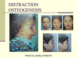
- 1. DISTRACTION OSTEOGENESIS PROF (Dr.) SAIBEL FARISHTA
- 2. -INTRODUCTION -HISTORICAL REVIEW -ILIZAROV PRINCIPLES - D O IN CRANIOFACIAL REGION -MERITS OF D O -DEMERITS OF D O - INDICATIONS -CONTRAINDICATIONS -BIOLOGIC BASIS OF D O - ORTHODONTIC TOOTH MOVEMENT INTO DISTRACTED BONE -IMPLANT PLACEMENT IN DISTRACTED BONE -BIOMECHANICAL EFFECTS OF DEVICE PLACEMENT -DEVICE SELECTION -VECTOR ORIENTATION -CLASSIFICATION OF REGENERATE BONE -D O OF MANDIBLE ,MAXILLA ,MIDFACE , CRANIOFACIAL SKELETON -PERIDONTAL DISTRACTION -EFFECTS OF D O on T M J -POTENTIAL MISTAKES -COMPLICATIONS -CONCLUSION -REFERENCES
- 3. INTRODUCTION For centuries many different techniques have been used in an attempt to modify growth ,both in terms of amount and direction .Orthodontists for example ,use intra oral and extra oral appliances to restrict growth of the maxilla in hope of accentuating mandibular saggital growth . Recently , a new technique of bone generation and reconstruction , termed Distraction Osteogenesis was introduced to clinicians of craniofacial region by orthopedic surgeons.
- 4. Distraction osteogenesis is the biologic process of new bone formation between bone segments gradually separated by incremental traction .This process begins when a distraction force is applied to the healing callus that joins the divided bone segments and continues as long as the tissue is stretched .It is of importance in cases where the traditional orthognathic procedures are found wanting to correct the growth deficiencies yielding unstable results and subsequent relapse. Bone is required for a variety of purposes –traumatic loss, tumor resection ,congenital deformities. The challenges that arise in craniofacial surgery due to inadequate bone volume or bone defects have led to many attempts at solutions in the past. CALLOTASIS -- Latin noun CALLUM-Scar tissue between bone segments Greek noun TAOIS –tension or extension
- 5. Distraction force applied to bone also creates tension in the surrounding soft tissues ,initiating a sequence of adaptive changes termed distraction Histogenesis.Under influence of tensional stresses produced by gradual distraction ,active histogenesis occurs in different tissues including skin ,fascia, tendon ,muscle ,cartilage, blood vessels ,peripheral nerves.
- 6. Historical review –Early forms of bone manipulation - Hippocrates more than 2000 years ago described the placement of traction forces on broken bones. Guy de Chauliac in 14th century applied traction with a pulley system that consisted of a weight attached to the leg by a chord .the weight was suspended over a pulley to create tension. John Barton in 1826 was the first to perform surgical division of bone or osteotomy . Joseph Malgaigne constructed an apparatus for external fixation of displaced transverse patellar fractures.
- 7. Early Techniques for Distraction Osteogenesis – At the turn of 20th century ALESSANDRO CODIVILLA first performed limb lengthening using external skeletal traction after an oblique osteotomy of the femur, later several surgeons modernized CODIVILLAS continuous extension procedure by modifying the osteotomy technique the distraction protocol or the device for bone fixation . Paul Magnuson recognized the biologic potential of periosteum and endosteum . Vittorio Putti designed the osteoton ,for femur lengthening .
- 8. Refinements of Distraction Osteogenesis Procedures . Summaries soon appeared reporting success and complication during limb lengthening. The complications included delayed healing , non unions ,deformities ,nerve palsy,and joint contracture . This prevented widespread acceptance of distraction osteognesis and required further improvements in osteotomy techniques ,distraction protocol ,appliance design. A significant contribution in the development of DO was made by the Russian surgeon ,GAVRIEL ILIZAROV .IN 1951 .He designed a new apparatus for bone .fixation . This fixation technique has two distinct advantages over other methods , Stable but not rigid fixation to provide axial micro movements Full control over the manipulation of bone segments ,regardless of their size shape or anatomic location ILIZAROV divided 2/3 rd of bony cortex with a narrow osteotome and completed the corticotomy by rotational osteoclasis , there by causing minimal trauma to the periosteum and bone marrow . His lengthening protocol utilized a 5-7 day latency period followed by a distraction of 1 mm per day performed in four increments of 0.25 mm.
- 9. Based on his clinical experience ,ILIZAROV discovered biologic principles of Distraction Osteogenesis,known as the ILIZAROV effects 1)The tension stress effect on the genesis and growth of tissues 2)the influence of blood supply and loading on the shape of bones and joints The third group of expts – the effect of the distraction on the orientation of newly formed tissues .He discovered that the regenerate within the distraction gap is always formed along the axis of applied traction. The next set of expts- the influences of the rate and rhythm of distraction on the formation of bone regenerate. Results proved that more frequent rates of distraction ,more favorable regenerate formation and cause fewer soft tissue problems
- 10. The last study looked at the relationship between blood supply and mechanical loading and its influence upon the shape and mass of the resulting bone .ILIZAROV stated that in order to maintain function the blood supply must be proportional to the mechanical load . D O IN CRANIOFACIAL COMPLEX In 1976 BELL and EPKER – RPE in patients with transverse deficiency . Osteo distraction of mandibular symphysis using principles of RPE was initially reported by Gurerro in 1990. IN 1992 Mc CARTHY reported results of clinical applications of Distraction Osteogenesis in patients with congenital cranio facial anomalies .Following the reports of Mc Carthy which demonstrated successful lengthening of human mandible by gradual bone distraction heralded the beginning of craniofacial distraction
- 11. MERITS OF DISTRACTION OSTEOGENESIS -Relatively simple operative technique -Lengthening of bone and soft tissues -Good long term stability -Avoidance of bone grafts -Multidimensional distraction -Feasible to distract bone grafts or irradiated bone -Results are apparently early -Reduces likelihood of relapse -Reduces likelihood of blood transfusion DRAWBACKS OF DISTRACTION OSTEOGENESIS -Skin scars with extra oral devices -Damage to Facial nerve -Damage To Tooth Germ -Premature consolidation -Transient changes in TMJ -Infection -Nonunion/inadequate bone formation -Device failure
- 12. CLASSIFICATION OF TREATMENT MODALITIES WITH D O D O in the field of craniofacial reconstruction is applicable for the following 6 Rx techniques 1) To lengthen the mandible 2) To advance the maxilla /midface or monobloc advancement 3) Bone segment transportation 4) Trifocal distraction treatment as in case of symphyseal defect 5) Distraction for alveolar augmentation 6) Distraction implantology
- 13. MANDIBULAR DISTRACTION OSTEOGENESIS Indications -Hemifacial Microsomia -Treacher Collin Syndrome -Congenital Micrognathia - Nager Syndrome -Pierre Robin Syndrome -Goldenhar Syndrome -Moebius Syndrome - Facial Cleft -Cranio Facial Microsomia -Trauma ,Pathologic defect -Alveolar defect -Transverse defect -Alveolar augmentation MAXILLARY DISTRACTION OSTEOGENESIS INDICATIONS -Orofacial clefts -Nasomaxillary dysplasia -Cranio synostosis -Maxillary atrophy -Alveolar augmentation
- 14. MIDFACIAL AND /OR CRANIAL DISTRACTION INDICATIONS =Craniosynostosis -Crouzon syndrome -Aperts syndrome -Sagittal synostosis -Midfacial cleft SIMULTANEOUS MANDIBULAR AND MAXILLARY DISTRACTION OSTEOGENESIS INDICATIONS -Hemi facial Microsomia =Treacher Collins syndrome CONTRAINDICATIONS OF DISTRACTION OSTEOGENESIS -Insufficient quantity or quality of bone which would inhibit fixation of the device such as osteoporosis -Inability to comply with the post operative distraction regimen and follow up schedule -Hypersensitivity to metals -Infection -Neuro psychiatric disorders -Immuno suppression
- 15. SELECTION OF PATIENT FOR DISTRACTION OSTEOGENESIS -Age of the patient -Severity of the anatomical malformations which can affect the soft tissues -Potential for bone growth depending on the etiology -Functional and esthetic effects -Secondary malformations -Psychological considerations BASIC PRINCIPLES OF DISTRACTION OSTEOGENESIS -Preservation of blood supply -Close apposition of cut bone surfaces to allow early bridge formation -Gradual distraction at regular rhythm. RATE OF DISTRACTION – number of millimeters per day at which the bone surfaces are stretched . The rate of 1mm a day is considered optimal. RHYTHM OF DISTRACTION – number of distractions per day usually in equally divided increments to total the rate. The rhythm may vary from one cycle per day of 1mm to 0.025 mm four times per day or 0.5 mm twice daily .
- 16. BIOLOGIC BASIS OF DISTRACTION OSTEOGENESIS D O begins with the formation of a reparative callus .Callus is placed under tension by stretching which generates new bone . D O consists of 3 sequential periods 1)LATENCY PERIOD –is the period from bone division to the onset of traction and represents the time allowed for callus formation 2)DISTRACTION PERIOD –interval during which gradual traction is applied and new bone or distraction regenerate is formed 3)CONSOLIDATION PERIOD –allows maturation and corticalization of the regenerate after traction forces are discontinued . LATENCY PERIOD – consists of 4 stages and is comparable to that during fracture healing 1)inflammation 2)softcallus 3)hard callus 4)remodelling Following the surgical separation of bone into 2 segments ,a cascade of vascular disruption and extravasations of blood from the damaged bone ends and the associated soft tissue ,a hematoma forms between and around the bone segments .The hematoma is converted to a clot and bone necrosis occurs at the ends of the fracture segments. There is in growth of vasoformative elements and capillaries to restore blood supply clot is replaced by granulation tissue
- 17. During soft callus stage granulation tissue becomes fibrous vascularity increases mesenchymal stem cells within the callus appear inside the terminal of the newly formed capillaries . Cartilage replaces the granulation tissues. This occurs more in the inter fragmentary space more towards the periphery This fibro cartilaginous tissue is transformed into immature woven bone The Cartilage calcifies as the capillaries invade the cartilage osteoblasts follow and lay down new bone. Woven Bone is converted to lamellar bone by a resorption replacement sequence and the medullary canal is reconstituted .
- 18. DISTRACTION PERIOD During D O normal process of fracture healing is disturbed by application of gradual traction Dynamic microenvironment New tissue is formed in a direction parallel to the vector of traction Gradually stretched tissue promotes angiogenesis ,increase tissue oxygenation and increased fibroblastic proliferation Between 3rd-7th day of distraction capillaries grow into the fibrous tissue and extends toward medullary cavity. Newly formed vessels have a spiral pathway and numerous circular folds suggesting growth rates 10 times higher than rates of vessel growth during normal fracture healing during two weeks of distraction primary osteons begin to form. The osteoid producing osteoblasts located among the collagen fibers lay down osteoid tissue on these collagen fibers . Osteogenesis is initiated at the existing bone walls and progresses toward the center of the distraction gap by the end of the second week the osteoid begins to mineralize. The elongation of the regenerate depends on the growth of primary osteons ,the length of which increases rapidly during the early stages of distraction.
- 19. CONSOLIDATION PERIOD After distraction ceases ,the fibrous inter zone gradually ossifies and one distinct zone of woven bone completely bridges the gap as the regenerate matures the zone of primary osteons significantly decreases and is later completely resorbed . Marrow cavity is restored Haversian remodelling takes place Maturation of distracted bone continues for a year or more before the structure of newly formed bony tissue is comparable to that of the pre existing bone Radiographically the first evidence of bone regenerate is usually observed at the end of the distraction period
- 20. MECHANISM OF NEW BONE FORMATION – It appears that 3 different modes of bone formation occur during Mandibular Distraction including Intramembranous ,Endochondral ,Transchondroid ossification . Although there is no direct evidence, we consider that the cells involved in transchondroid bone formation temporarily express cartilage tissue phenotype before differentiating directly into osteocytes which survive in the chondroid tissue until it is resorbed and remodelled into lamellar bone .The mechanism of new bone formation is dependent on both blood supply and the rate of distraction. The most appropriate rate of distraction is between 0.5 -1.0mm/day.
- 21. TOOTH MOVEMENT INTO REGENERATE BONE Possibility of moving teeth into regenerate bone is no longer disputed however the timing of initiating the tooth movement into newly created bone remains a topic of discussion . Gurerro ,Constasti ,Razdolsky ,Pensler ,Dessner – not until 8-12 weeks Liou ,Chen ,Huang – begin much earlier in the consolidation period Rather than basing clinical decisions on emperic supositions 3 Important factors are to be considered -Systemic Factors and Patients Health -Tooth/Bone Related Factors -Mechanical Factors SYSTEMIC FACTORS – healing capacity , general health, blood supply , calcium balance ,bone quality TOOTH BONE RELATED FACTORS –size shape of roots ,quantity of surrounding bone ,amount of bone between osteotomy cut and root ,root surface area MECHANICAL FACTORS- type of force to initiate tooth movement –tipping ,translatory ,intrusion ,extrusion . Gingival fibers that might stretch during distraction there by applying forces to teeth.
- 22. If little inter dental bone is left walking of tooth occurs before the regenerate is mineralized enough to support . Start with translatory force, this also adds up to the time taken for the regenerate to mineralize Start movement of the distal most tooth since many a times tooth distal to the osteotomy is a molar.
- 23. PLACEMENT OF IMPLANT INTO DISTRACTED BONE Sufficient osseointegration occurs with implants placed in the distraction regenerate in the consolidation period ,this shortens the treatment duration . The parallel columns of bone extending from the host margins were firm enough to maintain the orientation of the implants .Prosthetic loading should be delayed until 24 weeks.
- 24. BIOMECHANICAL EFFECTS OF DISTRACTION DEVICE ORIENTATION DURING MANDIBULAR LENGTHENING AND WIDENING Mandible is a complex 3D STRUTURE consisting of two halves that are fused in the midline at an acute angle . V shaped Mandible- anatomic axis of the right and left sides are not parallel to each other .
- 27. ORHTODONTICS IN INTRA ORAL MANDIBULAR DISTRACTION OSTEOGENESIS Careful Evaluation Of diagnostic records ---ideal treatment plan Diagnostic Records include – - detailed clinical examination - cephalometric examination - dental cast analysis FACTORS To Be CONSIDERED – Patients Age Future Growth Potential Oral hygiene Plane of deformity Amount of bone lengthening Psychosocial status Nutrition General health clinical examination – mandibular sagital defeciency convex facial profile lip incopetance large overjet transverse dimensiosn dark buccal corridors v shaped arch. scissor bite
- 28. CEHPALOMETRIC ANALYSIS S T A FRONTAL CEPHALOMETRIC ANALYSIS DENTAL CAST ANALYSIS OCCLUSOGRAM Arch perimeter analysis Boltons analysis Flatten curve of spee
- 29. SAGITTAL MANDBULAR DISTRACTION OSTEOGENESIS INDICATIONS 5 indications for distraction over orthognathic surgery major mandibular advancements > 7mm Pre surgical degenerative TMJdisease Sleep apnoea Inadeqate mandibular anatomy Secondary mandibular advancements PRE DISTRACTION ORTHODONTICS Maxillary teeth are leveled and aligned, Ideal arch form developed heavy rectangular surgical archwire with inter dental surgical hooks is placed If there is no need for expansion mandibular archwire is placed Pre surgical records are taken Prediction surgery is then performed on casts INTRA DISTRACTION AND POST DISTRACTION ORTHODONTICS DISTRACTION DEVICE SHOULD BE ACTIVATED AS PER SCHEDULE Monthly radigraphicc examinaton is carried out Stabilize the device for 3 months Donot remove device until radiographic outline is achieved Class 2 elastics 4-6 Oz worn 24/ hrs /day during distraction and consolidation .this is done to counteract softtissue forces at the time of bone healing If not done reciprocal changes take place leading to detrimental artrhitic changes in condyle ,fossa, disc .
- 30. DISTRACTION DEVICE SELECTION D O devices have been developed for both external and Internal applications Device should be selected based on mechanical capabilities and patient acceptance DEVICE REQUIREMENTS Any device should allow for Transfer of distraction forces directly to the desired bone ends Provide adequate rigidity until osseus consolidation occurs TYPES OF DISTRACTORS –EXTAORAL DISTRACTORS - INTRA ORAL DISTRACTORS -SUBCUTANEOUS ,INTERNAL BURIED DEVICES(CRANIAL /MIDFACE DISTRACTION) EXTRA ORAL DISTRACTOR DEVICE Mono directional appliance Bidirectional appliance Multi directional appliance Drawbacks –Pin loosening , pin tract infection External scars Damage to tooth buds Breakage of metal frame work Damage to facial nerve Non compliance
- 31. INTRA ORAL DEVICES TYPES -Bone borne -Tooth Bone borne -Tooth borne ADVANTAGES -No external scar Good patient compliance No Damage To Facial Nerve Superior psychological tolerance DRAWBACKS of INTRA ORAL second surgery required for removal
- 32. FACTORS CONSIDERED FOR LENGTHENING OF MANDIBLE BY INTRA/EXTRA ORAL APPLIANCES -amount of desired lengthening and angular correction -vector of distraction -psychosocial requirements of the patients LENGTHENING CAPABILITIES Select appropriate length of device The ratio between device activation and observed amount of distraction varies sometimes as high as 2:1 Angular correction further decreases linear correction DISTRACTION VECTOR PLANNING Distraction vector defines the DESIRED DIRECTION of that the distal segment has to move factors to be considered -osteotomy design ,location masticatory muscle influence occlusal interferences ,distraction device adjustment
- 33. DISTRACTION DEVICE ORIENTATION Place device parallel to the desired vector of distraction -device oriented parallel to ramus – oblique distraction -for vertical elongation of ramus –place device perp to maxillary occlusal plane -for mandibular corpus lengthening place device parallel to maxillary occlusal plane -oblique device orientation in ramus causes clockwise rotation of mandible -INFLUENCE OF MASTICATORY MUSCLES functional compensations are developed check for any purposeful anterior positioning compensate the masticatory forces with orthopedic forces OCCLUSAL INTERFERENCES POSTERIOR OCCLUSAL INTERFEENCES in mandibular lenghtening causes clockwise rotation ,anterior openbite ,use of bite plane is necessary DISTRACTION DEVICE ACTIVATION angular device activation reduces the antero posterior lengthening of mandible At least 10mm of lengthening should be done prior to angular correction Orthopedic force may be instituted to produce angular correction FUTURE GROWTH AND OVECORRECTION IF GROWTH IS EXPECTED OVER CORRECTION IS PERFORMED to reduce total number of surgeries degree of over correction should provide a patient with a socially acceptable appearance for the most years possible
- 34. ORTHODONTIC MANAGEMENT DURING DISTRACTION AND CONSOLIDATION distraction regenerate is a semi rigid structure that connects both the proximal and distal segments of the mandible to each other this allows the orthodontist to change the direction of the distal segments HOFFMEISTER and co workers technique involves the removal of the distraction device before the regenerate is consolidated and is there fore malleable. the regenerate is then manipulated with orthodontic force to the desired end treatment position and left to consolidate
- 35. DIRECTION OF DISTRACTION simultaneous ramus and corpus lengthening FORMULA – Pin placement angle= 180- gonial angle x ramus deficiency total defeciency AMOUNT OF DISTRACTION calculated by drawing a triangle ,two sides of which represent the amount of mandibular corpus and ramus shortening FORMULA – Distraction amount =Dc +Dr -2(Dc Dr ) cos A where Dc= corpus defeciency , Dr =ramus defecieny , A=gonial angle OVERCORRECTION calculated based on growth deficiency the distribution of over correction must be the same as that for growth defeciency for ex if distribution of growth deficiency between mandibular ramus and corpus is 3:1 the ratio of over correction should be 3:1
- 36. CLASSIFICATION OF REGENERATE BONE each regenerate is classified by 1)length of the forming regenerate from each host bone margin ,length of the radiolucent inter zone relative to the length of distraction 2)the width of the forming regenerate relative to the width of the host bone margin 3)the shape and density of forming regenerates and the radiolucent interzone 4)the presence or absence of corticalisation
- 38. Eleven types of mandibular regenerates are evident . Clinical stability is dependent on radigraphic appearance of the regenerate ,not the duration of consolidation period -every regenerate with obliterated interzone and equal to the width of the host bone margins was stable. -regenerates with an un obliterated inter zone and those with obliterated interzone but with unequal width to the host bone segments are questionable in their stability. 30 -60./. Of bone minerals should be gained to be evident radiographically corticalization is one of the last stages of regenerate consolidation and indicates that distraction device can be removed and that bone can undergo functional loading
- 39. Distraction begins after five days of latency at the rate of approximately 1mm/day the elongation is completed in 3-4 weeks , device is left in place for 6-8 weeks of consolidation until radiographic evidence of new cortical bone is seen during distraction the distance between the pins and soft tissue –external canthus to buccal commisure is measured weekly
- 40. OBSTRUCTIVE SLEEP APNOEA IN CHILDREN candidates should be critically evaluated so that the risks and benefits of distraction versus traditional treatment can be weighed pre operative planning requires the use of sterolithographic model based on C T scan of the infants the fixation plates are cut pre surgically to fit the existing contours of the model due to fast healing response of infants the distraction is initiated immediately after the surgery .Infant distraction proceeds at the rate of 1.5mm/day divided in three equal increments .This is followed by 3weeks of consolidation The introduction of lactosorb resorbable distraction devices have brightened the areas of applications
- 41. MAXILLARY DISTRACTION Midface deficiency manifests on the nasal and paranasal regions as well as at occlusal levels According to protocol photographs ,cephalograms ,dental models are obtained leveling aligning of the arches is performed , the day before the surgery a quad helix appliance is placed In the surgical procedure dissection of the lateral nasal process is done , an incomplete osteotomy is performed above the roots of the canine and molar tooth ,pterygo maxillary disjunction is not done On the 5 th day distraction is initiated using a Petit facial mask with force of 900 grams for total of 16-18 hours /day when the projected advancement is obtained the amount of force is decreased to 450 grams per side and is maintained for two months At 8-10 weeks post distraction there is radiographic evidence of increase in size of tuberosity at the pterygomaxillary junction Gradual maxillary distraction by means of incomplete osteotomies and external force systems is a minor ,relatively simple procedure with minimal morbidity
- 44. TRANSPORT D O Transport D O is the technique of regenerating bone and soft tissue in a discontinuity defect secondary to tumor or trauma. An osteotomy is made 1.5 cms from the end of the distal stump of bone adjacent to the discontinuity defect creating a transport disc . The transport disc is advanced through the discontinuity defect by means of a distraction device. New bone is created in the gap between the transport disc and original mandible the leading edge of the transport disc becomes enveloped by a fibrocartilagenous cap . If this cap is not removed prior to docking significant compressive forces must be applied in order to cause necrosis of the fibrocartilage there by allowing bony fusion Transformational osteogenesis can be induced by compressive forces .This leads to local necrosis of pathologic tissue followed by neovascularization of the fibrous and cartilagenous tissue with local resorption at the bone ends leading to progressive remodelling and fusion of the two segments .
- 45. MANDIBULAR SYMPHYSEAL WIDENING Narrow mandible is often associated with narrow maxilla therefore it is necessary to treat the maxillary transverse defeciency prior to mandibular correction The symphyseal distractor has an occlusal splint incrporated into the design ,the surface is ground flat to avoid any occlusal interferances during distraction Mandibular growth remains unaffected after distraction , condylar morphology remained un changed no posterior or lateral flatenning occurred
- 46. ALVEOLAR RIDGE AUGMENTATION Two Categories VERTICAL AUGMENTATION-the transport alveolar segment is translated vertically and the height of the ridge is increased HORIZONTAL AUGMENTATION-transport alveolar segment is translated horizontally thereby increasing the alveolar ridge width distraction implants are suitable for increasing the height of the anterior mandibular and maxillary alveolar ridge height alveolar ridgee height should be started 1 week after surgery and performed slowly at the rate of 0.5mm per day.
- 47. INTRA ORAL CUSTOM DISTRACTION DEVICES
- 49. EFFECT OF DISTRACTION OSTOGENESIS ON TMJ Although the distraction forces are applied locally the force of distraction could be distributed to the adjacent tissues including condyle a secondary beneficial effect appears to occur at the level of T M J. In an attempt to optimize the potential effect of functional remodeling ,patients are encouraged to masticate soft diet during entire distraction period . Distraction has additional beneficial effect on the TMJ region .in unilateral cases ,the affected side uprights in size and volume .the contralateral un operated side shows no apparent deleterious changes at the level of TMJ. In bilaterally affected and treated cases ,both condylar heads increase in size and volume , becoming symmetric .
- 50. POTENTIAL MISTAKES 2 major groups 1)iatrogenic mistakes –primary or strategic mistake - secondary or tactical mistakes - technical mistakes 2)patient related mistakes -
- 51. POTENTIAL COMPLICATIONS 1)REGENERATE MALFORMATION --HYPOTROPHIC REGENERATE --HYPERTROPHIC REGENERATE --REGENERATE FRACTURE HYPOTROPHIC REGENERATE –delay in bone tissue formation followed by an alteration in regenerate structure progresses to delayed consolidation pseudoarthrosis occurs Signs- lack of radiographic evidence of distraction gap mineralization Correction- decrease the rate of distraction temporary cessation of distraction HYPERTROPHIC REGENERATE excessive rate of bone formation leads to premature consolidation Signs –uniform tissue density throughout the intersegmentary gap Correction – if soft tissue permits 2-3mm of acute distraction followed by gradual distraction with increased rate
- 52. REGENERATE FRACTURE USUALLY OCCURS AFTER FRAME REMOVAL ,or progressively increasing soft tissue tension ,inadequate duration of consolidation period treatment – Rx same as that for fractures AXIAL DEVIATIONS eliminate the main cause ,it may include replacement of the device with a larger one ,reorientation of entire distraction vector repositioning using elastic traction SOFT TISSUE OVER STRETCHING leads to degenerative necrotic changes that negatively affect the outcome BLOOD VESSELS it is least resistant to compressive force , correction –releasing the tension followed by reduced rate of distraction PERIPHERALNERVES D O complication includes 2-15/. Of nerve injury occurs due to direct injury during osteotomy or indirectly by focal nerve injury by progressive edema or compression due to fixatures
- 53. In most cases lengthening by D O can be continued but at a slower rate . Rehabilitation process may take more than a year SKELETAL MUSCLES Signs-limited range of motion , tenderness, joint contracture . Complication – muscle atrophy physical therapy with active or passive joint motion should be done INFECTION Mainly associated with external distraction devices management should be started immediately antibiotics , releasing incisions should be given
- 54. CONCLUSION AS We become more comfortable with the mere application of this exiting technique ,we will most certainly find more novel uses for it ,as well as different iterations of previous use Future trends include – refinements of distraction protocols improvements in distraction devices enhancements of regenerate using growth factors modification of osteotomies the uses and applications of D O in treating both simple and complex deformities of craniofacial skeleton are restricted neither by mechanical configuration of distraction device nor by the biologic capacity of the human body but are actually only limited by the boundaries of our imagination .
- 55. REFERENCES : 1) The histology of distraction osteogenesis using different external fixators ;clinical orthop 241; 106; 1989 2)use of maxillary distraction osteogenesis for maxillary advancement ;J Oral Maxillofac surg 1994 ;52 ;282-6 3)force levels and strain patterns during distraction osteogenesis . J Oral Maxillofac Surg ;58 ;171-8 ;2000. 4) mandibular lengthening by gradual distraction ; AJODO 126 ;2004 5) T B OF CRANIOFACIAL DISTRACTION ; Cherakashin ,Cope ; Samuchkov . 6) Experimental mandibular growth ; Plastic Re Constru Surg 1997 ;506
- 56. -Effect of mandibular distraction surgery on T M J ; AJODO 2001 -Simultaneous mandibular and maxillary distraction in hemifacial microsomia in adults;Plast Reconstru Surg 1997 ;100 ;852 -61 -Distraction osteogenesis on irradiated mandible; Plastic Reconstru Surg 89;1-8 :1992.
- 57. THANK YOU