
Recommended
More Related Content
Similar to Respiratory disorders.pptx
Similar to Respiratory disorders.pptx (20)
More from RuchiPal10
Recently uploaded
Recently uploaded (20)
Respiratory disorders.pptx
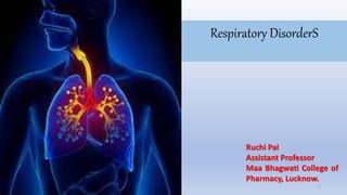
- 1. Respiratory DisorderS Ruchi Pal Assistant Professor Maa Bhagwati College of Pharmacy, Lucknow. 1
- 2. Respiratory system- Respiration is a type of metabolic process which involves energy production indirectly by the oxidation of complex substances. The respiratory system mainly consists of the upper respiratory tract, alveoli, bronchi, bronchioles, trachea, pleura, and pleural cavity. Disorder- A disorder is defined as a state of irregular functioning of the body. The respiratory system disorders or respiratory diseases are the medical terms used to study about the various types of infections, allergies and other diseases related to the different organs, tissues and specialized cells of the human respiratory system. Respiratory Diseases include Asthma, Chronic obstructive pulmonary disease(COPD), Tuberculosis, Pulmonary fibrosis, Pneumonia, & lung cancer. 2
- 3. 3
- 4. ASTHMA A condition in which a person's airways become inflamed, narrow and swell and produce extra mucus, which makes it difficult to breath. Asthma can be minor or it can interfere with daily activities. In some cases, it may lead to a life- threatening attack. Chronic inflammation is associated with airway hyper responsiveness that leads to recurrent episodes of wheezing breathlessness, chest tightness, and coughing, particularly at night or early morning. It can be controlled not cured. 4
- 5. Three-step problem- 1-Airway inflammation 2-Airways hyper-responsiveness to stimuli 3-Muscles within airways contract (Bronchospasm) 5
- 6. How does asthma works? 6
- 7. Types of Asthma (1) Allergic / Atopic / Extrinsic Asthma → Hyper- responsiveness to inhalation specific allergen such as house dust, pet dander, feathers, molds, pollen, food. (2) Non - allergic / Non-atopic / Intrinsic Asthma -Irritants in air not related to allergies such as air pollution, cold, heat, weather changes, fumes, smoke, room deodorants, RTI (Respiratory tract infections), emotions, NSAIDs, Preservatives, stress . ETA (Exercise-Induced Asthma). • Occupational Asthma (3) Mixed Asthma. (4) Cough variant Asthma - Persistent dry cough. (5) Nocturnal Asthma -Worsen at night. 7
- 8. Causes & Risk factors Combination of environmental & Genetic factors. Airborne allergens Respiratory infection Physical activity Cold air Air pollutants & irritants. Hay fever & other allergies. Certain medications. Strong emotions & stress. Food allergy. GERD (Gastro esophageal reflux diseases) Low birth weight Extreme changes in weather Eczema Family history Menstrual cycle 8
- 10. Sign & Symptoms of Asthma Shortness of breath. Chest tightness or pain. Wheezing when exhaling, which is a common sign of asthma in children. Trouble sleeping caused by shortness of breath, coughing or wheezing. Coughing or wheezing attacks that are worsened by a respiratory virus, such as a cold or the flu. Fatigue Mental confusion Extreme anxiety Fever Central cyanosis Nasal flaring Diaphoresis Mucus production Status asthmaticus. 10
- 11. Classification based upon severity 11
- 12. Complications Status asthmaticus is a sustained state of asthma which results when airways remain blocked for prolonged periods (days & weeks) & also do not respond to any treatment. Pneumonia is a bacterial or viral lung infection. Atelectasis occurs when a part of lung collapse due to airway obstruction with large amount of mucus. Underperformance & fatigue is a very common asthma complication. Absenteeism from workplace is also very common. 12
- 13. Diagnosis 1) Medical history 2) Physical examination. 3) Spirometry (spirometer) 4) Peak expiratory flow (PEF) 5) Pulse oximetry 6) Chest X-ray- Asthma or other respiratory condition occurs. To find out infection, foreign bodies & other conditions of lungs. 7)Allergy blood testing- Known allergens inhale by patients (allergens cause asthma attacks) Take blood sample IgE level high Find out the particular allergen that cause disease. 13
- 14. 8)Blood gases- Arterial blood gases analysis Blood sample collects Find pH Level, Oxygen level, carbon dioxide level, acid- base balance. 9)CBC (Complete blood count)- Eosinophils level Erythrocytes sedimentation rate WBCs count 10)Sputum culture- To find out/diagnose infection. 14
- 15. Treatment • Long term medications 1)Inhaled corticosteroids- Fluticasone propionate, budesonide, beclomethasone. 2)Leucotriene modifiers- Montelukast Zafirlukast 3)Long-acting beta agonist (LABA)- Formoterol Salmeterol 4)Combination inhalers. • Quick relief medications 1-Short acting beta agonists (SABA)- Albuterol Levalbuterol 2-Anticholenergics Ipratropium bromide 3-Oral & Intravenous corticosteroids Prednisolone Methyl prednisolone. 15
- 16. Bronchial thermoplasty- Bronchial thermoplasty is a treatment for severe asthma. It's a way to open your airways. Bronchial thermoplasty is an asthma treatment that targets the smooth muscle in the lungs. Bronchial thermoplasty is an innovative, new, non drug procedure developed for the treatment of severe persistent asthma. The treatment uses heat to shrink the smooth muscle so it can't tighten and cause asthma symptoms. The treatment involves three sessions, with three weeks between each session. 16
- 17. • Non-pharmacological (No medication) Oxygen therapy Postural drainage / chest physiotherapy To drain out mucus Coughing & deep breathing exercise (YOGA) Avoidance of known allergens. Allergy causing food or any other things. 17
- 18. COPD(Chronic Obstructive Pulmonary Disorder) • Also known as- Chronic obstructive lung disease (COLD) Chronic obstructive airway disease (COAD) Chronic airflow limitation (CAL) Chronic obstructive respiratory disease (CORD) A group of lung diseases that block airflow and make it difficult to breathe. Emphysema and chronic bronchitis are the most common conditions that make up COPD. COPD is a condition in which air flow is obstructed by emphysema, chronic bronchitis or both. 18
- 19. Causes Cigarette smoking Exposure to smoke from biomass. Exposure to dust of polluted air. Alpha antitrypsin deficiency can cause emphysema in non smokers. An association of low birth weight. Genetic effects. long-term exposure to irritating gases or particulate matter. Types of COPD Chronic bronchitis It involves long term cough with mucus. Emphysema It involves damage to lungs over time. 19
- 20. Pathogenesis of COPD Noxious gases inhalation Abnormal inflammatory response in airways, parenchyma, pulmonary vacuoles Body attempts to repair the chronic inflammation Narrowing in the air ways Over time injury & repair process causes scar tissue formation Permanent narrowing of airway lumen Parenchymal destruction (Emphysema) Airflow obstruction resulting in- Muscular weakness Increased circulatory inflammatory markers. 20
- 21. Sign & Symptoms of COPD Shortness of breath Wheezing (whistling sound during breathing) Chest tightness (effort in breathing) Ongoing (chronic) cough (sputum) Difficulty with routine activities Fatigue Weight loss Muscle loss Frequent colds or flu Dyspnea- Difficulty in breathing Enlarged alveoli 21
- 22. Advanced symptoms (Severe COPD) Cyanosis-bluish discoloration of skin (lips, nails appears blue) Morning headache ( due to high level of carbon dioxide) Hemoptysis- Blood in cough / mucus. Swollen feet & ankle. Lungs not properly functioning More efforts by heart to pumps blood Complications Respiratory infections ( flu, cold, pneumonia) Heart problems ( Heart attack) Lung cancer Pulmonary hypertension Depression 22
- 23. Diagnostic Evaluation of COPD Medical history Physical examination Chest X-ray CT Scan Lung function test (LFT)/ Pulmonary function test (PFT) • Spirometry • Lung volumes ABG Analysis ( Arterial blood gas analysis) Take blood from arteries Observe level of oxygen, carbon dioxide & pH of blood Pulse oximetry ( Oxygen saturation level) 23
- 24. Treatment Pharmacological treatment 1)Nicotine replacement therapy- • Smoking • Exposure to smoke 2)Bronchodilators- • Beta 2 agonist (salmeterol, albuterol) • Anticholinergics • Theophyline 3)Anti-inflammatory agents • Fluticasone • Budesonide 4)Mucolytics • Guaifenesin • N-acetylcysteine 5) Antibiotics • Amoxicillin 6)Alpha-1-Antitrypsin deficiency treatment • Replacement 7)Vaccination • Flu & pneumonia vaccines 24
- 25. Non-Pharmacological treatment 1) Nebulization ( Inhalers) 2) Oxygen therapy 3) Exercise 4) Pulmonary rehabilitation 5) Surgery Bullectomy Lung volume reduction surgery Lung transplant A bullectomy is a surgical procedure to remove bullae— air-filled spaces in the lungs that can compress healthy lung tissue and cause symptoms such as dyspnea (shortness of breath), repeated infections, and pneumothorax (lung collapse). 25
- 26. Bronchiectasis • Bronchiectasis is a disease in which there is permanent enlargement of parts of the airways of the lung. • It is a condition in which the bronchial tubes of lungs get permanently destroyed, and become wide and thick. • Caused by recurrent airway infection & inflammation. • Persistent, irreversible, pathological dilation (& out pouching) of the bronchi & bronchioles secondary to destruction of airway cartilage and elastic tissue. • Could be primary (lung was normal) or secondary (lung had a problem before the Bronchiectasis). Primary: e.g. Adenovirus Secondary: cystic fibrosis-prevent clearance of organisms from the lungs. 26
- 27. Causes of bronchiectasis 1.Cystic fibrosis (CF)- An inherited life-threatening disorder that damages the lungs & digestive system. It affects the cells that produce mucus, sweat & digestive juices( fluids become thick & sticky). 2.Infection: Tuberculosis (TB) Severe pneumonia • Viral pneumonia: Adenovirus or influenza virus. • Bacterial pneumonia: Staphylococcus aureus & Hemophilus influenzae. 3.Immotile ciliary syndrome (aka Kartagener or primary ciliary dyskinesia) • The immotile-cilia syndrome is a congenital disorder characterized by all the cilia in the body being either immotile or showing an abnormal and inefficient beating pattern. Most symptoms come from the ciliated airways (nose, paranasal sinuses, and bronchs) and from the middle ear. 4.Toxin inhalation Chemical fumes Aspiration (e.g. gastric content) 5.GERD(Gastro esophageal reflux diseases) 6.Yellow nail syndrome. 27
- 29. 29
- 30. Sign & Symptoms • Bronchiectasis develops symptoms in a few months or years, and includes: Chronic cough, Blood with cough, Thick mucus with cough, Wheezing sounds during breathing, Breathlessness, Pain in chest, Loss in weight, Fatigue, Clubbing ( thickening of skin under nails and toes ), & Recurring infections in airways. 30
- 31. Diagnosis of bronchiectasis 1)Pathology Gross examination Usually in the lower lobes Bronchi & bronchioles: dilated, cylindrical & saccular. The dilations extend to the lung periphery. Could be tubular, cylindrical, varicose Filled with pus Microscopic ( Histopathology) Dilation of bronchi & bronchioles Inflammatory cells and inflammatory debris. 2) Radiologically CXR: Usually nonspecific. Might show some infiltrates “tram-tracking”: dilation of airways. Crowded bronchial markings extending to the periphery. CT Crowded bronchial markings extending to the periphery. HRCT- (High-Resolution Composed Tomography Scanning is the standard test for confirming the diagnosis.) 3) Sputum analysis, gram stain & culture. 4) Sweat chloride test for CF. 5)Test for HIV/ AIDS 6)Immunoglobulin (gamma-globulin) levels. 31
- 32. Treatment of bronchiectasis Antibiotics-for recurrent pneumonias Remove mucus • Percussion • Postural Drainage Surgery Chest Physiotherapy (CPT) Oxygen therapy Bronchodilators Antibiotics Vaccinations 32
- 33. THANK YOU Inhale the future, exhale the past 33