Deadenylase Expression in Small Cell Lung Cancer Related To Clinical Characte...
Poster_CBCD_2014
1. Aversa R.1,*, Sorrentino A.1,*, Ambrosio M.R.1,2,*, Zambelli A.3, D'Apice L.4, Ciccodicola A.1, 5,*, and Costa V.1,*
1Institute of Genetics and Biophysics "A. Buzzati-Traverso", CNR, Naples; 2Department of Translational Medical Sciences, University of Naples “Federico
II”, Naples; 3Oncology Department, IRCCS S. Maugeri Foundation, Pavia; 4Institute of Protein Biochemistry, CNR, Naples, Italy; 5Department of Science
and Technology, University of Naples “Parthenope”; *Computational & Biology Open laboratory - ComBOlab: www.combolab.it
Breast cancer is the most common tumor in women and the second leading cause of death, mainly caused by metastasis1. Tumor cells invasiveness is due to an alteration of cell-cell
and cell-matrix connections, in which adhesion molecules have a key role. Moreover, it’s known that cancer cells manipulate the alternative splicing pattern of adhesion/motility
genes to escape immune system cells and to initiate epithelial-mesenchymal transition. Recent studies revealed that, among adhesion molecules, Semaphorins, a large family of
transmembrane or secreted molecules, are of peculiar interest. Semaphorins, through the interaction with their receptors – neuropilins and plexins – can be involved in different
biological processes as axon guidance, angiogenesis, cell migration and adhesion2. A growing number of studies - and our recent work3 on SEMA6B in breast cancer among them -
has demonstrated their involvement in cancer progression, often with divergent functions depending on the activated pathway in a cell-type specific manner4,5.
Background
RNA-Seq experiment
On going and future works
Contact information
References
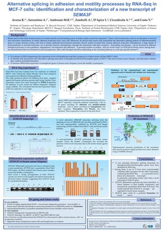
Total RNA has been isolated from two replicates of cultured
MCF-7 cells. Paired-end cDNA libraries have been prepared
and sequenced on Illumina Hi-Seq platform.
The reads produced (about 150 millions) have been mapped
against the human reference genome hg19 using TopHat. Gene
expression values have been measured in FPKM (Fragments
Per Kilobase of exon model per Million fragments mapped)
using Cufflinks. The correlation between the expression values
of the two replicates is very high.
To evaluate, by RNA-Sequencing, the expression levels of Semaphorins and their receptors in a breast cancer cell line (MCF-7 cells)
To identify and characterize new alternative splicing transcripts of Semaphorins/Plexins/Neuropilins genes in MCF-7 cells and in breast cancer biopsies, and determine whether
they could be potential biomarker
To measure differential expression of semaphorin genes in breast cancer biopsies versus the healthy counterparts
Aims
About 14000 expressed genes have been detected in the two
MCF-7 replicates. Using the database GeneCards, a list of
89 genes encoding for adhesion and motility-related
molecules and 21 genes encoding for Semaphorins and
their receptors, Neuropilins and Plexins, has been
obtained. Their expression is shown in the plot above.
0
20
40
60
80
100
MCF-7 Rep. 1 MCF-7 Rep. 2
66
86
56
73
Million
Sequenced reads Uniquely mapped reads (UMRs)
R=0.99
Differential expression analysis of
SEMA3F in breast cancer biopsies
SEMA3F differential expression has been evaluated using
a semi-quantitative RT-PCR and a quantitative Real-Time
PCR assays, on RNA isolated from breast tumor samples
versus the healthy counterparts.
Data reveal a strong up-regulation of both SEMA3F
canonic (as shown in the graph) and spliced transcripts in
all breast tumor samples, suggesting that SEMA3F could
be a potential biomarker.
Conclusions
11 new potential alternative splicing transcripts for
Semaphorin/Plexins/Neuropilins genes have been
identified by RNA-Seq
Among them, 4 out of 11 have been experimentally
validated (still in progress for the other transcripts),
confirming that RNA-Seq is a powerful tool to
detect new alternative transcripts.
In particular, interesting results come from a novel
transcript of SEMA3F gene.
SEMA3F differential expression has been identified in
tumor biopsies vs their healthy counterpart, and
measured by quantitative Real-Time PCR assay. The
results indicate that SEMA3F is strongly up-
regulated in breast tumor samples.
SEMA3F gene could be a potential biomarker for
breast cancer.
1. DeSantis et al., Breast Cancer Statistics 2013, CA Cancer J Clin (2014)
2. Choi et al., Dynamic control of β1 integrin adhesion by the plexinD1-sema3E axis, Proc Natl Acad Sci U.S.A. (2014)
3. D’Apice et al., Analysis of SEMA6B gene expression in cancer: Identification of a new isoform, Biochim. Biophys. Acta
(2013)
4. Tamagnone, Emerging role of semaphorins as major signals and potential therapeutic targets in cancer, Cancer Cell
(2012)
5. Gu and Giraudo, The role of semaphorins and their receptors in vascular development and cancer, Exp Cell Res (2013)
6. Mendes-da-Cruz et al., Semaphorin SEMA3F and Neuropilin-2 control the migration of human T-cell precursors, PLoS ONE
(2014)
In vivo analysis:
ELISA on nipple aspirate fluid (NAF) – non invasive diagnostic procedure – from healthy vs
breast cancer affected individuals to assess the potential use of SEMA3F protein as a biomarker
immunohistochemistry on paraffin embedded breast tissues (healthy versus tumor)
In vitro analysis:
SEMA3F cloning and overexpression in MCF10 cells
SEMA3F silencing – by siRNA – in MCF-7 and MDA-MB-231 cells
Since it has been recently demonstrated that SEMA3F has chemorepulsive effects towards immune cells6, we
are planning to perform:
Migration assays using breast cancer cells and lymphocytes co-cultures
Identification of a novel
SEMA3F transcript
SEMA3F
NM_004186
CD36 SEMA3F GNAT1
Chr. 7q21.11
3’5’
32 4 5 6 7 8 9 10 11 12 13 14 15 165’ 171 1819 3’1718
G A AG A C TC
20
TG CCT G AT G G
30
G G TT T CC G T G
40
C C G G AC G T C C
50
T G C C G G C G G C
60
T C C G C C C T C T
70
TA G AA G A GAT
80
G G T C AT G G T C
90
T T G AC G G G T G
100
C T G G AT CC T T
110
G AA
G A AG A C TC
20
TG CCT G AT G G
30
G G TT T CC G T G
40
C C G G AC G T C C
50
T G C C G G C G G C
60
T C C G C C C T C T
70
TA G AA G A GAT
80
G G T C AT G G T C
90
T T G AC G G G T G
100
C T G G AT CC T T
110
G AA
exon 17exon 15
UTR
Intron
X Skipped exon
Exon
A novel alternative SEMA3F transcript, deriving from the
skipping of exon 16 (SEMA3FΔ16), has been predicted by RNA-
Seq and experimentally validated by RT-PCR and Sanger
sequencing on RNA isolated from MCF-7 cells and breast tumor
biopsies.
SEMA
DOMAIN
R/K rich
DOMAIN
Ig-like C2
type
DOMAIN
PSI
DOMAIN
Prediction of SEMA3F
3D structures
Tridimensional structure predictions of the annotated
(left) and the putative (right) proteins have been obtained
using I-TASSER software.
The authors declare that no conflict of interests existed.
1 2 3 4 5 6 7 8
H T H T H T H T H T H T H T H T
307 bp
149 bp
1 2 3 4 5 6 7 8
H T H T H T H T H T H T H T H T
307 bp
149 bp
Log2 (FPKM>1) MCF-7 Rep. 1
Log2(FPKM>1)MCF-7Rep.2
0
10
20
30
40
50
60
70
80
1 2 3 4 5 6 7 8 9 10 11 12 13 14 15 16 17
193 288 242
45
2578 229
49
8
16
1045
3
73
48
10 12 13
208
Relativeexpression
tumor
0%
10%
20%
30%
40%
50%
60%
70%
80%
90%
100%
1 2 3 4 5 6 7 8 9 10 11 12 13 14 15 16 17
Relativeexpression
tumor
healthy
healthy
307 bp
149 bp
T 1 T 2 T 3
307 bp
149 bp
MCF-7
A semi-quantitative RT-PCR performed on breast tumor
samples versus the healthy counterparts has revealed the
presence of the novel transcript only in the tumor tissues (green
arrows).
RNA-Seq datasets
Uniquely mapped
reads (hg19)
Transcriptome
quantification
Reads mapping on
adhesion/ motility
genes
Junction
identification
Exon skipping in
adhesion/ motility
genes
Junction
filtering
Experimental
in vitro
validation
In silico analysis of
the new transcripts
In silico protein
prediction
Workflow of the computational and experimental
approach used to identify and validate new transcripts.
SEMA6B
SEMA4G
SEMA3E
SEMA6A
SEMA4F
PLXNC1
SEMA4C
SEMA3F
SEMA4D
PLXNA3
PLXNB1
PLXNB2
SEMA3C
SEMA3B
SEMA4A
SEMA4B
PLXNB3
PLXNA1
PLXND1
PLXDC2
NRP1
Rosanna Aversa -PhD student in Cellular and Molecular Biotechnologies at Second University of Naples (SUN)
E-mail: rosanna.aversa@igb.cnr.it
Website: www.combolab.it
Phone: +390816132258 Fax: +390816132617