1. ALSO IN THIS ISSUE:
High Blood Pressure Prevention
Pathology Today
A Publication of the Robert J. Tomsich Pathology & Laboratory Medicine Institute | Summer 2016
Feature Story
Lung cancer diagnoses on small samples
using minimally invasive techniques
Cystic Fibrosis:
Updates in Diagnosis
and Treatment | p 6
New Multiplex Molecular
Detection of Enteric
Pathogens | p 10
Alumni Connect | p 14
News | p 16
2. 2
Pathology Today SUMMER | 2016
Lung cancer continues to be the leading cause of cancer
death in the world, responsible for 1.6 million deaths every
year. Non-small cell lung carcinoma (NSCLC) comprises
up to 80% of lung cancers. Though patients with early
lung cancers, Stage I and 2, have a survival rate of 50-
80%, most patients (80%) are diagnosed at an advanced
stage with metastases to peripheral sites and mediastinal
lymph nodes, accounting for the high mortality rate. The
increased use of targeted cancer screening with CT will
increase the diagnosis of these tumors at an earlier stage
and improve prognosis, as will the improvement in the
systemic therapy for advanced disease.
The diagnosis of NSCLC is treated with conventional
chemotherapies that have significant debilitating side
effects with only limited improvement of survival. However,
in the past five to seven years, new molecular drivers for
non-small cell lung carcinoma are being discovered leading
to the development of more effective therapies that target
the mutations specific to each patient’s cancer. Because
of this new approach, it has become imperative to sample
each patient’s tumor and have sufficient tissue to perform
these diagnostic molecular tests so that the appropriate
target therapy is used.
Historically these diagnoses were made on core needle
biopsies or surgical resection specimens; however,
currently over 50% of diagnoses come from small samples,
including cytology samples. Tumors located in the
peripheral lung parenchyma may be sampled by CT-guided
transthoracic fine needle aspiration biopsies. During this
procedure, an interventional radiologist locates the tumor
using a CT scan to more accurately localize the needle.
With more centrally located lesions an endobronchial
ultrasound guided fine needle aspiration may be used. In
this setting an interventional pulmonologist uses ultrasound
localization to more accurately localize the lesions in
real time. The samples, obtained by a transbronchial
fine needle technique, are evaluated on site by the
cytopathology team and provide information that is vital to
patient care. Based on the interpretation of these cytologic
smear preparations, the cytopathologist provides feedback
regarding adequacy for diagnosis, directs triaging the
specimen for appropriate testing and can be helpful to
pulmonologists in providing rapid staging information.
In patients with more advanced stage disease, peripheral
fine needle aspiration of distant metastases to the skin,
subcutaneous tissue or palpable lymph nodes provides an
even less invasive method for diagnosis. Finally, patients
who do not show evidence of metastatic disease and are
healthy enough to undergo surgery may go directly to
the operating room for a resection of the tumor. In this
scenario, the tissue from the surgically resected tumor will
provide the needed tissue for further molecular testing.
Overall, a myriad of procedures is available to procure
adequate tissue for appropriate molecular testing in
patients regardless of the extent of the disease. From
fine needle aspiration of localized and distant metastatic
disease, guided by ultrasound or CT imaging, to complete
resection of an early stage tumor in the OR, the pathologist
must be ready to perform both confirmatory diagnostic
testing for tumor type as well as testing for possible
molecular targets on small tissue samples.
One such set of mutations is on the ALK gene. Presence
of this oncogenic fusion renders the tumor susceptible
to tyrosine kinase inhibitors (TKIs), but is only present
in 3-8% of lung adenocarcinomas. The typical patient
is younger and is usually a non-smoker. Most mutations
involve fusion with a portion of the echinoderm
microtubule associated protein like 4 (EML4) gene. ALK-
EML4 rearrangements are activating mutations that confer
susceptibility to crizotinib, a specific TKI. Other oncogenic
fusions such as ROS and RET also confer susceptibility to
Lung Cancer Diagnoses on Small Samples
Using Minimally Invasive Techniques
By Jordan Reynolds, MD, and Carol Farver, MD
3. 3
Pathology Today SUMMER | 2016
crizotinib. ROS1 encodes a receptor tyrosine kinase that
is related to the insulin receptor. These translocations are
found in 1.5% of lung adenocarcinomas, also typically
in non-smoking younger patients.1
These mutations
also show response to the ALK inhibitor crizotinib. RET
rearrangements are identified in ~2% of patients with
lung adenocarcinoma, again in young non-smokers.
RET rearranged tumors may respond to tyrosine kinase
inhibitors such as vandetanib and cabozantinib.
Detection of these oncogenic mutations may be
accomplished in many ways. Immunohistochemistry
(IHC) can be used, but preanalytic conditions such as
fixation time or media (paraffin versus alcohol as typically
used in cell blocks) may affect staining. Fluorescence in
situ hybridization (FISH) allows for correlation of fusion
positive cells with the tumor morphology. This test may be
performed on either paraffin-embedded sections or cytology
smears.2, 3
Currently, ALK testing for FISH is performed on
the ThinPrep slide on all cytology specimens with sufficient
tumor cells. For core biopsies or resections, IHC testing
is performed on the paraffin block. For ROS and RET
mutations, we offer testing on surgical specimens (core
biopsy and resection) using FISH.
EGFR encodes a tyrosine kinase that, when activated,
results in cellular growth and survival via the RAS/RAF/
MEK and Pi3K/AKT/mTOR pathways. EGFR mutations
are present in 15-25% of lung adenocarcinomas, and are
typically found in females, non-smokers and Asians. Since
these mutations involve missense insertions and deletions,
sequencing of the tumor with either PCR or next generation
sequencing is necessary. Immunohistochemistry testing is
not acceptable because while the protein may be expressed
on the surface, knowledge of the specific mutation is
needed. Most EGFR mutations confer susceptibility to the
TKIs, but exon 20 insertions and T790M point mutations
are associated with resistance to TKIs.4
Testing can be performed on surgical specimens as well
as cytology samples. With our in-house samples, we are
able to readily extract DNA from the residual fluid from
each FNA. The advantages of adapting cytology in NSCLC
molecular diagnosis is because optimal quality nucleic
acids may be reliably isolated. Another benefit is the
preservation of the original cytopathology slides. Sparing
of the cell block is possible in case future diagnostic testing
is needed.
For those DNA samples that were insufficient for NGS
libraries, which account for 4% or less liquid-based
cytology (LBC) specimens, a reflexive real-time PCR assay
can determine the EGFR mutation hotspot status in a
timely manner.
The main advantage of real-time PCR over NGS is its
quick turnaround time (2-3 days), which could be crucial
in a clinical setting. Since EGFR-mutated cancers account
for 10-35% of NSCLC and may be treatable with TKI,
we currently use Therascreen as an alternative to NGS to
ensure the timely delivery of actionable molecular results
to oncologists.
All patients with small biopsy specimens, resections
and cytology samples that demonstrate differentiation
of adenocarcinoma should also undergo testing. Small
samples may be inconclusive of tumor subtype due to
missed sampling of adenocarcinoma (i.e. adenosquamous
carcinoma, unsampled adenocarcinoma in a squamous
cell carcinoma). Small cell carcinomas or squamous cell
carcinoma in the proper clinical context (heavy smokers)
should probably not undergo testing.
While there are many targetable genes, the most
commonly found are EGFR and ALK, and these should be
prioritized, since these are the most common genes that
have a targetable mutation. Knowledge of the presence of
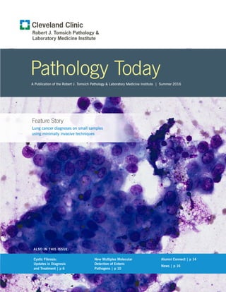
Lung adenocarcinoma, HE stain 20x. (Cover photo is lung
adenocarcinoma, Diff-Quik 40x.)
4. 4
Pathology Today SUMMER | 2016
KRAS mutation can be useful, but the priority should go
to EGFR and ALK. Sequencing to detect the specific type
of EGFR mutation is recommended. EGFR, gene copy
number or immunohistochemistry staining simply tells the
status of the EGFR receptor. At Cleveland Clinic, all lung
cancer patients that fit into the above criteria undergo next
generation sequencing. In the event that the DNA content
is insufficient for NGS testing, allele-specific PCR testing
for EGFR and KRAS will be performed.
While paraffin-embedded tissue from surgical specimens
has been the classically validated method, cytology smears
and cellient cell blocks may also be used to perform
sequencing studies. We have found that the residual
cytology fluid can be used for next generation sequencing.
The supernatant fluid and cell pellet left after the FNA is
obtained can be used for DNA extraction and sequencing
studies. At our institution, cases that have tumor cellularity
greater than 20% on the ThinPrep slide may be selected
for NGS on the residual fluid. This is possible because of
the willingness of the clinician obtaining the sample to
perform additional passes just for testing.
We can perform ALK, ROS and RET testing on all surgical
tissue including biopsies and resections. Next generation
sequencing can be performed from extraction of DNA
from selected blocks. Cytology specimens are currently
validated for ALK testing. This can be performed on
either the ThinPrep or cell block slide. ROS and RET
testing is currently available on surgical specimens, and
is undergoing validation and will hopefully be available
for testing in the near future. Next generation sequencing
can be performed on all cases that have residual fluid
remaining in the tumor sample. It may also be performed
from scraping tumor cells on slides with greater than
2000 tumor cells (Both Pap or Wright stained slides
are commonly used in adequacy assessment). Before
sacrificing any slides, we would archive a digital image
using our ePathology Department to preserve a record of
what was on the slide. In fact, a single FNA can be used to
diagnose the tumor, subtype it with immunohistochemistry
and provide valuable theranostic information. All of this
information can be provided on small samples, using
minimally invasive techniques.5
References
1. Scheffler M, Schultheis A, Teixido C, et al. ROS1
rearrangements in lung adenocarcinoma: prognostic
impact, therapeutic options and genetic variability.
Oncotarget. 2015;6: 10577-10585.
2. Takeuchi K, Soda M, Togashi Y, et al. RET, ROS1
and ALK fusions in lung cancer. Nat Med. 2012;18:
378-381.
3. Wang W, Tang Y, Li J, Jiang L, Jiang Y, Su X.
Detection of ALK rearrangements in malignant pleural
effusion cell blocks from patients with advanced
non-small cell lung cancer: A comparison of Ventana
immunohistochemistry and fluorescence in situ
hybridization. Cancer Cytopathology. 2015;123:
117-122.
4. Lozano MD, Labiano T, Echeveste J, et al. Assessment
of EGFR and KRAS mutation status from FNAs and
core-needle biopsies of non-small cell lung cancer.
Cancer Cytopathology. 2015;123: 230-236.
5. Rooper LM, Nikolskaia O, Carter J, Ning Y, Lin M-T,
Maleki Z. A single EBUS-TBNA procedure can support
a large panel of immunohistochemical stains, specific
diagnostic subtyping, and multiple gene analyses in the
majority of non-small cell lung cancer cases. Human
Pathology. 2016; 51: 139-145.
5. 5
Pathology Today SUMMER | 2016
About the Authors
Jordan P. Reynolds, MD
Jordan P. Reynolds, MD, joined
Cleveland Clinic in 2011 as a staff
pathologist in the departments of
Anatomic Pathology and Molecular
Pathology in the Pathology and
Laboratory Medicine Institute.
Dr. Reynolds served fellowships in
cytopathology and surgical and medical pathology at the
Mayo Graduate School of Medicine at Mayo Clinic. He
completed his residency in anatomic and clinical pathology
at the University of Cincinnati, where he also served
as chief resident in the Department of Pathology and
Laboratory Medicine.
Dr. Reynolds received his medical degree from
Northeastern Ohio Universities College of Medicine and
his bachelor of science degree from Kent State University.
He is a Diplomate of the American Board of Pathology in
anatomic and clinical pathology, and cytopathology. He
also is a member of the National Medical Association,
American Society of Cytopathology, and the United States
and Canadian Academy of Pathology.
Dr. Reynolds has contributed to numerous publications
and scientific presentations, and is the recipient of several
awards. His interests are cytology with an emphasis on
genitourinary pathology. Dr. Reynolds can be reached at
216.444.4833 or reynolj4@ccf.org.
A
Carol F. Farver, MD
Carol Farver, MD, is a lung
pathologist and has served as the
Director of Pulmonary Pathology
in the Department of Pathology at
Cleveland Clinic since 1995 and
is Professor of Pathology at the
Cleveland Clinic Lerner College of
Medicine of Case Western Reserve
University School of Medicine. She received her MD
degree from Yale University School of Medicine and did her
subsequent residency and fellowship/research training in
pulmonary pathology at Brigham and Women’s Hospital
and the Harvard School of Public Health in Boston. She
has authored over 115 scientific publications and 30
chapters and is co-editor of two major textbooks in the
field of pulmonary pathology. She has been included in the
“Best Doctors in America” list since 2009. She is the past
President of the Cleveland Clinic Medical Staff and served
on the Cleveland Clinic Board of Governors.
6. 6
Pathology Today SUMMER | 2016
Cystic fibrosis (CF) is a multisystem genetic disease
caused by mutations, also known as pathogenic variants,
in the cystic fibrosis transmembrane conductance
regulator (CFTR) gene. The gene encodes the CFTR
protein, which acts as a chloride channel and regulates
ion transport across cell membranes. Defects in the
assembly and function of the CFTR protein lead to highly
viscous secretions from epithelial cells, causing clinical
manifestations in the lungs, pancreas, intestines, liver,
sweat glands and male reproductive system.1
Pulmonary
disease is the leading cause of death among individuals
with CF, with median predicted survival age of 39 years.2
Over 33,000 individuals in the United States have CF
and 1,000 new cases are diagnosed each year.2
It is most
common among Caucasians but can affect people of any
ethnic or racial background. The condition has autosomal
recessive inheritance, meaning that a person develops
symptoms only if they are homozygous, that is, they have
two CFTR gene copies with pathogenic variants. Since
heterozygous carriers are unaffected, most people diagnosed
with the condition have no known family history. When
both partners in a couple are carriers, each of their children
has a 25% chance to inherit two variants and be affected
with CF. There is also a 50% chance for each child to
inherit just one variant and be an unaffected carrier for CF
and a 25% chance of being neither affected nor a carrier.
Individuals with CF are now primarily detected at birth
through mandatory newborn screening (NBS). CF
screening is now included on the NBS panels in all states.3
In most state programs, immunoreactive trypsinogen (IRT)
enzyme testing is performed as the initial screen.4,5
This
screen is sensitive but not specific for CF so when positive,
molecular testing of the CFTR gene may be performed as
a second tier screen prior to reporting results to clinicians
and families.
While sweat chloride testing remains the standard
diagnostic test for patients with clinical symptoms of CF,
genetic testing is routinely performed as confirmation of
clinical findings.6
With the advent of targeted treatment
for CF, this confirmation is critical to patient care and
management.
New Treatment Options
For many years, the management of CF focused solely on
supportive treatment of symptoms, most often of airway
obstructions and infections caused by viscous pulmonary
secretions. However, in 2012 the FDA approved a
medication in a new category that targets defects in the
Cystic Fibrosis: Updates in Diagnosis and Treatment
By Jacquelyn D. Riley, MS, LGC
Table 1. CF Carrier Frequency in Different Ethnic Groups
Ethnic Group Observed Carrier Frequency
African American 1 in 84
Ashkenazi Jewish 1 in 29
Asian 1 in 242
Caucasian 1 in 28
Hispanic 1 in 59
Jewish 1 in 32
Middle Eastern 1 in 91
Native American 1 in 70
South Asian 1 in 118
Other Ethnicity 1 in 111
1 Ethnicity 1 in 34
Part African American 1 in 56
Part Caucasian 1 in 32
Part Hispanic 1 in 51
Not Provided 1 in 37
All Individuals 1 in 38
Source: Rohlfs EM, Zhou Z, Helm RA, Nagan N, Rosenblum LS, et al. (2011)
Cystic fibrosis carrier testing in an ethnically diverse U.S. population. Clin
Chem. 57: 841-848.
7. 7
Pathology Today SUMMER | 2016
protein function.7
The first CFTR modulator, called ivacaftor
(Kalydeco), was found to be beneficial to individuals with
the G551D variant and other CFTR gating abnormalities.8
For these patients, the CFTR protein is correctly assembled
and located at the cell membrane but the chloride channel
is closed.9
The medication binds to the protein and restores
its function. Patients who took the medication as part of
a randomized controlled trial had significantly improved
pulmonary function, decreased pulmonary symptoms
and increased body weight (patients with CF can be
underweight and malnourished due to abnormal intestinal
absorption). However, gating variants are present in a
minority of CF patients and ivacaftor was not found to be
as effective in patients with F508del, which represents
70% of CFTR pathogenic variants among Caucasians.10
F508del interferes with both channel gating and protein
folding. In July 2015, the FDA approved a combination
drug, lumacaftor-ivacaftor (Orkambi), for patients with
homozygous F508del.11
Patients on this combination
CFTR modulator have experienced moderate improvements
in pulmonary health.12
This development represents an
exciting application of personalized molecular medicine,
where a patient’s genotype is used to guide specific
treatments. Therefore, molecular testing of the CFTR gene
in patients with cystic fibrosis is more important than ever.
New CFTR Test Platform
Last year, the Cleveland Clinic Molecular Pathology Lab
transitioned the CFTR mutation assay to a FDA-approved
next generation sequencing (NGS) platform. The new
test detects 139 variants in the CFTR gene, versus 32
variants that were detected with the previous test. The
139 variants were selected from the CFTR2 (Clinical and
Functional Translation of CFTR) database as representing
the full set of clinically validated variants classified as
CF-causing.13
CFTR2 is a consortium project that gathers
clinical and molecular information from patients around
the world, which is reviewed by a team of international CF
experts. The assay includes all 23 pathogenic variants that
are recommended for pan-ethnic carrier screening by the
American College of Medical Genetics and the American
College of Obstetricians and Gynecologists.14
Using this
set of variants ensures that the test has high sensitivity for
both carrier screening and diagnostic testing but minimizes
the risk of identifying variants of uncertain significance.
Since the NGS technology not only sequences multiple
genomic regions across multiple samples at the same time,
it also allows for simultaneous sequencing of the same
nucleotide multiple times, the test has high accuracy,
minimizing the need for repeat testing and ensuring reliable
turnaround times. This assay also includes detection of the
PolyTG/PolyT intronic region, a repetitive sequence that
is challenging to assay. Previously, the region had to be
examined separately, an extra step that delayed results.
This region can modify the significance of the R117H
variant so it is clinically relevant for select patients.
The ability to accurately and quickly obtain molecular
diagnosis in patients with cystic fibrosis, coupled with
the development of new treatments that target the protein
abnormalities caused by specific pathogenic variants,
have changed the future for this population of patients.
Undoubtedly, they will have better quality of life and
longer survival due to these advances in personalized
molecular medicine.
A Note on Nomenclature
As use of molecular testing has become widespread
over the last several years, efforts have been made
to standardize the language that is used to describe
genomic changes. New discoveries have challenged
old assumptions – common variants are not always
benign and rare changes are not always harmful.
Terms like “mutation” and “polymorphism” have
not been clearly or consistently applied and have
developed connotations that may be inaccurate.
New guidelines recommend that the term “variant”
be used to describe any change in the genomic
sequence, with modifiers such as “pathogenic,”
“benign” or “uncertain significance.” There are also
detailed guidelines to describe what type of sequence
change has occurred, such as nucleotide substitution,
deletion or insertion. If you are interested, you can
learn more at the Human Genome Variation Society
website at http://varnomen.hgvs.org/.
8. 8
Pathology Today SUMMER | 2016
References
1. Cutting GR. Cystic fibrosis genetics: from molecular
understanding to clinical application. Nat Rev Genet.
2015;16(1):45-56.
2. Cystic Fibrosis Foundation Patient Registry: Annual
Data Report 2014. Available at http://www.cff.org/
[Online].
3. National Newborn Screening and Genetics Resource
Center. National newborn screening status report.
Austin (TX) Available at http://genes-r-us.uthscsa.
edu/sites/ genes-r-us/files/nbsdisorders.pdf [Online].
Updated Nov 02, 2014.
4. Comeau AM, Parad RB, Dorkin HL, Dovey M,
Gerstle R, Haver K, Lapey A, O’Sullivan BP, Waltz
DA, Zwerdling RG, Eaton RB. Population-based
newborn screening for genetic disorders when multiple
mutation DNA testing is incorporated: a cystic fibrosis
newborn screening model demonstrating increased
sensitivity but more carrier detections. Pediatr.
2004;113(6):1573-1581.
5. Sontag MK, Hammond KB, Zielenski J, Wagener JS,
Accurso FJ. Two-tiered immunoreactive trypsinogen
(IRT/IRT)-based newborn screening for cystic fibrosis in
Colorado: screening efficacy and diagnostic outcomes.
J Pediatr. 2005;147(Suppl):S83-8.
6. Farrell PM, Rosenstein BJ, White TB, Accurso FJ,
Castellani C, Gutting GR, Burie PR, LeGrys VA, Massie
J, Parad RB, Rock MJ, Campbell, III, PW. Guidelines
for diagnosis of cystic fibrosis in newborns through
older adults: Cystic Fibrosis Foundation consensus
report. J Pediatr. 2008;153:S4-S14.
7. Food and Drug Administration News Release.
FDA approves Kalydeco to treat rare form of cystic
fibrosis. 2012 Jan 31. Available at http://www.fda.
gov/NewsEvents/Newsroom/PressAnnouncements/
ucm289633.htm [Online].
8. Ramsey BW, Davies J, McElvaney NG, Tullis E, Bell
SC, D˘revínek P, Griese M, McKone EF, Wainwright
CE, Konstan MW, Moss R, Ratjen F, Sermet-Gaudelus
I, Rowe SM, Dong Q, Rodriguez S, Yen K, Ordoñez
C, Elborn JS; VX08-770-102 Study Group. A CFTR
potentiator in patients with cystic fibrosis and
the G551D mutation. N Engl J Med. 2011 Nov
3;365(18):1663-72.
9. Sloane PA, Rowe SM. Cystic fibrosis transmembrane
conductance regulator protein repair as a therapeutic
strategy in cystic fibrosis. Curr Opin Pulm Med. 2010
Nov; 16(6): 591–597.
10. Kerem BS, Rommens JM, Buchanan JA, Markiewicz
D, Cox TK, Charavarti A, Buchwald M, Tsui LC.
Identification of the cystic fibrosis gene: genetic
analysis. Science. 1989;245:1073-1080.
11. Food and Drug Administration News Release. FDA
approves new treatment for cystic fibrosis. 2015
Jul 2. Available at http://www.fda.gov/NewsEvents/
Newsroom/PressAnnouncements/ucm453565.htm
[Online].
12. Wainwright CE, Elborn JS, Ramsey BW, Marigowda G,
Huang X, Cipolli M, Colombo C, Davies JC, De Boeck
K, Flume PA, et al. Lumacaftor-ivacaftor in patients
with cystic fibrosis homozygous for Phe508del CFTR.
N Engl J Med. 2015;373:220–231.
13. The Clinical and Functional Translation of CFTR
(CFTR2). Available at http://www.cftr2.org/ [Online].
14. American College of Obstetricians and Gynecologists
Committee on Genetics. ACOG committee opinion No.
486: Update on Carrier Screening for Cystic Fibrosis.
Obstet Gynecol. 2011;117(4):1028-1031.
15. Cutting GR. Cystic fibrosis genetics: from molecular
understanding to clinical application. Nat Rev Genet.
2015;16(1):45-56.
16. Cystic Fibrosis Foundation Patient Registry: Annual
Data Report 2014. Available at http://www.cff.org/
[Online].
17. National Newborn Screening and Genetics Resource
Center. National newborn screening status report.
Austin (TX) Available at http://genes-r-us.uthscsa.
edu/sites/ genes-r-us/files/nbsdisorders.pdf [Online].
Updated Nov 02, 2014.
18. Comeau AM, Parad RB, Dorkin HL, Dovey M,
Gerstle R, Haver K, Lapey A, O’Sullivan BP, Waltz
DA, Zwerdling RG, Eaton RB. Population-based
newborn screening for genetic disorders when multiple
9. 9
Pathology Today SUMMER | 2016
mutation DNA testing is incorporated: a cystic fibrosis
newborn screening model demonstrating increased
sensitivity but more carrier detections. Pediatr.
2004;113(6):1573-1581.
19. Sontag MK, Hammond KB, Zielenski J, Wagener JS,
Accurso FJ. Two-tiered immunoreactive trypsinogen
(IRT/IRT)-based newborn screening for cystic fibrosis in
Colorado: screening efficacy and diagnostic outcomes.
J Pediatr. 2005;147(Suppl):S83-8.
20. Farrell PM, Rosenstein BJ, White TB, Accurso FJ,
Castellani C, Gutting GR, Burie PR, LeGrys VA, Massie
J, Parad RB, Rock MJ, Campbell, III, PW. Guidelines
for diagnosis of cystic fibrosis in newborns through
older adults: Cystic Fibrosis Foundation consensus
report. J Pediatr. 2008;153:S4-S14.
21. Food and Drug Administration News Release.
FDA approves Kalydeco to treat rare form of cystic
fibrosis. 2012 Jan 31. Available at http://www.fda.
gov/NewsEvents/Newsroom/PressAnnouncements/
ucm289633.htm [Online].
22. Ramsey BW, Davies J, McElvaney NG, Tullis E, Bell
SC, D˘revínek P, Griese M, McKone EF, Wainwright
CE, Konstan MW, Moss R, Ratjen F, Sermet-Gaudelus
I, Rowe SM, Dong Q, Rodriguez S, Yen K, Ordoñez
C, Elborn JS; VX08-770-102 Study Group. A CFTR
potentiator in patients with cystic fibrosis and
the G551D mutation. N Engl J Med. 2011 Nov
3;365(18):1663-72.
23. Sloane PA, Rowe SM. Cystic fibrosis transmembrane
conductance regulator protein repair as a therapeutic
strategy in cystic fibrosis. Curr Opin Pulm Med. 2010
Nov; 16(6): 591–597.
24. Kerem BS, Rommens JM, Buchanan JA, Markiewicz
D, Cox TK, Charavarti A, Buchwald M, Tsui LC.
Identification of the cystic fibrosis gene: genetic
analysis. Science. 1989;245:1073-1080.
25. Food and Drug Administration News Release. FDA
approves new treatment for cystic fibrosis. 2015
Jul 2. Available at http://www.fda.gov/NewsEvents/
Newsroom/PressAnnouncements/ucm453565.htm
[Online].
26. Wainwright CE, Elborn JS, Ramsey BW, Marigowda G,
Huang X, Cipolli M, Colombo C, Davies JC, De Boeck
K, Flume PA, et al. Lumacaftor-ivacaftor in patients
with cystic fibrosis homozygous for Phe508del CFTR.
N Engl J Med. 2015;373:220–231.
27. The Clinical and Functional Translation of CFTR
(CFTR2). Available at http://www.cftr2.org/ [Online].
28. American College of Obstetricians and Gynecologists
Committee on Genetics. ACOG committee opinion No.
486: Update on Carrier Screening for Cystic Fibrosis.
Obstet Gynecol. 2011;117(4):1028-1031.
For a disease summary, see: Moskowitz SM, Chmiel JF,
Sternen DL, Cheng E, Cutting GR. CFTR-related disorders.
Pagon RA, Bird TC, Dolan CR, Stephens K, editors.
GeneReviews. Seattle (WA): University of Washington;
2008. Available at www.ncbi.nlm.nih.gov/books/
NBK1250. [Online] Updated Feb 19, 2008.
About the Author
Jacquelyn Riley, MS, LGC
Certified Genetic Counselor
Jacquelyn Riley is a genetic
counselor in the Cleveland Clinic
Pathology Laboratory Medicine
Institute. She received a masters
in science degree in medical
genetics from the University of
Wisconsin-Madison. Before joining Cleveland Clinic in
June 2011, she worked for Spectrum Health in Grand
Rapids and Kalamazoo, Michigan, where she specialized
in prenatal genetic counseling and also worked in the
adult general genetics clinic. She is Cleveland Clinic’s first
laboratory genetic counselor, where she supports molecular
genetics, cytogenetics and prenatal screening labs. Her
interests include appropriate genetic test utilization and
the variety of roles that genetic counselors can play in
clinical laboratories. She is an active member of the
National Society of Genetic Counselors where she co-
chairs the Test Utilization Sub-committee. Riley can be
reached at rileyj2@ccf.org or through Client Services at
800.628.6816.
10. 10
Pathology Today SUMMER | 2016
The World Health Organization estimates that enteric
pathogens cause about 1.7 billion cases of diarrheal
disease worldwide each year.1
In the United States
diarrheal disease is most commonly associated with
consumption of contaminated food, but may also be
spread from person-to-person. Centers for Disease Control
and Prevention surveillance assessments estimate that
47.8 million food-borne illnesses and 4-17 million
episodes of traveler’s diarrhea occur annually.2-4
Although
very common, most diarrheal illness lasts only a few days
and symptomatic individuals often do not seek medical
care, are not treated, and are not tested to determine
the etiology of their illness. According to recent guidance
from the American College of Gastroenterology, fluid and
electrolyte replacement are the most important intervention
and mild illness typically resolves without further
intervention. Empiric antibiotic therapy is recommended
for travelers with moderate to severe illness; and
microbiologic assessment is recommended prior to therapy
for those with moderate to severe watery diarrhea, those
with bloody diarrhea, and individuals with diarrhea lasting
more than one week.2
A variety of bacterial, viral and parasitic pathogens
may cause gastroenteritis and microbiologic analysis
has traditionally involved several test formats including
bacterial culture, enzyme immunoassays for organisms
or toxins, microscopy and, more recently, polymerase
chain reaction (PCR). Performing all of these methods
to detect the causative pathogen(s) requires many hours
of technical time and a number of different media and
laboratory reagents or kits. These traditional test methods
have been shown to lack sensitivity and an etiology is often
not determined despite significant effort.5-8
Importantly, the
time to achieve all of these results is typically two or three
days. By this time, decisions about therapy may have been
determined based upon symptoms, duration of illness and
patient history, not based on microbiologic data.
Detection of enteric pathogens is the ideal laboratory
setting for application of a molecular test. A distinct set
of etiologic agents can be defined and traditional tests are
slow, insensitive, and not inexpensive if multiple tests are
ordered. The FilmArray™ Gastrointestinal (GI) Panel (Biofire
Diagnostics, Inc. Salt Lake City, UT) is a comprehensive,
molecular test that has been approved by the Food and
Drug Administration for use in diagnostic laboratories. The
panel detects 13 bacterial species, four parasites and five
viruses using the PCR method (Table 1). Test components
are contained in a single test “pouch” to which a hydration
New Multiplex Molecular Detection of Enteric Pathogens
By Susan Harrington, PhD
Table 1. Enteric pathogens detected by the FilmArray GI Panel
Campylobacter (jejuni, coli, upsaliensis)
Salmonella spp.
Shigella spp./EIEC
Shiga toxins (STEC)
E. coli O157
ETEC (LT/ST)
EPEC
EAEC
Yersinia enterocolitica
Plesiomonas shigelloides
Vibrio spp. (parahemolyticus, vulnificus, cholerae)
V. cholerae
C. difficile toxin A/B
Giardia duodenalis
Cryptosporidium spp.
Entamoeba. histolytica
Cyclospora cayatenensis
Norovirus
Rotavirus A
Adenovirus 40/41
Astrovirus
Sapovirus
11. 11
Pathology Today SUMMER | 2016
solution and the sample are added prior to insertion
into the FilmArray instrument. DNA or RNA extraction,
purification, reverse transcription, nested PCR and detection
are all performed inside the test pouch. Results are
available from the instrument in one hour. PCR products
are contained within the pouch, minimizing the risk of
cross-contamination within the laboratory. To improve
throughput, up to 12 instrument units can be combined to
achieve rapid detection of pathogens with high sensitivity
and specificity.6, 8
Ease of specimen collection and ordering is another
advantage of testing with the FilmArray GI panel. After
passing the specimen into a clean container, it should be
transferred to Cary Blair transport medium for submission
to the laboratory. Only the FilmArray GI test needs to be
ordered. Although this test will be more expensive than
individual tests for viral, parasitic or bacterial pathogens,
savings may be achieved with fewer test orders and
rapid results.
We recommend testing with the FilmArray GI panel
based upon guidance derived from the American College
of Gastroenterology.2
Patients with community-acquired,
watery diarrhea of less than seven days duration, with no
or low-grade fever do not need to be tested. Diagnostic
assessment is recommended for those with dysentery,
moderate-to-severe disease and symptoms lasting more
than seven days. FilmArray testing is also appropriate
for those with severe abdominal pain, dehydration,
hospitalization or immunocompromised state. Separate
testing for Clostridium difficile toxin is recommended for
health care-associated diarrhea and for patients who have
recently received antibiotics.
The FilmArray panel has a few limitations. Aeromonas
species are not included. Stool culture for Aeromonas
spp. may be ordered if FilmArray is negative. Similarly,
diarrheal disease in immunocompromised patients may
be due to microsporidia and microscopic examination for
microsporidium requires a special stain that is ordered
separately. Although the FilmArray detects the most
common parasitic pathogens in the United States, an ova
and parasite microscopic examination for helminths and
modified acid fast staining for Cystoisospora, Cyclospora
and Cryptosporidium may be helpful if FilmArray does not
detect a pathogen or for those with traveler’s diarrhea.
Intermittent shedding of parasites may require testing of
multiple specimens with specific tests.
Molecular methods detect DNA and/or RNA. They will
detect dead organisms and free nucleic acids from toxins or
organisms. In general tests of cure are not recommended
– instead resolution of clinical symptoms should be
monitored. In the limited settings when a test of cure is
needed, molecular methods should not be used due to
detection of nonviable DNA. Culture would be preferred.
Similarly, a specific test for a bacterial, viral or parasitic
pathogen may be ordered if the differential is known to be
limited such as if a patient is involved in an outbreak or
has a specific exposure.
With detection of many different pathogens comes the
need to interpret complex and possibly, unexpected
results. Of note is the presence of the C. difficile toxin
target on the FilmArray panels. Toxigenic C. difficile may
colonize the human host, especially children less than
two years old. Positive results determined from patients
not recently exposed to antibiotics should be interpreted
with caution. The FilmArray GI panel incorporates a
number of lesser known targets such as pathogenic E. coli
(enteroaggregative E. coli (EAEC), enteroinvasive E. coli
(EIEC), enteropathogenic E. coli (EPEC), enterotoxigenic
E. coli (ETEC)), astrovirus and sapovirus. Although
associated with outbreaks and known to be present in
patients in developed countries, pathogenic E. coli, other
than Shiga-toxin producing strains, are mostly associated
with diarrhea in children in the developing world.9
Until
Table 2. Guidance for testing for enteric pathogensa
Testing not indicated:
Community-acquired, watery diarrhea for 7 days duration,
with no or low-grade fever
Diagnostic assessment with FilmArray is recommended:
dysentery
moderate-to-severe illnessb
and fever 72 hrs
diarrhea symptoms 7 days
severe abdominal pain or dehydration, hospitalization
immunocompromised state
C. difficile toxin assay is recommended:
health care-associate diarrhea
recent antibotics
a
Guidance derived from reference #2
b
Moderate illness: forced change in activities; Severe illness: total disability
12. 12
Pathology Today SUMMER | 2016
recently, detection of these pathogens was limited to
research laboratories and epidemiological assessments.
Relevance in clinical samples in the U.S. is uncertain
and hasn’t been well-studied with this new generation of
multiplex assays, such as the FilmArray. In a small study of
230 specimens collected prospectively in the United States
Khare, et al. found 33% of samples to be positive by
FilmArray, but only 8% by conventional test methods (8).
The most common pathogens detected were Clostridium
difficile toxin (14%), norovirus (6%) and sapovirus (6%).
A few samples each were positive for Campylobacter
spp., Salmonella spp., Shigella spp., adenovirus and
astrovirus, as well as, EAEC, EPEC and ETEC. Pathogenic
E. coli were among the most common species detected
by FilmArray in a study conducted among ten European
countries and were frequently found in mixed infections.10
As expected, other prevalent infecting species determined
were Campylobacter spp., C. difficile, norovirus, rotavirus
and Salmonella spp. Another interesting finding from this
study was variability from country to country with regard
to the most common pathogens detected. Thus, regional
differences may impact test ordering or empiric therapy as
data on circulating pathogens are compiled.
The impact of multiplex molecular testing for enteric
pathogens on the diagnosis and treatment of diarrheal
disease remains to be seen, but the prospect is intriguing.
Certainly, an etiologic agent will be identified more
frequently than might have been with conventional testing.
This should lead to appropriate therapy directed at the
pathogen(s) detected and is likely to result in a reduction
in inappropriate antibiotic therapy for infections with viral
pathogens and some bacterial species. Investigation of
the clinical relevance of mixed infections and pathogenic
E. coli in patients in the U.S. is needed, but is now
possible with comprehensive molecular panels. Knowing
the pathogen quickly may lead to other benefits such as a
reduction in phone calls and office visits and an increase in
satisfaction for both the patient and provider. If utilized in
the appropriate patient population, the information gained
should help bring an overall improvement in patient care.
References
1. Henderson P. 2013. End Preventable deaths: global
action plan for prevention and control of pneumonia
and diarrhoea. World Health Organization/The United
Nations Children’s Fund (UNICEF).
2. Riddle MS, DuPont HL, Connor BA. 2016. ACG
Clinical Guideline: Diagnosis, Treatment, and
Prevention of Acute Diarrheal Infections in Adults. The
American Journal of Gastroenterology 111:602-622.
3. Scallan E, Hoekstra RM, Angulo FJ, Tauxe RV,
Widdowson MA, Roy SL, Jones JL, Griffin PM. 2011.
Foodborne illness acquired in the United States--major
pathogens. Emerging Infectious Diseases 17:7-15.
4. Steffen R, Hill DR, DuPont HL. 2015. Traveler’s
diarrhea: a clinical review. JAMA 313:71-80.
5. Buchan BW, Olson WJ, Pezewski M, Marcon MJ,
Novicki T, Uphoff TS, Chandramohan L, Revell P,
Ledeboer NA. 2013. Clinical evaluation of a real-time
PCR assay for identification of Salmonella, Shigella,
Campylobacter (Campylobacter jejuni and C. coli),
and shiga toxin-producing Escherichia coli isolates
in stool specimens. Journal of Clinical Microbiology
51:4001-4007.
Table 3. Supplemental tests for specific enteric pathogens:
Bacteria:
Culture for Salmonella, Shigella, Campylobacter, Shiga Toxins
(Stool culture) (STCUL)
Culture for Vibrio spp. (VIBCUL)
Culture for Yersinia spp. (YERCUL)
Culture for Aeromonas/Plesiomonas spp. (AERPLE)
C. difficile Toxin PCR (CDPCR)
Parasites:
Cryptosporidium Giardia Antigens by EIA (OVAPSC)
Cystoisospora/Cryptosporidium/Cyclospora examination (CRYSPO)
Ova and Parasite (microscopic) Examination (OVAP)
Fungi:
Microsporidia examination (MICSPO) Test performed in parasitology.
Virus:
Rotavirus Antigen Detection (EROTA)
Rotavirus and Adenovirus 40/41 Antigens (ROTAD)
Norovirus Group 1 and 2 Detection by PCR (NORPCR)
13. 13
Pathology Today SUMMER | 2016
6. Buss SN, Leber A, Chapin K, Fey PD, Bankowski MJ,
Jones MK, Rogatcheva M, Kanack KJ, Bourzac KM.
2015. Multicenter Evaluation of the BioFire FilmArray
Gastrointestinal Panel for the Etiologic Diagnosis
of Infectious Gastroenteritis. Journal of Clinical
Microbiology 53:915-925.
7. Harrington SM, Buchan BW, Doern C, Fader R,
Ferraro MJ, Pillai DR, Rychert J, Doyle L, Lainesse
A, Karchmer T, Mortensen JE. 2015. Multicenter
evaluation of the BD max enteric bacterial panel PCR
assay for rapid detection of Salmonella spp., Shigella
spp., Campylobacter spp. (C. jejuni and C. coli),
and Shiga toxin 1 and 2 genes. Journal of Clinical
Microbiology 53:1639-1647.
8. Khare R, Espy MJ, Cebelinski E, Boxrud D, Sloan
LM, Cunningham SA, Pritt BS, Patel R, Binnicker MJ.
2014. Comparative evaluation of two commercial
multiplex panels for detection of gastrointestinal
pathogens by use of clinical stool specimens.
Journal of Clinical Microbiology 52:3667-3673.
9. Croxen MA, Law RJ, Scholz R, Keeney KM,
Wlodarska M, Finlay BB. 2013. Recent advances in
understanding enteric pathogenic Escherichia coli.
Clinical microbiology reviews 26:822-880.
10. Spina A, Kerr KG, Cormican M, Barbut F, Eigentler
A, Zerva L, Tassios P, Popescu GA, Rafila A, Eerola
E, Batista J, Maass M, Aschbacher R, Olsen KE,
Allerberger F. 2015. Spectrum of enteropathogens
detected by the FilmArray GI Panel in a multicentre
study of community-acquired gastroenteritis. Clinical
Microbiology and Infection: the Official Publication
of the European Society of Clinical Microbiology and
Infectious Diseases 21:719-728.
About the Author
Susan Harrington, PhD
Susan Harrington, PhD, is a staff
microbiologist at the Cleveland
Clinic Pathology and Laboratory
Medicine Institute. She directs the
Mycobacteriology and Specimen
Processing sections of the main campus laboratory and
serves to integrate microbiology services across the
Cleveland Clinic Health System. She is actively involved in
teaching Medical Laboratory Science students, residents,
medical students and fellows and is the medical director
for the Cleveland Clinic School of Medical Laboratory
Science. Her research interests include molecular detection
of enteric pathogens, the diagnosis and susceptibility
testing of mycobacteria and optimization of specimen
processing procedures.
Dr. Harrington received her undergraduate degree from the
University of Pittsburgh and completed a master’s degree
in public health at the Johns Hopkins Bloomberg School of
Public Health. She earned a PhD in bacterial pathogenesis
at the University of Maryland and completed a clinical
microbiology fellowship at the NIH Clinical Center. Her first
position as a laboratory director was at Albany Medical
Center in Albany, NY.
Dr. Harrington serves on the Board of Governors for the
ASCP Board of Certification, and actively participates on
committees for the Board of Certification. She has been a
member of the American Society for Microbiology (ASM)
since 1984 and currently volunteers on the editorial board
of the Journal of Clinical Microbiology. Dr. Harrington
speaks at regional meetings and has authored many peer-
reviewed journal articles. She is certified as a diplomate of
the American Board of Medical Microbiology.
Dr. Harrington can be reached at harrins2@ccf.org.
14. 14
Pathology Today SUMMER | 2016
We are thrilled to feature one of
our distinguished alumni, James
Richard, DO, in this installment
of Alumni Connect. Dr. Richard
graduated medical school from
Ohio University College of
Osteopathic Medicine and did a
rotating internship at Grandview
Hospital in Dayton, Ohio. He was
a resident in anatomic and clinical pathology at Cleveland
Clinic from 1983 to 1987 and then joined the medical
staff at Doctors Hospital in Massillon, Ohio, near his
hometown. Dr. Richard moved to Lansing, Michigan, in
1991 for a staff pathologist position at Sparrow Hospital.
In 1996 he founded CAP-Lab, a private practice AP
laboratory serving various hospitals and physician offices
in mid-Michigan. He was laboratory director of McLaren
Medical Center in Lansing from 2000 to 2010 and served
on that hospital’s board of directors for 10 years. In 2014,
Sparrow purchased CAP-Lab and Dr. Richard returned to
Sparrow to begin his next adventure as chairman of the
Pathology Department and laboratory director of their
laboratories. He continues to serve in that capacity.
Dr. Richard and Robin, his wife of 40 years, reside in the
Greater Lansing area and their three children live in Texas,
Ohio and North Carolina. Their eight grandchildren are a
great source of joy for them, not to mention a great source
of stories with adventures of their own.
We invited Dr. Richard to tell us about his current position,
reflect on his training at Cleveland Clinic, and provide
current residents and fellows with valuable advice:
“While at Cleveland Clinic I was encouraged
to pursue my passion in Pathology, wherever
it may lead me. I was not told what I could or
could not attempt. Some of my choices raised
eyebrows, like when I chose to do malpractice
in pathology as my grand rounds talk. But I was
always encouraged to do my best. The wonderful
professional staff were a great resource to my
practice even after I completed my residency. They
were there for me, both during and after training.
During my second year of residency I was having
a challenge of faith about whether or not I was cut
out to be a pathologist. When I went to the staff
member in charge of the residents, I was told to
stick it out a little longer, as I was performing as
an average pathology resident. Nothing motivated
me more than being told I was “average!” Later
in practice, I got tired of being told I was “just
a doctor and really didn’t understand business,”
so I got an MBA to speak the administrators’
language and effectively communicate my value
as a pathologist to the health care system. We,
as pathologists, do much more to contribute to
patient care than reading slides and managing the
laboratory. Our role on hospital committees, such
as blood bank utilization and infection control,
leads to better care for all hospital patients.
During my third year of residency, I was elected
president of the Clinic house staff, and that
catalyzed my efforts to become involved in
organized medicine. I was later elected county
medical society president, and then served on the
board of the state medical society and the board
of the hospital. I also served as state pathology
society president and delegate to the CAP House
of Delegates, and I am currently serving as
Speaker of the House of Delegates for the CAP.
Get involved as a pathologist and stay with it.
You can make a difference in our specialty. Every
time I heard it was somebody else’s job to do, I
just figured that I was that somebody else.
I have been a laboratory director of small
hospitals, large hospitals, publicly traded
reference labs, and have owned and operated
Alumni Connect
“We, as pathologists, do much more
to contribute to patient care than
reading slides and managing the
laboratory. Our role ... leads to
better care for all hospital patients.”
15. 15
Pathology Today SUMMER | 2016
New Staff
Kathryn Dyhdalo, MD
Cytopathology
Gynecologic Pathology
Keith Lai, MD
Gastrointestinal
Pathology
Please send us your news and accomplishments to be
featured in this “Alumni Connect” section in future issues
of Pathology Today. If you prefer to receive an electronic
version, please let us know by providing your preferred
email address to ClientServices@ccf.org.
Fadi W. Abdul-Karim, MD, MEd
Vice-Chair, RT-PLMI
Center for Pathology Education
Jonathan L. Myles, MD
Pathology and Laboratory
Medicine Specialty Director
Alumni Connect Steering Commitee
Fadi Abdul-Karim, MD, MEd, Jonathan Myles, MD,
Karl Theil, MD, Paul Suchy, PhD, Daniel Kelly and
Kathy Leonhardt
The second annual Multispecialty Pathology Symposium
will be held at the Monte Carlo in Las Vegas Jan. 20-22,
2017. Hosted by John Goldblum, MD, and Cristina
Magi-Galluzzi, MD, PhD, the program will feature 2½
days of the expert analysis in the fields of soft tissue,
GI, GU, head and neck, cytology and lung.
This symposium will help practicing pathologists,
fellows and residents maintain, develop,and increase
their knowledge, competence, and professional
performance with the intent to improve patient care
and reduce disease impact.
For more information and registration,
visit www.ccfcme.org/Pathology2017.
We want to hear from you
Cleveland Clinic Multispecialty
Symposium in Las Vegas
my own AP lab with 50 employees. I have been
an employee and a private practice partner. I
even put together a mobile frozen section lab in
the back of an old ambulance to service plastic
surgeons operating at a surgery center. Use your
training and your imagination to be able to bring
something to the table. I have held a number
of professional positions over the years, but
each time I went in with the attitude that I
had something they needed – even if I had to
convince them of it.
I found Cleveland Clinic to be a great place to
train and came away with good memories and a
sense of purpose. I feel that I have the opportunity
to make a difference every day, whether at the
microscope, in the clinical laboratory, or in the
various meetings and efforts I make to help the
patients and physicians we serve. I am proud to
be a member of the Cleveland Clinic family.”