Radiolarian micropalaeontology: collection and examination
DAnjou_JoPL_2014 (1)
1. NOTE
Locating cryptotephra in lake sediments
using fluid imaging technology
Robert M. D’Anjou • Nicholas L. Balascio •
Raymond S. Bradley
Received: 28 February 2012 / Accepted: 23 July 2014 / Published online: 1 August 2014
Ó Springer Science+Business Media Dordrecht 2014
Abstract We report a new approach to locate and
quantify cryptotephra in sedimentary archives using a
continuously-imaging Flow Cytometer and Micro-
scope (FlowCAMÒ
). The FlowCAM rapidly photo-
graphs particles flowing in suspension past a
microscope lens and performs semi-automated ana-
lysis of particle images. It has had primarily biological
applications, although the potential sedimentological
applications are numerous. Here we test the ability of
this instrument to image irregularly shaped, vesicular
glass shards and to screen sediment samples for the
presence of cryptotephra. First, reference samples of
basalt and rhyolite tephra (sieved 63 lm) were
analyzed with the FlowCAM, demonstrating the
ability of the instrument to photograph individual
tephra shards. The highest-quality images were used to
create a reference library of tephra particles, against
which other particle morphologies could be automat-
ically compared. Lake sediment samples with known
concentrations of tephra were then analyzed. The
tephra image library was used to perform pattern
recognition calculations, automatically distinguishing
tephra-like images from other particles in the sediment
samples. The number of tephra shards identified by the
FlowCAM technique was compared to manual count-
ing using a polarizing light microscope, demonstrating
that this rapid approach is capable of determining the
relative concentrations of tephra in a given sediment
sample. However, the FlowCAM systematically
underestimates tephra concentrations relative to man-
ual counts. We conclude that with a well-developed
image library, the FlowCAM can be an effective tool
for cryptotephra and sedimentological applications,
but it may be inappropriate for large volume samples
or if particle morphologies are outside the range of the
image library.
Keywords Tephra Á Cryptotephra Á Lake sediment Á
Fluid imaging Á Particle recognition Á FlowCAM
Introduction
Tephrochronology plays a valuable role in paleoenvi-
ronmental studies by providing time markers in the
form of volcanic ash (tephra) deposits. Applications of
tephrochronology have seen significant expansion
with the use of cryptotephra—low-concentration
tephra deposits that are not visible in the sediment
record (Lowe and Hunt 2001). The actual concentra-
tion of glass shards within cryptotephra horizons can
R. M. D’Anjou Á N. L. Balascio (&) Á R. S. Bradley
Department of Geosciences, Climate Systems Research
Center, University of Massachusetts Amherst, Amherst,
MA 01003, USA
e-mail: balascio@ldeo.columbia.edu
Present Address:
N. L. Balascio
Lamont-Doherty Earth Observatory of Columbia
University, Palisades, NY 10964, USA
123
J Paleolimnol (2014) 52:257–264
DOI 10.1007/s10933-014-9786-2
2. vary widely, from thousands to just a few per cubic
centimeter in distal areas. Cryptotephra from Icelandic
eruptions, for example, are being identified in increas-
ingly distal locations throughout the North Atlantic
(Abbott et al. 2011; Dugmore et al. 1995; Gro¨nvold
et al. 1995; Turney et al. 1997; Hall and Pilcher 2002;
Jennings et al. 2002; Pilcher et al. 2005; Lowe 2011;
Coulter et al. 2012; Balascio et al. 2011a, b; D’Andrea
et al. 2012). Their wide distribution and occurrence in
marine, terrestrial and glacial records represents a
powerful tool for Holocene correlation in the North
Atlantic.
While methodological improvements have enabled
detection and isolation of very low concentrations of
cryptotephra in distal areas, many of these techniques
are destructive and/or labor intensive. Non-destructive
approaches to searching sediment cores for tephra
include magnetic susceptibility (Calanchi et al. 1998;
Carter et al. 2002), X-ray radiography (Dugmore and
Newton 1992), X-ray fluorescence (Kido et al. 2006;
De Vleeschouwer et al. 2008; Balascio 2011), X-ray
diffraction (Andrews et al. 2006), and reflectance and
luminescence (Carter and Manighetti 2006). These
techniques highlight textural or compositional con-
trasts between tephra deposits and the background
sediment. Although they rapidly identify target zones,
reducing the number of samples requiring further
processing with extraction-microscopy procedures,
these scanning methods have proven unsuitable for
detecting very low concentrations of cryptotephra
typical of distal deposits (Gehrels et al. 2008). Here,
we investigate an alternative and novel method of non-
destructively measuring cryptotephra concentrations
using fluid imaging.
In this study, we test the ability of FlowCAMÒ
technology to detect and quantify the abundance of
fine volcanic glass shards in a variety of sediment
samples. Our objectives are to establish the strengths
and limitations of the method’s ability to locate
cryptotephra and measure their relative concentration
and to explore its use as a potential alternative to labor-
intensive, manual methods of heavy-liquids separation
and microscopy.
Fluid imaging background
The Flow Cytometer and Microscope (FlowCAMÒ
)
combines the capabilities of flow cytometry, micros-
copy, and particle image analysis (Brown 2004;
Sieracki et al. 1998; Sterling et al. 2004; Tauxe et al.
2006). These instruments have been used primarily in
biological applications to measure the size and
abundance of cells (A´ lvarez et al. 2011 and references
within; Barofsky et al. 2010; Brown 2011a, b; Buskey
and Hyatt 2006; Ide et al. 2008). However, in this study
we use the instrument to process sediment samples.
To process sediment samples, particles are sus-
pended in a viscous heavy-liquid and transferred to the
FlowCAM by pipette to the top of the flow cell
apparatus. A peristaltic pump pulls the sample through
the flow cell, where digital images of the sample are
acquired at a constant rate (10 frames s-1
) as particles
flow past a microscope lens. Images of individual
grains are automatically isolated and extracted from
the background image by the FlowCAM software
(VisualSpreadsheetTM
) according to user-defined
detection thresholds (Brown 2011b). Each particle
image is then automatically characterized using 26
morphological properties, such as area-based spheri-
cal diameter, circularity, elongation, roughness, trans-
parency, edge gradient, and aspect ratio (Brown
2010a). The software allows users to build a library
of reference images that are most representative of the
targeted particles. During sample analysis, the statis-
tical filter assigns a score to each particle based on
morphological similarity to reference examples in the
library (Brown 2008). Subsequent visual inspection of
these results can be performed quickly by viewing the
sorted images (Brown 2010b). This semi-automated
identification process reduces the time involved in
standard microscopy techniques and improves repro-
ducibility (the same libraries can be used as a
reference for every sample).
In this study, we evaluate the FlowCAM’s perfor-
mance in distinguishing fine tephra (63 lm diame-
ter) from a non-volcanic sedimentary matrix. Volcanic
glass particles that compose cryptotephra horizons can
have a range of morphologies, but are typically
distinct from other minerogenic grains (e.g. Enache
and Cumming 2006). In general, the morphology of
volcanic tephra shards is related to both magmatic
composition and eruption style (Ersoy et al. 2007;
Heiken 1972, 1974; Heiken and Wohletz 1985). They
are frequently characterized by angular, vesicular
shapes with concavities and troughs on grain surfaces,
although the vesicles may vary in shape and density,
and particle shapes can be blocky, round, or elongate.
Smaller particles are often fragments of vesicle walls
258 J Paleolimnol (2014) 52:257–264
123
3. and may consist of slightly curved or needle-like
grains. In our application, a library of tephra images
was created using samples of volcanic ash. The tephra
library built for this experiment does not contain the
full range of shard morphologies, but does include a
wide range of angular, blocky, elongate, and vesicular
grains that provides a starting point for evaluation of
the method.
Methods
Instrument and software setup
Analyses in this study were performed using a
Benchtop model FlowCAM at 100X magnification
with a 100-lm flow cell. Before each analysis, the
FlowCell was cleaned, then primed, with 15 ml of a
non-fibrous and non-reactive heavy-liquid (5 % PVP,
polyvinylpropyl [Sigma-Aldrich, Mw, 130,000]) to
remove any air bubbles or residual particles. Camera
gain and flash duration were adjusted to target an
average intensity of 160–180. Samples were sus-
pended in *20 ml of PVP and pipetted into the
FlowCam with a pump setting of ‘‘4-Slow.’’ Samples
were processed using ‘‘AutoImage’’ mode at a
constant imaging rate of 10 frames s-1
. At the
beginning of each analysis, the first *0.25 ml of
suspended sample was used to tune the instrument
focus, and define the background for the automated
background image subtraction. When *1 ml of sam-
ple remained in the top of the flow chamber, *10 ml of
PVP were added to ensure that all particles had been
imaged and the re-collection rate was *100 %. After
analysis, samples were re-collected in their original
containers for later use. Total time for each analysis
and cleaning between samples ranged from 5 to
10 min.
Pure rhyolitic and basaltic tephra samples were
processed to test the FlowCAM’s ability to image
cryptotephra particles 63 lm in diameter and gen-
erate a library of tephra images for use as the reference
in the statistical filters (Fig. 1). Basaltic tephra sam-
ples from the Grı´msvo¨tn AD 2004 eruption (Jude-Eton
et al. 2012) and rhyolitic tephra from the Askja AD
1875 eruption (Meara 2011) were used as standards.
Both tephra samples were sieved to63 lm to focus
on the particle sizes typical of distal tephra deposits.
Although these samples did not capture the full range
of tephra morphologies found in nature, they demon-
strated the value of even a limited reference database,
and provided an initial step toward building a more
comprehensive database of tephra shard images that
can be used for future applications.
Artificially mixed samples were also analyzed with
the FlowCAM. These contained quartz-rich lake
sediment (approximately 10–20 lm) and varying
concentrations of tephra from the rhyolitic and basaltic
sample standards. However, the abundance of fine-
grained sediment decreased the FlowCAM’s ability to
image all particles flowing past the lens. To correct for
this, we defined minimum particle size thresholds of
15 lm diameter and a 2 lm minimum distance
between particles (these settings were applied to all
of our analyses). This enabled the software to avoid:
(i) saturating the detector with the finest particles, and
(ii) imaging clusters of particles in the same frame.
However, this can result in an underestimate of the
final number of tephra particles identified. Applying
this statistical filter, the software was able to identify
basaltic and rhyolitic tephra compositions in the mixed
samples. This indicated that our library could be used
to identify a range of tephra compositions in sediment
when particle morphologies are similar to those in the
library.
Application to natural lake sediment
We performed FlowCAM analysis on a suite of lake
sediment samples from a lake in Norway (69.22°N,
16.01°E) known to contain late Pleistocene finely
dispersed tephra shards attributed to the Vedde Ash
(rhyolitic) and Saksunarvatn tephra (basaltic) (Hafl-
idason et al.2000). Prior to analysis, the lake sediment
samples were acidified using concentrated nitric acid
to remove organic matter (Pilcher et al. 1996) and
rinsed in triplicate using deionized water. Samples
were then washed in deionized water over a 20-lm
sieve to remove sub-20 lm particles. Heavy-liquid
separations were performed to concentrate tephra
shards, typically 2.3–2.5 g cm-3
, from the remaining
mineral grains using standard methods (Turney 1998).
The concentrated lake sediment samples were
processed using the FlowCAM, and re-collected into
their original containers. After an initial statistical
analysis using the tephra image library developed
from our tephra standards, the highest-quality tephra
images (i.e. in focus with well-defined edges) were
J Paleolimnol (2014) 52:257–264 259
123
4. selected and added to the library. Samples were then
re-analyzed using this updated image library. For each
sample, images were filtered using the image library, a
cutoff value was established based on inspection of the
particle images, and the number of images that were
determined to be tephra were summed. To determine
the accuracy of this method relative to conventional
methods, the re-collected samples were then mounted
onto glass slides and examined under a polarized light
microscope for manual counts of tephra grains.
Results
Seventeen lake sediment samples were analyzed span-
ning two tephra horizons previously identified by
conventional methods at depth intervals of 243–250
and 283–291 cm. FlowCAM analyses show tephra
concentrations ranging from 2 to 90 grains per sample,
with distinct peaks of 89 and 90 grains occurring at 247
and 284 cm, respectively (Fig. 2a). Samples re-col-
lected and examined using standard microscopy
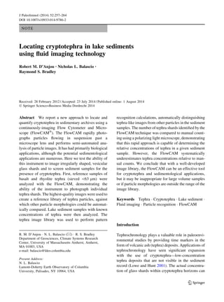
Fig. 1 Representative tephra images from the tephra image library produced by the FlowCAM
260 J Paleolimnol (2014) 52:257–264
123
5. methods indicated concentrations ranging from 24 to
317 tephra shards per sample, with distinct peaks (317
and 312 shards) at the same depths of 247 and 284 cm
that were identified by the FlowCAM analysis (Fig. 2a).
Although the absolute number of grains counted on the
FlowCAM was consistently lower than the microscopy
counts, the relative concentrations were consistent at all
depths and exhibit a linear relationship (Fig. 2b). The
FlowCAM identified an average of 22.8 ± 9.7 % of the
grains counted under a microscope.
To estimate the uncertainty of microscope counts,
sample depths with the highest concentrations were
counted under the microscope in triplicate. Average
counts for the two peaks at 247 and 284 cm were
317 ± 11 and 312 ± 14, respectively. By contrast,
the VisualSpreadsheet software uses an algorithm
based on quantitative particle values to determine the
likelihood of a particle actually being tephra, so that
identification is standardized and variability in the
software based identification of tephra images
between multiple runs of the same sample was tested
and determined to be negligible. However, the Flow-
CAM images only the portion of the sediment sample
that flows in the camera’s limited field of view, which
at 1009 magnification is one-third of the sample
volume (A´ lvarez et al. 2011). Therefore, changes in
the homogeneity of the sample and very sparse
quantities of tephra grains can negatively affect the
reproducibility of FlowCAM based tephra counts.
Tephra counts from the FlowCAM method identi-
fied a greater percentage of shards in the samples
containing basaltic tephra, 30 ± 9 %, compared with
17 ± 6 % for the rhyolitic tephra (Fig. 3a). However,
for both compositions, a strong linear correlation
exists between FlowCAM and traditional microscopy
counts (Fig. 3b).
Discussion
The image-based, FlowCAM approach to identifying
cryptotephra is capable of providing insight into
relative concentrations of tephra shards. The method
was effective at imaging tephra shards from 20 to
100 lm diameter, which were the limits of samples we
analyzed, and at concentrations approaching zero. In
lake sediment samples, comparison of FlowCAM
tephra counts with standard microscopy show that the
instrument was able to identify samples with the
highest concentration of tephra as determined from
microscope counts. However, the FlowCAM was only
capable of measuring 23 ± 10 % of the microscope
counts in each sample. Interestingly, in a previous
study A´ lvarez et al. (2011) found that using the
FlowCAM at 1009 in autoimage mode, only 31.49 %
of the total sample was being captured within the field
of view of the camera lens. When taking this into
account, our ability to image 23 ± 10 % of the total
0
200
400
600
800
0
20
40
60
80
100
243
244
245
246
247
248
249
250
283
284
285
286
287
288
289
290
291
Depth (cm.)
Microscope
FlowCAM
A
y = 3.4117x
R² = 0.8939
0
50
100
150
200
250
300
350
0 10 20 30 40 50 60 70 80 90 100
MicroscopyTephraGrainCounts
B
MicroscopeTephraGrainCounts
FlowCAMTephraGrainCounts
FlowCAM Tephra Grain Counts
Fig. 2 a Grain counts for the 17 lake sediment samples using the FlowCAM (light grey) and traditional microscopy methods (black).
b Scatter plot of these values displaying a linear relationship
J Paleolimnol (2014) 52:257–264 261
123
6. tephra concentration not only makes sense, but also
bolsters the idea that the FlowCAM is in fact imaging
and correctly identifying a high percent of the tephra
flowing through the field of view. When this 31.49 %
field of view is corrected for in the results of this study,
the FlowCAM consistently imaged and accurately
identified 74.6 % of the tephra contained in each
sample. Additional sources of error in the absolute
number of tephra particles counted by the FlowCAM
may include: the software settings used to extract
particle images, particle clumping, the focus of
images, and the range of tephra morphologies within
the user-defined tephra image library.
The ultimate goal of this study was to test the
suitability of this instrument and method as a replace-
ment for the laborious and destructive extraction-
microscopy technique. This was under the assumption
that an unprocessed sample of lake sediment could be
rapidly analyzed by the FlowCAM, and subsequently
re-collected, preserving the sample for later analyses.
However, our study showed that ratios of tephra to
background sediment had to exceed *1:200 to be
processed in a reasonable amount of time (20–30 min
per sample). In contrast, distal cryptotephra deposits
occur in very low concentrations and may not be
suitable for FlowCAM analysis. In sedimentary
archives where cryptotephra horizons contain very
high concentrations of tephra shards this method
would be advantageous. However, analysis of small
sediment volumes (*1 cm3
) with low tephra concen-
trations limit the number of particles counted in a
reasonable amount of time. Thus, pre-processing of
the sediment core is still required to reduce the sample
to a manageable volume prior to FlowCAM analysis.
Nevertheless, the FlowCAM can provide an indication
of where tephra is likely to be more abundant in a
sediment core—making subsequent isolation of tephra
shards using conventional density-separation methods
more efficient by focusing attention on specific depth
horizons. In other types of records (peat, or sedimen-
tary records with visible/high concentrations of
tephra), the FlowCAM’s possibility as an effective
replacement to standard microscopy seems very
possible, and warrants further investigation in future
studies.
Limitations and recommendations
Our study demonstrates that the FlowCAM is able to
detect tephra shards and may be useful in cryptotephra
applications. However, the ability to distinguish
tephra in sediment is dependent on the quality of the
user-defined library of tephra images and the differ-
ence in the morphology of tephra from the background
0%
5%
10%
15%
20%
25%
30%
35%
40%
45%
243
244
245
246
247
248
249
250
283
284
285
286
287
288
289
290
291
PercentTephraImagedbyFlowCAM
Depth (cm.)
Basaltic Tephra
Rhyolitic Tephra
A
y = 3.2728x + 29.039
R² = 0.9833
y = 3.0535x + 5.1502
R² = 0.9246
0
50
100
150
200
250
300
350
0 10 20 30 40 50 60 70 80 90 100
MicroscopyTephraGrainCounts
Rhyolitic (Vedde) Tephra
Basaltic (Saksunarvatn)
Tephra
B
FlowCAM Tephra Grain Counts
Fig. 3 a Percent of total tephra grains imaged by the FlowCAM
(FlowCAM counts/microscope counts 9 100) for the rhyolitic
(light grey) and basaltic (black) populations. b Scatter plot of
FlowCAM versus microscope counts for each population
displaying linear relationships
262 J Paleolimnol (2014) 52:257–264
123
7. sediment. In applying this method, we recommend
that care be taken to build a library of tephra images
that captures a range of tephra morphologies predicted
for a particular environment or region. If the library is
inadequate, this method may fail to detect tephra even
if it is present. For example, diagenetically altered,
crystal-rich, lithic-rich or phreatomagmatic tephras
containing fewer of the distinctive bubble wall shard
morphologies may be challenging to account for, but
present an interesting area for future work in fine-
tuning the FlowCAM technique. Additionally, uncer-
tainties in particle counts may be reduced with
additional preparation steps to disaggregate samples,
such as the use of a dispersant (Calgon) and/or
sonication prior to FlowCAM analysis.
Conclusions
The results from this study indicate a strong potential
for the FlowCAM to provide a valuable method of
particle discrimination in a wide variety of sedimen-
tological applications. Further investigation of the
utility and reproducibility of the results is necessary.
However, the instrument was able to efficiently
capture high-quality images of tephra shards within a
sample, and that the images clearly depicted the
distinct morphological characteristics of the targeted
tephra shards. These distinct particle properties
allowed the visual pattern recognition software to
identify and separate the tephra images within a
sample’s data file. We were able to accurately
determine the relative concentrations of tephra within
a sediment sample, although the absolute tephra
concentrations were consistently underestimated.
The primary reason for lower tephra counts using the
FlowCAM is because of the camera’s limited field of
view, which has been established by others and can be
used to correct particle counts. Further investigation of
the applications and limitations of the instrument in
cryptotephra studies could address: (i) a greater
number of tephra standards, (ii) additional background
sediment types, and (iii) particle grain sizes. The
FlowCAM may also provide an alternative approach
to traditional microscopy methods when the identifi-
cation and enumeration of any microscopic ‘‘group’’
of particles in a mixed sediment sample can be defined
by a set of distinct set of particle characteristics.
Acknowledgments This project was funded by National Sci-
ence Foundation grant ARC-0909354 and National Oceanic and
Atmospheric Administration grant NA09OAR4600215. We
would like to thank Jon Woodruff, Kinuyo Kanamaru and
Lucien von Gunten for their input early on in this study, as well
as Alexa Van Eaton and two anonymous reviewers for
comments on earlier drafts.
References
Abbott PM, Davies SM, Austin WEN, Pearce NJG, Hibbert FD
(2011) Identification of cryptotephra horizons in a North
East Atlantic marine record spanning marine isotope stages
4 and 5a (60,000–82,000 a b2k). Quat Int 246:177–189
A´ lvarez E, Lopez-Urrutia A´ , Nogueira E, Fraga S (2011) How to
effectively sample the plankton size spectrum? A case
study using the FlowCAM. J Plankton Res 33:1119–1133
Andrews JT, Eberl DD, Kristjansdottir GB (2006) An explor-
atory method to detect tephras from quantitative XRD
scans: examples from Iceland and east Greenland marine
sediments. Holocene 16:1035–1042
Balascio NL (2011) Lacustrine records of Holocene climate and
environmental change from the Lofoten Islands, Norway.
Ph.D. Dissertation. University of Massachusetts Amherst
Balascio NL, Wickler S, Narmo LE, Bradley RS (2011a) Distal
cryptotephra found in a Viking boathouse: the potential for
tephrochronology in reconstructing the Iron Age in Nor-
way. J Archaeological Sci 38:934–941
Balascio NL, Zhang Z, Bradley RS, Perren B, Dahl SO, Bakke J
(2011b) A multi-proxy approach to assessing isolation
basin stratigraphy from the Lofoten Islands, Norway. Quat
Res 75:288–300
Barofsky A, Simonelli P, Vidoudez C (2010) Growth phase of
the diatom Skeletonema marinoi influences the metabolic
profile of the cells and the selective feeding of the copepod
Calanus spp. J Plankton Res 32:263–272
Brown L (2004) Continuous imaging fluid particle analysis—a
primer. Fluid imaging technologies white paper. http://
fluidimaging.com
Brown L (2008) Particle image understanding—a primer. Fluid
Imaging Technologies, Yarmouth, ME
Brown L (2010a) VisualSpreadsheetÓ: intelligent pattern rec-
ognition for particle analysis. Fluid Imaging Technologies
VisualSpreadsheetÓ Particle Analysis Software Product
Literature. http://fluidimaging.com
Brown L (2010b) VisualSpreadsheetÓ: interactive, intuitive
particle analysis software. Fluid Imaging Technologies
VisualSpreadsheetÓ Particle Analysis Software Product
Literature. http://fluidimaging.com
Brown L (2011a) Characterizing biologics using dynamic
imaging particle analysis. BioPharm Int 24:4–9
Brown L (2011b) FlowCAM tech brief: proper thresholding of
transparent particles. Fluid imaging technologies tech
briefs. http://fluidimaging.com
Buskey EJ, Hyatt CJ (2006) Use of the FlowCAM for semiau-
tomated recognition and enumeration of red tide cells
(Karenia brevis) in natural plankton samples. Harmful
Algae 5:685–692
J Paleolimnol (2014) 52:257–264 263
123
8. Calanchi N, Cattaneo A, Dinelli E, Gasparotto G, Lucchini F
(1998) Tephra layers in late quaternary sediments of the
central Adriatic Sea. Mar Geol 149:191–209
Carter L, Manighetti B (2006) Glacial/interglacial control of
terrigenous and biogenic fluxes in the deep ocean off a high
input, collisional margin: a 139 kyr-record from New
Zealand. Mar Geol 226:307–322
Carter L, Manighetti B, Elliot M, Trustrum N, Gomez B (2002)
Source, sea level and circulation effects on the sediment
flux to the deep ocean over the past 15 ka off eastern New
Zealand. Glob Planet Change 33:339–355
D’Andrea WJ, Vaillencourt DA, Balascio NL, Werner A, Roof
SR, Retelle M, Bradley RS (2012) Mild little ice age and
unprecedented recent warmth in an 1800 year lake sedi-
ment record from Svalbard. Geology 40:1007–1010
De Vleeschouwer F, van Vlie¨t-Lanoe´ B, Fagel N, Richter T,
Boe¨s X (2008) Development and application of high-res-
olution petrography on resin-impregnated Holocene peat
columns to detect and analyse tephras, cryptotephras, and
other materials. Quat Int 178:54–67
Dugmore AJ, Newton AJ (1992) Thin tephra layers in peat
revealed by X-radiography. J Archaeol Sci 19:163–170
Dugmore AJ, Larsen G, Newton AJ (1995) Seven tephra is-
ochrones in Scotland. The Holocene 5:257–266
Enache MD, Cumming BF (2006) The morphological and
optical properties of volcanic glass: a tool to assess density-
induced vertical migration of tephra in sediment cores.
J Paleolimnol 35:661–667
Ersoy O, Gourgaud A, Aydar E, Chinga G, Thouret J-C (2007)
Quantitative scanning-electron microscope analysis of
volcanic ash surfaces: application to the 1982–1983 Ga-
lunggung eruption (Indonesia). Geol Soc Am Bull
119:743–752
Gehrels MJ, Newnham RM, Lowe DJ, Wynne S, Hazell ZJ,
Caseldine C (2008) Towards rapid assay of cryptotephra in
peat cores: review and evaluation of various methods. Quat
Int 178:68–84
Gro¨nvold K, O´ skarsson N, Johnsen SJ, Clausen HB, Hammer
CU, Bond G, Bard E (1995) Ash layers from Iceland in the
Greenland GRIP ice core correlated with oceanic and land
sediments. Earth Planet Sci Lett 135:149–155
Haflidason H, Eiriksson J, Van Kreveld S (2000) The tephro-
chronology of Iceland and the North Atlantic region during
the Middle and Late Quaternary: a review. J Quat Sci
15:3–22
Hall VA, Pilcher JR (2002) Late-Quaternary Icelandic tephras in
Ireland and Great Britain: detection, characterization and
usefulness. Holocene 12:223–230
Heiken G (1972) Morphology and petrography of volcanic
ashes. Geol Soc Am Bull 83:1961–1988
Heiken G (1974) An atlas of volcanic ash. Smithson Contrib
Earth Sci 12:1–101
Heiken G, Wohletz KH (1985) Volcanic ash. University of
California Press, Berkeley, CA
Ide K, Takahashi K, Kuwata A, Nakamachi M, Saito H (2008) A
rapid analysis of copepod feeding using FlowCAM.
J Plankton Res 30:275–281
Jennings AE, Gronvold K, Hilberman R, Smith M, Hald M
(2002) High resolution study of Icelandic tephras in the
Kangerlussuaq trough, southeast Greenland, during the last
deglaciation. J Quat Sci 17:747–757
Jude-Eton T, Thordarson T, Gudmundsson MT, Oddsson B
(2012) Dynamics, stratigraphy and proximal dispersal of
supraglacial tephra during the ice-confined 2004 eruption
at Grı´msvo¨tn Volcano, Iceland. Bull Volc 74:1057–1082
Kido Y, Koshikawa T, Tada R (2006) Rapid and quantitative
major element analayis method for wet fine-grained sedi-
ments using and XRF microscanner. Mar Geol 229:209–
225
Lowe DJ (2011) Tephrochronology and its application: a
review. Quat Geochron 6:107–153
Lowe DJ, Hunt JB (2001) A summary of terminology used in
tephra-related studies. Les Dossiers de l’Archeo-Logis
1:17–22
Meara R (2011) Climatic and environmental impact of Holo-
cene silicic explosive eruptions in Iceland. Ph.D. Thesis.
University of Edinburgh, p 324
Pilcher JR, Hall VA, McCormac FG (1996) An outline tep-
hrochronology for the Holocene of the north of Ireland.
J Quat Sci 11:485–494
Pilcher J, Bradley RS, Francus P, Anderson L (2005) A Holo-
cene tephra record from the Lofoten Islands, Arctic Nor-
way. Boreas 34:136–156
Sieracki C, Sieracki ME, Yentsch CS (1998) An imaging-
in-flow system for automated analysis of marine micro-
plankton. Mar Ecol Prog Ser 168:285–296
Sterling MC Jr, Bonner JS, Ernest ANS, Page CA, Autenrieth
RL (2004) Characterizing aquatic sediment–oil aggregates
using in situ instruments. Mar Poll Bull 48:533–542
Tauxe L, Steindorf JL, Harris A (2006) Depositional remanent
magnetization: toward an improved theoretical and
experimental foundation. Earth Planet Sci Lett 244:515–
529
Turney CSM (1998) Extraction of rhyolitic component of
Vedde microtephra from minerogenic lake sediments.
J Paleolimnol 19:199–206
Turney CSM, Harkness DD, Lowe JJ (1997) The use of mic-
rotephra horizons to correlate Late-glacial lake sediment
successions in Scotland. J Quat Sci 12:525–531
264 J Paleolimnol (2014) 52:257–264
123