1. Development of Robust Antibody Purification by
Optimizing Protein-A Chromatography in
Combination With Precipitation Methodologies
Srinivas Chollangi, Ray Parker, Nripen Singh, Yi Li, Michael Borys, Zhengjian Li
Bristol-Myers Squibb, Biologics Development, Global Manufacturing & Supply,
Hopkinton, Massachusetts; telephone: þ9784 784 6513; fax: 978-784-6639; e-mail: Yi.
Li@bms.com
ABSTRACT: To be administered to patients, therapeutic
monoclonal antibodies must have very high purity, with process
related impurities like host-cell proteins (HCPs) and DNA reduced
to <100 ppm and <10 ppb, respectively, relative to desired product.
Traditionally, Protein-A chromatography as a capture step has been
the work horse for clearing a large proportion of these impurities.
However, remaining levels of process and product related impurities
still present significant challenges on the development of polishing
steps further downstream. In this study, we have incorporated high
throughput screening to evaluate three areas of separation: (i)
Harvest treatment; (ii) Protein-AChromatography; and (iii) Low pH
Viral Inactivation. Precipitation with low pH treatment of cell
culture harvest resulted in selective removal of impurities while
manipulating the pH of wash buffers used in Protein-A
chromatography and incorporating wash additives that disrupt
various modes of protein–protein interaction resulted in further
and more pronounced reduction in impurity levels. In addition, our
study also demonstrate that optimizing the neutralization pH post
Protein-A elution can result in selective removal of impurities.
When applied over multiple mAbs, this optimization method
proved to be very robust and the strategy provides a new and
improved purification process that reduces process related
impurities like HCPs and DNA to drug substance specifications
with just one chromatography column and open avenues for
significant decrease in operating costs in monoclonal antibody
purification.
Biotechnol. Bioeng. 2015;112: 2292–2304.
ß 2015 Wiley Periodicals, Inc.
KEYWORDS: high-throughput screening; monoclonal antibody;
precipitation; protein-a chromatography
Introduction
Monoclonal antibodies and their derivatives are currently a major
source of revenue generation in global biotechnology market
(Lawrence, 2007a,b). However, process economics can be greatly
influenced by the choice of expression and purification steps.
Recent advances in cell line selection, growth media, and feeding
strategies have led to significant improvement in cell densities and
expression levels are consistently reaching 5–10g/L across the
industry in a 14 day fed-batch process (Huang et al., 2010).
Consequently, process bottlenecks have shifted downstream and
purification costs are now outweighing the upstream cell culture
costs (Follman and Fahrner, 2004; Gagnon, 2012; Guiochon and
Beaver, 2011). Implementation of platform approach has greatly
reduced downstream operation costs but challenges associated with
meeting final drug substance specifications still remain (Guiochon
and Beaver, 2011; Shukla et al., 2007).
For monoclonal antibodies purification platform, Protein-A
chromatography is still the most robust step in removing process
and product related impurities (Vunnum et al., 2009a). Owing to its
specific affinity towards Fc region containing antibodies and fusion
proteins and its ability to tolerate high conductivities, Protein-A
chromatography allows direct loading of harvested cell culture fluid
(HCCF) and enables removal of a vast majority of process and
product related impurities while enriching the antibody pool
(Vunnum et al., 2009a). Nevertheless, remaining impurities after
Protein-A still present a significant challenge to the purification
steps downstream in order to achieve the drug substance
specifications suitable for patient administration (Guiochon and
Beaver 2011; Liu et al., 2010). In addition, it has been consistently
shown that Protein-A chromatography alone constitutes more than
a quarter of the total raw materials costs in downstream purification
of the antibodies (Follman and Fahrner, 2004). Thus, optimal and
efficient usage of Protein-A affinity resin is critical to achieve high
product quality and reduce the costs of antibody production.
During Protein-A chromatography, post load wash step is a key
means to achieve impurity clearance. Manipulating the pH of wash
buffer is usually the first choice and conventionally, a wash step with
a pH between the load and elution conditions is employed for
Protein-A chromatography (Group 2009; Vunnum et al., 2009a).
Correspondence to: Y. Li
Received 18 February 2015; Revision received 23 April 2015; Accepted 28 April 2015
Accepted manuscript online 6 May 2015;
Article first published online 31 July 2015 in Wiley Online Library
(http://onlinelibrary.wiley.com/doi/10.1002/bit.25639/abstract).
DOI 10.1002/bit.25639
ARTICLE
2292 Biotechnology and Bioengineering, Vol. 112, No. 11, November, 2015 ß 2015 Wiley Periodicals, Inc.
2. The pH of this intermediate wash is lowered as low as possible
without initiating premature elution of the product and this
requires molecule specific effort. With this approach, HCP levels
post Protein-A are usually in the range of 3,000 to 12,000 ppm as
reported in a case-study conducted by a consortium of biotech
industry specialists (Group, 2009). Typically, this requires an
additional of at least two chromatography steps to further purify
and polish the product to achieve drug substance specifications
(Group, 2009). In 2007, Shukla and Hinckley (2008) reported one of
the first successful attempts to enhance the performance of
Protein-A chromatography by evaluating a number of wash
additives like sodium chloride, urea, propylene glycol, ethanol,
isopropanol, tween-80, spermine, and sodium sulfate during
Protein-A chromatography. Based on the results, they have
identified urea as the best wash additive to achieve effective
removal of HCPs. The levels of HCPs reported upon incorporating
urea wash were in the range of 1,000–3,500 ppm where recovery of
the product varied between 70 and 100%. However, it is also to be
noted that at high concentrations, agents like urea, ethanol, and
isopropanol can destabilize the structure of proteins through
formation of hydrogen bonds with peptide groups and exposing
hydrophobic residues (Herskovits and Jaillet 1969; Herskovits et al.,
1970a, b; Lim et al., 2009). On the other hand, presence of small
amounts of sodium chloride, sodium sulfate, propylene glycol, and
tween-80 are shown to help stabilize the protein structure by
interacting with the polar residues on the surface of the molecule
(Agarkhed et al., 2013; Damodaran and Kinsella 1981; Kerwin et al.,
1998; Thurow and Geisen 1984). In addition, like other agents
mentioned above, L-Arginine is known to help stabilize monoclonal
antibodies by breaking non-specific interactions between proteins
and preventing aggregation (Lange and Rudolph 2009; Schneider
et al., 2011; Shukla and Trout 2010). Studies by Yumioka et al.
(2010) and Sun (2013) have shown that L-Arginine can also be used
as a wash reagent to reduce impurity levels during Protein-A
chromatography (Sun, 2013; Yumioka et al., 2010). HCP levels were
reported to be reduced by about 90–95% in Protein-A product pool
using this strategy. However, in both of these cases, the HCP levels
are still high compared to final drug substance specifications and
require two more steps for further polishing. Also, with the lack of
high-throughput automation technologies at the time, all of these
experiments were carried out in a single case by case manner and
limited the scope of identifying the most optimal concentration and
combinations of the additives that can yield better results.
Following the above works, significant efforts were focused on
identifying the HCPs that are binding to Protein-A column and
co-eluting with the product during elution (Hogwood et al., 2013b;
Jin et al., 2010; Levy et al., 2014; Sisodiya et al., 2012; Tait et al.,
2012; Tarrant et al., 2012). These studies demonstrated that the
type of HCPs co-eluting with product varied from molecule to
molecule and from one expression system to the other, making the
development of effective platform purification process for Protein-A
chromatography highly challenging (Sisodiya et al., 2012). More
recently, Aboulaich et al, (2014) have covalently cross-linked
monoclonal antibodies to NHS-activated sepharose resin to study
their interaction with various host-cell proteins. Again, these
studies confirmed that the type of HCPs associating with the
product during Protein-A chromatography varies from molecule to
molecule. In addition, they showed that wash additives like arginine
can help reduce the HCP levels in product pool. However, the study
is limited by the fact that the full extent of interactions between
process related impurities and the antibody is not captured because,
unlike in cell culture broth, the complete surface of antibody is
inaccessible to the HCPs after its prior immobilization to the
sepharose resin. This limits the number of sites on the monoclonal
antibody available for HCP interaction and thereby not all HCP
populations that normally interact with the antibody are accounted
for. Also, the HCP interaction with Protein-A ligand and co-elution
with product during low pH treatment is not captured in such
setting. In addition, in all the above studies, the focus has been
primarily on understanding HCP interaction and removal during
Protein-A chromatography and did not evaluate the effects of HCP
clearance during Protein-A chromatography in context with other
steps (e.g., harvest clarification and low pH viral inactivation)
involved in typical purification platform.
Recent advancements in high-throughput automation technol-
ogies have enabled rapid acquisition of large datasets under several
different operating conditions (Coffman et al., 2008; Kelley et al.,
2008; Kramarczyk et al., 2008). This enables a practical strategy to
establish a framework where customized wash steps for a particular
mAb is optimized together with harvest clarification and viral
inactivation. Exploiting this, in this study we incorporated
high-throughput screening to investigate the effects of pH, buffer
composition, and a range of wash additives (caprylic acid,
propylene glycol, triton x-100, arginine, urea, isopropanol, EDTA,
and sodium chloride) to identify the most optimal conditions for
effective HCP removal and further improve the performance of
Protein-A chromatography. The wash additives employed and the
high-throughput design of the study was aimed at not only
identifying an effective wash condition, but also understand the
nature of interactions between the HCPs and the antibody during
Protein-A chromatography. This method of screening can serve as a
template for all therapeutic antibodies coming in the pipeline to
rapidly identify an effective wash strategy during Protein-A
chromatography. In addition, combining this strategy with
additional screening on separation techniques like precipitation
during cell culture harvest and optimizing the neutralization pH
post Protein-A chromatography yielded highly improved impurity
clearance essentially meeting drug substance specifications for HCP
and DNA clearance with just one column process. When tested
across multiple molecules, this method proved very robust and can
be effectively used to significantly improve the product quality while
minimizing the load and cost on subsequent operations.
Materials and Methods
Cell Culture and Harvest Treatment
Proprietary CHO cell lines engineered from either DG44 parental
cells or GS parental cells were used to produce the null harvest or
harvests containing recombinant monoclonal antibodies (mAb1,
mAb2, mAb3, and mAb4). All cells were cultured using a chemically
defined, animal component-free media. The bioreactors were
operated in a fed-batch mode with continuous feeds based on
maintaining glucose concentration between 2 and 4 g/L. Titers were
Chollangi et al.: Impurity Clearance During Antibody Purification 2293
Biotechnology and Bioengineering
3. measured using a Protein-A HPLC system (Agilent 1100, Waters
Corporation, Milford, MA) with an established reference standard.
During the harvest, cell culture suspensions were collected and then
were either left untreated or treated using acid titrants (acetic acid
or citric acid) to lower the pH to desired range. Treatment was
carried out for 30 min under gentle mixing. Following treatment,
the harvest material was centrifuged and filtered using a depth filter
(60SP05A, 3M, St. Paul, MN) followed by 0.8/0.2 mm filter capsule
(Pall Corporation, Port Washington, NY). Post filtration, pH of the
clarified harvest was adjusted to a range between 7.0 and 7.2 using
2 M Tris and subjected to analytical analyses to assess impurity
reduction.
Protein-A Chromatography
GE healthcare’s MabSelect Protein-A resin was used for all capture
chromatography experiments. Preparative scale column-based
chromatography experiments were carried out using an €AKTA
Avant instrument (GE Healthcare, Uppsala, Sweden) controlled by
Unicorn 6.3 software. All columns were packed in the lab according
to the resin manufacturer’s recommendations. Unless noted,
equilibration of the resin was carried out using phosphate-buffered
saline (PBS) at pH 7.4 followed by loading with harvested cell
culture fluid (HCCF). The resin was then subjected to PBS wash
followed by an acidic pH wash (pH 5.0–6.0) before eluting the mAb
using buffers at pH < 4.0.
Viral Inactivation and Filtration
Following elution, mAb pool from Protein-A chromatography was
subjected to viral inactivation by holding at low pH (3.4–4.0) for 1 h
at room temperature followed by neutralization to desired pH in a
range between 4.0 and 9.0. Titrationwas carried out using 0.1 HCl or
2 M Tris. Post neutralization, filtration was carried out using
0.8/0.2 mm filter capsule or a 0.8/0.2 mm syringe filter (Pall
Corporation).
High Throughput Liquid Handling and Chromatography
Batch-mode high-throughput chromatography experiments were
carried out using PreDictor plates packed with 50 mL MabSelect
resin (GE healthcare). Liquid handling was conducted in a high
throughput manner using Tecan Freedom-Evo system equipped
with a high-precision Liquid Handler arm (LiHa) and Robotic
Manipulator (RoMa) (Tecan Group, Ltd. Mannedorf, Switzerland).
Method creation and execution was carried out using Tecan
freedom-evoware software (Tecan Group Ltd.). Wash buffers
containing various additives were prepared using respective stock
solutions and diluting them as needed on the tecan. These buffers
are dispensed and stored in plate format compatible for tecan liquid
handling. For a typical batch mode Protein-A chromatography
experiment, the MabSelect resin is equilibrated with PBS followed
by loading with HCCF. The resin is then subjected to either PBS
wash or wash buffers containing various additives. The target
protein is then eluted using a low pH buffer (pH < 4.0) and
collected into a 96-well plate. The product pool is then neutralized
using 2 M Tris followed by filtration using 0.2 mm filter plate (Pall
Corporation). The filtrate is then subjected various analytical assays
to assess product quality and impurity clearance.
Analytical Characterization
Post capture chromatography and filtration, protein concentration
was assayed by high-throughput UV-Vis spectroscopy using the
DropSense96polychromaticmicroplatereader(Trinean,Gentbrugge,
Belgium). UV-Vis spectra were quantified using DropQuant software
(Trinean). Size exclusion chromatography (SEC) was conducted
using Waters’ Acquity H-Class Bio UPLC
1
to measure the monomer
and aggregate content in the product pool. Acquity UPLC
1
BEH200
SEC 1.7 mm column (4.6 Â 150mm) was utilized to perform this
assay where the mobile phase consisted of phosphate buffered saline
(10 mM Phoshphate, 137 mM Sodium Chloride, 2.7 mM Potassium
Chloride) at pH 6.8 and was run at a flow rate of 1 mL/min.
Quantification of monomer, low molecular, and the high molecular
weightspecieswasperformedusingEmpowersoftware(WatersCorp.
Orlando, FL). In addition, secondary structure, charge profile, and
thebindingactivity wereconfirmedusingcirculardichroism(JascoJ-
715spectropolarimeter), iso-electric focusing (ProteinSimple iCE3),
and custom ELISA with a reference standard. For hydrophobicity
estimation, primary sequences of the four antibodies in study are
aligned using ClustalW2 (Larkin et al., 2007) and identified that the
differences between the sequences largely lied in complementarity
determining regions (CDRs). 3D structures of the Fab regions of these
four antibodies were built using homology modeling tool SWISS-
Model (Arnold et al., 2006). Amino acid sequences of CDR loops
containing all sequence differenceswere identified inthesestructures
and the hydrophobicity scores of these CDR loops were calculated
using Kyte-Doolittle hydrophobicity score (Kyte and Doolittle, 1982).
ELISA for quantification of residual CHO host cell proteins (CHO-
HCP) and residual Protein-A was conducted in a high throughput
manner using the Tecan liquid handling system. Host cell proteins
were quantified using the CHO Host Cell proteins 3rd generation kit
(Cat # F550, Cygnus Technologies, Southport, NC) while residual
Protein-A was quantified using Repligen Protein-A ELISA kit (Cat #
9000-1, Repligen Corporation, Waltham, MA) according to
manufacturer’s protocol. Absorbance was measured at 450/650 nm
using EnVision Multilabel reader (PerkinElmer. Waltham, MA). Data
quantification and analysis was carried out using JMP software (SAS
Institute Inc. Cary, NC). Residual CHO DNA in the samples were
measured using real-time quantitative PCR (RT-PCR). RT-PCR was
carried out with the 2 Â TaqMan Universal PCR Master Mix kit (Life
Technologies, Carlsbad, CA) and 7900 HT Real Time PCR system
(Life Technologies) according the manufacturer’s instructions.
Results and Discussion
HCPs and DNA Co-Elution With Antibody
Three different load materials: (i) mAb1 expressing CHO cell HCCF;
(ii) null-CHO cell HCCF; and (iii) null-CHO cell HCCF spiked with
purified mAb1 were subjected to Protein-A purification process.
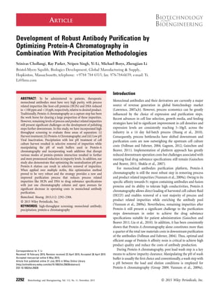
Figure 1 shows the amount of mAb1, HCPs, and DNA being loaded
and eluted from the Protein-A column. Consistent with the results
reported by Shukla and Hinckley (2008) our results show that there
2294 Biotechnology and Bioengineering, Vol. 112, No. 11, November, 2015
4. is an increase in the HCP and DNA levels in the elution pools when
the antibody is present. This phenomenon is attributed to the
non-covalent interactions between the impurities and the antibody
while the interaction between impurities and Protein-A resin is
minimal, particularly in the case of agarose based matrix (Levy
et al., 2014; Nogal et al., 2012; Shukla and Hinckley 2008; Sisodiya
et al., 2012; Tarrant et al., 2012). Specifically, Tarrant et al. (2012)
have compared the adsorption behavior of host-cell proteins onto
agarose based and glass based Protein-A resin matrices (Tarrant
et al., 2012). Using ELISA and SELDI-TOF mass-spectrometric
analysis they have identified that the hydrophilic agarose based
matrices exhibit low degree of interaction with the CHO host-cell
proteins while the glass based ProSep Ultra Plus Affinity (UPA)
resin exhibited high degree of non-specific interaction with the
host-cell proteins. This was attributed to the hydrophobic nature of
the controlled pore glass (CPG) back-bone of the ProSep UPA resin.
However, in the presence of antibody, both agarose and glass based
resins started exhibiting adsorptive behavior towards CHO host-cell
proteins; indicating non-specific interactions between the antibody
and the host-cell proteins. Previous results have also shown that the
degree of these antibody-HCP interactions is highly variable and
depends on the antibody present in the HCCF (Sisodiya et al., 2012).
HCPs Associate With mAb Through a Complex Mode of
Interactions
While the association of HCPs with antibody during Protein-A
chromatography is well established, the mode of this interaction is
not well understood yet. A number of groups have used proteomic
based approaches to investigate the profiles of HCPs present in
HCCF and the eluates from Protein-A column leading to
identification of specific populations that exhibit strong interaction
with antibodies (Hogwood et al., 2013a, b; Jin et al., 2010; Levy
et al., 2014; Nogal et al., 2012; Shukla and Hinckley 2008; Sisodiya
et al., 2012; Tait et al., 2012; Tarrant et al., 2012). While progress was
made to disrupt these interactions between HCPs and antibody, the
remaining levels of impurities are still high and require at least two
additional steps to bring the impurity levels down to drug substance
specifications (Group, 2009; Shukla and Hinckley, 2008). To assess
improved ways that can disrupt these interactions, batch mode
high-throughput Protein-A chromatography experiments were
carried out on mAb1 (an IgG4 with a pI between 6.5 and 7.0)
using wash buffers at various pH values and incorporating salts and
other wash additives into them. Figure 2 shows the effect of wash
buffer pH on recovery of antibody and removal of HCPs from the
product. As shown in panel A, the experiment was carried out both
in the presence and absence of 1 M NaCl. In both cases, as expected,
results show that there is pronounced loss of antibody when the pH
of wash buffer is lower than 5.0 compared to basic wash buffers with
pH > 7.0. In addition, the presence of 1 M NaCl (high conductivity)
made the loss of antibody more pronounced even up to pH 7.0 and
starts to show diminishing effect at pH greater than 7.0. Within this
range, effect of wash buffer pHonthe removal of HCPs from product
pool is however much more dramatic than the effect on recovery.
Traditionally, an acidic buffer (pH in the range of 6.0 to 5.0) is often
employed to wash off impurities from the Protein-A column and
Figure 1. CHO host cell proteins (HCP) and host cell DNA associate with the monoclonal antibodies (mAbs) and co-elute as impurities during low pH elution. Top panel shows
the levels of mAb, HCPs, and DNA loaded onto the Protein-A column while bottom panel shows the levels of mAb, HCPs, and DNA present in the Protein-A eluate. The loading
material came from either CHO cell supernatant expressing mAb, null cell supernatant, or null cell supernatant spiked with purified mAb.
Chollangi et al.: Impurity Clearance During Antibody Purification 2295
Biotechnology and Bioengineering
5. also serve as a transition phase buffer to reduce the pH and help
keep the elution column volumes (CVs) low. However, the results
here show that a basic buffer (pH ! 8.0) is instead much more
effective in reducing the HCPs from product pool. The presence of
NaCl helps in realizing this benefit even at pH 7.0 or greater.
Combined with the recovery data shown in panel A, these results
suggest that basic wash buffers with pH ! 9.0 are effective in
keeping the recoveries high and obtain selective removal of HCPs
from product pool.
Considering the results shown in Figure 1 and keeping the pI of
mAb1 (6.5–7.0) in view, these results strongly suggest that
electrostatic interactions do play an important role in antibody-HCP
interactions. Shown in Table I is the list of iso-electric points of
various molecular entities found in HCCF and Protein-A elution
pools (Aboulaich et al., 2014; Gottschalk 2009; Levy et al., 2014).
Levy et al. (2014) have shown that the diverse population of CHO
host cell proteins has a wide range of iso-electric points but a
majority of this population has a pI in the range of 4.5 to 7.0. At pH
values between 4.5 and 7.0 mAbs are typically neutral or cationic
while DNA is anionic and a good number of HCPs are either neutral
or anionic. This results in a mix of strong electrostatic interactions
and hydrophobic interactions between the mAbs and the impurities
thus co-purifying during elution. However, when the resin
is subjected to washes with buffers at high (pH ! 8.0), mAbs
become either neutral or more anionic and with DNA and a vast of
majority of HCPs being strongly anionic they start dissociating from
other. With mAbs strongly bound to the Protein-A resin owing to
their specific affinity, HCPs and DNA are washed off the column
during these high pH washes resulting in product pool enriched
with pure mAb. However, it is also to be noticed that in the pH range
of 7.0 to 10.0 there is still some population of HCPs that is charged
or neutral and can bind to the mAbs non-specifically by range of
forces like hydrogen bonding, Van der Waals forces or hydrophobic
interactions. To assess whether these interactions can be
dissociated using wash additives, a high throughput batch-mode
chromatography experiment was carried out using wash buffers
containing a combination of salts and various wash additives.
Figure 3A and B show the contour maps of residual HCPs present
in Protein-A elution pools and the recovery after subjecting the
Figure 2. High pH washes aid in reducing the HCP levels in Protein-A eluate without a loss of recovery. (A) Recovery of mAb in Protein-A eluate is plotted against the pH of
wash buffer used during Protein-A chromatography. (B) HCP (ppm) levels in Protein-A eluate are plotted against the pH of wash buffer used during Protein-A chromatography. All
experiments were conducted either in the presence or absence of 1 M NaCl in the wash buffers. Error bars represent standard deviation of the analytical results. (* & **, P < 0.005).
Table I. Iso electric points of various class of molecules commonly
found in CHO cell harvests and Protein-A elution pools (Aboulaich et al.,
2014; Levy et al., 2014; Gottschalk, 2009).
Molecule class pI range
HCPs 2–11
DNA 2–3
Viruses 4–7.5
Protein-A 4.8–5.2
Endotoxins 1–4
2296 Biotechnology and Bioengineering, Vol. 112, No. 11, November, 2015
6. HCCF loaded resin to washes with various buffers containing
additives: sodium chloride, arginine, EDTA, iso-propyl alcohol
(IPA), propylene glycol, sodium octanoate (sodium caprylate/
caprylic acid), triton x-100, and urea. In Figure3A, the color
progression represents the levels of HCPs remaining in the elution
pool while in Figure 3B, the color progression represents recovery of
the monoclonal antibody in elution pool. As expected,
Figure 3B shows that the recoveries of the mAb1 are good at
high pH and lower when we use low pH wash buffer in combination
with high concentrations of NaCl and wash additives particularly,
urea. As observed in Figure 2, the control panel shown in
Figure 3A demonstrates that high pH buffer is more effective in
Figure 3. High pH washes and additives like Arginine and Urea help reduce HCP levels in Protein-A eluates. Shown in the figure are contour plots representing (A) HCP levels in
Protein-A eluates and (B) recovery of the antibody in eluate. CHO cell harvest was loaded equally onto a 96-well plate containing Protein-A resin followed by washes with buffers at
3 pH values (pH 5.5, 7.0, and 9.0) containing a range of wash additives in the presence or absence of NaCl. For each wash additive, the contour plot panel was generated using 12 data
points; four NaCl concentrations in combination with three excipient concentrations. (A) The deep blue color represents low levels of HCPs in the eluate while the deep red color
represents very high amounts of HCPs levels still remaining in the Protein-A eluate. (B) The deep blue color represents high recoveries while the deep red color represents low
recoveries.
Chollangi et al.: Impurity Clearance During Antibody Purification 2297
Biotechnology and Bioengineering
7. removing HCPs from the product pool. In addition, the results also
show that supplementing wash buffers with various wash additives
can have a positive effect in reducing the HCPs levels from product
pool. Particularly, urea and arginine demonstrate excellent
efficiency in disrupting the interactions between mAb1 and the
HCPs. In both cases, increasing the amount of the wash additive and
increasing the pH enhanced the ability of the buffers to disrupt the
antibody-HCP interactions while maintaining good recoveries
(Fig. 3A and B, bottom panels). It has been shown previously that
arginine can stabilize proteins by breaking non-specific protein-
protein interactions (Arakawa et al., 2006, 2007; Arakawa and Kita
2014; Arakawa and Tsumoto 2003; Borders et al., 1994; Schneider
et al., 2011; Shukla and Trout 2010; Tsumoto et al., 2005). The
guanidinium group on arginine was shown to be primarily
responsible for this ability as it can affect not only the electrostatic
and hydrophobic interactions but also hydrogen bonds between
proteins. On the other hand, urea is shown to interact with proteins
both directly, by forming hydrogen bonds with the protein and
indirectly by altering the solvent environment thereby mitigating
the hydrophobic effects that lead to protein–protein interactions.
However, it is also to be noted that at high concentrations, urea can
start to destabilize the protein structures by forming hydrogen
bonds with peptide groups present in the hydrophobic core of the
molecule and unraveling the tertiary structure of the protein
(Herskovits et al., 1970b; Lim et al., 2009). Thus a careful balance
needs to be maintained in case of urea to retain the protein structure
but disrupt non-specific protein–protein interactions. Combined
with the effects of pH described above, both of these wash additives
thus appear to be very effective in disrupting non-specific
mAb1-HCP interactions.
A wide variety of metals are often added to cell culture media
to boost cell viability and promote high titers. During the course
of cell culture, these metals can bind to antibodies and Gagnon
et.al., have shown that in addition to altering antibody charge
and hydrophobicity, metal ions can cause local conformational
changes and lead to formation of secondary complexes with
contaminants like HCPs, DNA, and endotoxins (Gagnon, 2010).
However, in the case of mAb1, our results show that addition of
EDTA to the wash buffer only has a modest effect in terms of
HCP removal. At pH 7.0, EDTA has slightly improved effect on
HCP removal compared to the control while at pH 9.0, it appears
to behave similar or inferior to the control. Albeit better at pH
7.0, organic solvent IPA exhibited similar performance to EDTA
at pH 9.0. In contrast, propylene glycol and non-ionic surfactant
Triton X-100 appear to be much more effective than the control
buffer in breaking down thenon-specific interactions between
mAb1 and HCPs at all pH values. Both of these agents are
hydrophilic and known to stabilize proteins by altering solvent
environment. Finally, sodium octanoate (also known as sodium
caprylate/caprylic acid) is an eight-carbon saturated fatty-acid
chain that is shown in the literature to selectively precipitate
HCPs and other impurities in HCCF (Brodsky et al., 2012).
When added to the wash buffers, this agent proved very effective
compared to the control wash, particularly at pH 9.0, and
selectively disrupted the interactions between HCPs and the
antibody. Combined together, the effect of high pH, salt, and the
effects of urea, arginine, propylene glycol, and triton x-100 on
strongly disrupting the interactions between HCPs and mAb1
suggest that the HCPs associate with antibodies through a
complex mode of interactions which may include electrostatic
interactions, hydrophobic interactions and hydrogen bonds.
HCPs can be Selectively Precipitated by Optimizing the
Neutralization pH Post Protein-A
The ICH Q5A guidance document requires the use of at least two
orthogonal steps for viral clearance to ensure the safety of
products produced using mammalian cell culture processes (FDA,
1998). In addition to nanofiltration, incubating product pools in
low pH environment (pH 3.0–4.0) is often used as a robust
method to inactivate enveloped viruses (Brorson et al., 2003;
Gagnon 2012; Omar et al., 1996). Given the requirement of low pH
buffers to elute antibodies from Protein-A column, this capture
step is often utilized as both a separation as well as transition for
viral inactivation step. However, Protein-A elution pools are often
reported to be associated with turbidity and the extent of this
turbidity was shown to be highly variable from antibody to
antibody (Tobler et al., 2006; Yigzaw et al., 2006). Depending upon
the operating pH of the second chromatography step downstream
of Protein-A, the low pH viral inactivated pool needs to be
neutralized to a pH suitable for loading onto the next column.
During this process, a significant increase in turbidity is often
reported (Tobler et al., 2006; Yigzaw et al., 2006). This
precipitation and turbidity was viewed as a risk for downstream
process as it poses problems of clogging inline sterile filters. The
risk is usually mitigated by using depth filters to remove the
precipitants and higher order aggregates (Yigzaw et al., 2006). In
our studies with mAb1, we observed similar precipitation
behavior when we used the control washes without any additives.
To further characterize the phenomenon, we have titrated the viral
inactivated pool to various pH values between 4.0 and 8.5 in
increments of 0.5 and then filtered the pools using a 0.2 mm filter.
Figure 4A shows the visual representation of turbidity in the
samples pools after titration while panel 4B shows that
the turbidity can be removed by filtration. Panel 4C shows the
quantitative measurement of absorbance reading at 410 nm before
and after filtration. As it can be noticed, the turbidity peaked
between the pH range of 5.0 and 7.5 and as the pool was titrated
to pH values further high, the turbidity went down. When the
filtered pools were subjected to analysis for antibody and HCP
contents, results showed that the precipitation strongly correlated
with selective removal of HCPs (Fig. 5). In contrast to what one
might expect for an affinity chromatography step, the HCP levels
in the elution pool from Protein-A chromatography have been
reported to be as high as 2,000–50,000 ppm (Group, 2009;
Sisodiya et al., 2012; Yigzaw et al., 2006). As described above in
section 3.2, a vast majority of CHO host cell proteins have a pI in
this pH range of 4.5–7.5 and can become less soluble during the
neutralization process leading to aggregation and precipitation.
However, as the pH moves further away from this range i.e.,
pH > 7.5 or pH < 4.0, the HCPs become polar and stay solubilized
in the solution leading to a decrease in turbidity. Thus, insoluble
aggregate formation during neutralization post viral inactivation
2298 Biotechnology and Bioengineering, Vol. 112, No. 11, November, 2015
8. is not necessarily an undesirable phenomenon and one can design
a process where the neutralization pH is optimal to get selective
precipitation of HCPs while minimizing any product loss. This
optimal pH would vary from one expression system to the other
and from one antibody to another depending up on its surface
properties and requires empirical characterization.
Enhancing Impurity Clearance by Optimizing Purification
Train
The most common purification schemes for monoclonal antibodies
utilize Protein A affinity chromatography as a capture step followed by
2to3chromatographicstepsforpolishing(Kelleyetal.,2009;Vunnum
et al., 2009b). Even though these chromatography steps are able to
meet the stringent purification requirements, they are expensive and
often the downstream purification train contributes to about 50–80%
of the total costs involved in antibody purification (Guiochon and
Beaver, 2011). With rapidly rising demand for therapeutic antibodies,
significant attention is thus being focused on reducing manufacturing
costs and improving process efficiency for industrial scale production.
Based on the findings described so far, we have tested various
purification schemes to evaluate the robustness of early separation
steps and the most effective and economical purification train.
As described in sections 3.2 and 3.3, majority of CHO host cell
proteins are unstable in acidic pH range between 4.5 and 7.0 and can
be precipitated out by optimal adjustments to the pH. This can be
performed post Protein-A as well as before Protein-A during cell
culture harvest. Lydersen et al. (1994) have previously shown that
acidification of fermentation broths can successfully induce
precipitation of host cell proteins and cellular debris (Lydersen
et al., 1994). In case of mAb1, we have observed similar behavior (See
Fig. 6). Acidification of the cell culture broth below pH 5.0 using
either citric acid or acetic acid, lead to precipitation and removal of
host cell proteins while maintaining antibody recovery above 95%.
mAb1 was then subjected to preparative scale purification using
various trains (see Table II) where acid precipitation was used either
with control Protein-A process or with optimized Protein-A washes
identified in section 3.2 and optimized neutralization pH identified
in section 3.3. Table III shows the results achieved using these
purification trains. As noted in train 1, purification of the antibody
without harvest treatment and control washes inProtein-A resulted
in high levels of host cell proteins while the DNAwas reduced to 10–
26 ppb. In contrast, using either of the enrichment techniques i.e.,
harvest treatment or enhancedProtein-A washes, lead to significant
decrease in both HCP and DNA levels. However, among the two,
enhanced Protein-A washes lead to a much greater decrease in the
impurity levels compared to acid precipitation (Train 2 vs. Train 3).
When combined with optimization of neutralization pH post viral
Figure 4. Neutralization to optimal pH after low pH hold for viral inactivation leads to increase in turbidity of the pool. (A) Pool turbidity of samples as a function of pH. VI hold is
at pH 3.5. (B) Pool turbidity of samples shown in panel A after filtration with 0.2 mm filter. (C) Absorbance at 410 of the samples shown in panels A and B.
Figure 5. Neutralization to optimal pH after low pH hold for viral inactivation leads
to selective precipitation of HCPs. Shown above are the recovery of mAb and HCP
levels in viral inactivated and neutralized bulk samples post filtration. Error bars
represent standard deviation of the analytical results.
Chollangi et al.: Impurity Clearance During Antibody Purification 2299
Biotechnology and Bioengineering
9. inactivation, enhanced Protein-A washes lead to reduction of HCPs
and DNA to 10–33 ppm and <1 ppb, respectively, essentially
meeting the drug substance specifications for HCP and DNA
clearance with just one column step. In train 5, addition of the acid
precipitation step prior to Protein-Achromatography lead to a slight
improvement in the product quality, but the gains were at the
expense of antibody recovery. Considering these results train 4 was
identified as the most optimal for mAb1 purification. In addition,
secondary structure analysis using circular dichroism, charge
profile analysis using iso-electric focusing and binding activity
analysis using ELISA confirmed that the antibody subjected to
enhanced washes still retained its structural integrity and
comparable to reference standard (data not shown).
Purification Efficiency Varies by Molecule
Based on the results obtained above, further tests were carried out
on additional antibodies to check the effects of wash pH during
Protein-A chromatography and the effect of neutralization pH post
viral inactivation on HCP removal. Shown in Table IV is the IgG
classification, iso-electric points and estimated hydrophobicity for
each of the mAbs chosen. For hydrophobicity estimation, 3D model
structures of the Fab regions of these four antibodies were built
using homology modeling tool SWISS-Model (Arnold et al., 2006)
and the hydrophobicity scores of the CDR loops were calculated
using Kyte-Doolittle hydrophobicity score (Kyte and Doolittle,
1982). Results suggest that mAb3 is the most hydrophobic while
mAb1 is the least hydrophobic. Different surface charge and
hydrophobicity from the four mAbs serve the purpose of a broad
screening for common mechanisms.
Consistent with the observations made for mAb1, results in
Figure 7A show that high pH washes are more effective in removing
the HCPs from the product pool. However, the extent of removal
clearly varies from antibody to antibody. Particularly, in comparison
to IgG4s (mAb1 and mAb2), the IgG1s (mAb3 and mAb4) required a
wash buffer with a pH of almost 10.0 to achieve similar percentage
reduction in HCPs. This is not unexpected because of the differences
in the pI of the molecules and a higher pH is required to make mAb3
and mAb4 anionic and dissociate the HCPs. However, it is also to be
notedthat thepHof buffersolutioncanhavesignificant impactonthe
stability of antibodies and alkaline pH, particularly pH > 10 can lead
to deamidation of asparagine residues on the antibody (Pace et al.,
2013; Patel and Borchardt 1990a, b). Thus, one has to exhibit caution
on choosing the appropriate pH for wash buffers.
Neutralization pH studies on mAb2, mAb3, and mAb4 post viral
inactivation yielded results similar to mAb1. In all cases, there was
an increase in turbidity of the pools with an increase in pH and the
peak turbidity started decreasing when the pH was increased above
7.5 (data not shown). Strongly correlating with this turbidity,
filtration of the samples resulted in removal of HCPs from the pool
(Fig. 7B). However, unlike other antibodies in the study, mAb2
exhibited significant loss of recovery in the pH range of 6.0–7.0.
This pH range coincided with the iso-electric point of the antibody
which might have resulted in aggregation/precipitation of the
product and removal by filtration. Similar observation was not
made for mAb1 although the pI was between 6.5 and 7.0. This
suggests that the stability of molecule is highly variable from
antibody to antibody and needs empirical experiments to
determine the optimum pH for neutralizing the Protein-A pool.
Having identified the optimal Protein-Awash conditions and the
neutralization pH post viral inactivation, mAb2, mAb3, and mAb4
were subjected to purification using Train 4 (see Table II) and
compared against the control purification process using Train 1. In
all cases, implementation of the enhanced Protein-A washes along
with optimization of neutralization pH yielded vastly improved
product quality with three out of four mAbs meeting the drug
substance specifications for HCP and DNA removal using just one
column process. Even though there was significant reduction in
HCPs and DNA compared to the control process, there was
essentially no removal of high molecular weight (HMW) aggregates
in mAb2. For antibodies that have high aggregates in the HCCF,
Figure 6. CHO host cell protein (HCP) levels present in the clarified harvest after
acid precipitation. Acid precipitation was carried out using either Acetic acid or Citric
acid as the titrant.
Table II. Conditions employed in various purification trains tested.
Step Train 1 Train 2 Train 3 Train 4 Train 5
Harvest treatment Control Low pH Control Control Low pH
Protein A chromatography Control Control Enhanced washes Enhanced washes Enhanced washes
Neutralization to optimal pH No No No Yes Yes
Table III. Recovery and purity levels of the final mAb pool obtained by
purification using various trains shown in Table II.
Train 1 Train 2 Train 3 Train 4 Train 5
Recovery (%) >95 >90 >95 >95 >90
HCP (ppm) 7,100–25,500 940–2,400 180–275 10–33 3–12
DNA (ppb) 10–26 7–18 <5 <1 <1
2300 Biotechnology and Bioengineering, Vol. 112, No. 11, November, 2015
10. further optimization is needed to get efficient removal of HMW
species. A new generation of Protein-A resins have recently come
into the market that claim to offer resolution between HMW species
and the monomer (Eshmuno-A
1
, EMD Millipore). In addition,
recent studies have also demonstrated removal of HMWaggregates
by harvest treatments using flocculation (Kang et al., 2013). These
need to be evaluated in combination with the above Protein-A
washes to gain further improvements in product quality where
aggregation of the antibody poses a problem.
Conclusions
The most common purification schemes for monoclonal antibodies
and Fc fusion proteins utilize Protein-Achromatography for capture
followed by two to three additional steps for intermediate
purification and polishing (Kelley et al., 2009; Vunnum et al.,
2009b). While Protein-A removes a vast majority of those
impurities, remaining levels of process and product related
impurities present significant challenges downstream. Previous
studies have demonstrated that a bulk of those impurities that
co-purify with the antibody do so by associating with the antibody
itself (Levy et al., 2014; Shukla and Hinckley, 2008). Proteomic
based approaches to investigate the profiles of HCPs present in
HCCF and the eluates from Protein-A column lead to identification
of specific populations that exhibit strong interaction with
antibodies (Hogwood et al., 2013a, b; Jin et al., 2010; Levy et al.,
2014; Nogal et al., 2012; Shukla and Hinckley 2008; Sisodiya et al.,
2012; Tait et al., 2012; Tarrant et al., 2012). However, common
framework to selectively disrupt these interactions between HCPs
and antibody are still lacking.
In this study, using batch mode high-throughput experiments,
effect of pH was tested to selectively disrupt interactions between
the antibody and the HCPs. In contrast to the conventional
operation where a wash buffer pH was always chosen to be
Figure 7. (A) Comparison of the effect of Protein-A wash pH on HCP removal from various mAb harvest pools. (B) Comparison of the effect of neutralization pH on HCP removal
from various mAb Protein-A eluate pools. Error bars represent standard deviation of the analytical results.
Table IV. Chemical characteristics of various mAbs used in purification
evaluation.
Molecule Class pI range Hydrophobicitya
mAb1 IgG4 6.5–7.0 À70.0
mAb2 IgG4 6.0–7.0 À56.8
mAb3 IgG1 8.0–8.5 À26.4
mAb4 IgG1 8.0–8.5 À41.4
a
3D structures of the Fab regions were built using homology modeling tool
SWISS-Model (Arnold et al., 2006) and hydrophobicity scores of the CDR loops were
calculated using Kyte-Doolittle hydrophobicity score (Kyte and Doolittle, 1982) as
shown in the last column. According to the surface models, mAb3 is the most
hydrophobic while mAb1 is the least hydrophobic molecule.
Chollangi et al.: Impurity Clearance During Antibody Purification 2301
Biotechnology and Bioengineering
11. between the load pH and elution pH (Vunnum et al., 2009b),
results from these studies suggested that a high pH wash buffer is
more effective in removing process related impurities and this
was demonstrated across multiple antibodies. In addition, the
high throughput screening technique used in this study also
provided an effective template to comprehensively screen a
variety of buffers and wash additives in a very short time frame.
This has lead to rapid identification of optimal concentrations of
wash additives like arginine, urea, and caprylic acid that result in
getting additional removal of impurities. These results also
suggested that the HCPs associate with antibodies through a
complex mode of interactions employing a wide variety of forces
like electrostatic interactions, hydrogen bonds, hydrophobic
interactions, and/or van der waal’s forces. Extended studies on
proteome and the protein interactions are underway to categorize
the residual HCPs across harvest clarification, Protein-A, and
low pH inactivation respectively.
Further, our studies also demonstrate that turbidity associated
with Protein-A pools and neutralization thereafter is not necessarily
an undesirable phenomenon. This is usually a consequence of HCPs
becoming less soluble whenthe product poolpH is at orclose to their
iso-electric points and thus provide a way to further reduce the
impurities by filtration. When used in combination with other
separation techniques like acid precipitation during cell culture
harvest, new wash incorporated Protein-A chromatography and
optimal neutralization of product pool post Protein-A can lead to
removal of HCPs and DNA to drug substance specifications with just
one column. This would essentially reduce the need for additional
polishing and leave opportunities for the subsequent steps to add
robustness and focus on objectives such asviral clearance. Integral to
purificationprocessofany therapeuticmoleculeisthedemonstration
of robust viral clearance along with other impurity clearance (Bray
and Brattle, 2004; CPMP/BWP/269/95, 2001; Shi et al., 2004; Zhou
and Dehghani, 2007). Initial studies have demonstrated that
compared to Protein-A chromatography with phosphate buffer
wash only, incorporation of the enhancedProtein-Awash led to two to
three additional logs of clearance for retroviruses, and between one
and three additional logs of clearance for parvoviruses (data not
shown).Acomprehensiveanalysisofimprovedvirus clearanceacross
multiple purification steps (i.e., harvest clarification, protein-a
chromatography,polishing chromatography, and virus filtration) is
ongoing and will be presented as a part of future work. To be
acknowledged is also the fact that current conditions identified here
in Protein-A chromatography are not optimal for separating high
molecular weight aggregates or various glycoforms of the proteins.
Those areas are currently being explored together with new
generationProtein-A resins as well as novel flocculation techniques.
In summary, with high-throughput framework we can rapidly
develop a robust Protein-A purification strategy and co-optimize
with harvest treatment and viral inactivation. Such screening can
employ common strategies for most mAbs (Figs. 3 and 4, Table II),
as well as identify molecule specific strategies (Fig. 7, Table V) to
accomplish the robustness. When combined with recent advance-
ments in depth filtration and flocculation techniques, the
empowered Protein-A can represent a significant leap in process
economics while meeting the drug substance specifications and
maintaining critical product attributes.
We would like to acknowledge the contributions from Bristol-Myers Squibb
Process Analytics department for supporting us with various assays and the
upstream department for providing us with the harvest material. We also
thank Dr. Yuanli Song for performing molecular simulation.
References
Aboulaich N, Chung WK, Thompson JH, Larkin C, Robbins D, Zhu M. 2014. A novel
approach to monitor clearance of host cell proteins associated with monoclonal
antibodies. Biotechnol Prog 30(5):1114–1124.
Agarkhed M, O’Dell C, Hsieh MC, Zhang J, Goldstein J, Srivastava A. 2013. Effect of
polysorbate 80 concentration on thermal and photostability of a monoclonal
antibody. AAPS PharmSciTech 14(1):1–9.
Arakawa T, Ejima D, Tsumoto K, Obeyama N, Tanaka Y, Kita Y, Timasheff SN. 2007.
Suppression of protein interactions by arginine: A proposed mechanism of the
arginine effects. Biophys Chem 127(1–2):1–8.
Arakawa T, Kita Y. 2014. Multi-faceted arginine: Mechanism of the effects of arginine
on protein. Curr Protein Pept Sci 15(6):608–620.
Arakawa T, Kita Y, Ejima D, Tsumoto K, Fukada H. 2006. Aggregation suppression of
proteins by arginine during thermal unfolding. Protein Pept Lett 13(9):921–927.
Arakawa T, Tsumoto K. 2003. The effects of arginine on refolding of aggregated
proteins: Not facilitate refolding, but suppress aggregation. Biochem Biophys
Res Commun 304(1):148–152.
Arnold K, Bordoli L, Kopp J, Schwede T. 2006. The SWISS-MODEL workspace: A
web-based environment for protein structure homology modelling. Bioinfor-
matics 22(2):195–201.
Borders CL, Jr., Broadwater JA, Bekeny PA, Salmon JE, Lee AS, Eldridge AM, Pett VB.
1994. A structural role for arginine in proteins: Multiple hydrogen bonds to
backbone carbonyl oxygens. Protein Sci 3(4):541–548.
Bray J, Brattle K. 2004. Monoclonal antibody production: Minimizing virus safety
issues. New York, NY: Plenum Publishers. p 199–225.
Brodsky Y, Zhang C, Yigzaw Y, Vedantham G. 2012. Caprylic acid precipitation
method for impurity reduction: An alternative to conventional chromatography
for monoclonal antibody purification. Biotechnol Bioeng 109(10):2589–2598.
Brorson K, Krejci S, Lee K, Hamilton E, Stein K, Xu Y. 2003. Bracketed generic
inactivation of rodent retroviruses by low pH treatment for monoclonal
antibodies and recombinant proteins. Biotechnol Bioeng 82(3):321–329.
Table V. Recovery and impurity clearance levels in various mAb pools after purification using Train 1 (Control) or Train 4 (enhanced Protein-A washes)
described in Table II.
Load Control purification train Optimized purification train
Molecule HCP (ppm) DNA (ppb) Recovery (%) HCP (ppm) DNA (ppb) Monomer (%) Recovery (%) HCP (ppm) DNA (ppb) Monomer (%)
mAb1 5.8Eþ 05 7.7E þ 05 >95 9,851 18 >98 >95 13 <1 >98
mAb2 6.8E þ 05 5.0E þ 05 >95 17,486 2417 >94 >95 146 <5 >94
mAb3 1.7E þ 06 3.1E þ 05 >95 7,085 834 >98 >90 68 <1 >98
mAb4 3.6E þ 05 1.5E þ 05 >95 1,362 1,054 >99 >95 74 <1 >99
2302 Biotechnology and Bioengineering, Vol. 112, No. 11, November, 2015
12. Coffman JL, Kramarczyk JF, Kelley BD. 2008. High-throughput screening of
chromatographic separations: I. Method development and column modeling.
Biotechnol Bioeng 100(4):605–618.
CPMP/BWP/269/95. 2001. Note for guidance on virus validation studies. Wharf,
London: Canary.
Damodaran S, Kinsella JE. 1981. The effects of neutral salts on the stability of
macromolecules. A new approach using a protein-ligand binding system. J Biol
Chem 256(7):3394–8.
FDA. 1998. FDA Q5A Guidance Document: Viral Safety Evaluation of Biotechnology
Products Derived from Cell Lines of Human or Animal Origin.Administration
FaD, editor: Federal Register.
Follman DK, Fahrner RL. 2004. Factorial screening of antibody purification
processes using three chromatography steps without protein A. J Chromatogr A
1024(1–2):79–85.
Gagnon P. 2010. Production of biobetter IgG with enhanced wash and elution of
Protein A; 2010 March 24. Beijing, China.
Gagnon P. 2012. Technology trends in antibody purification. J Chromatogr A
1221:57–70.
Gottschalk U. 2009. Process scale purification of antibodies. Hoboken, NJ: John Wiley & Sons.
Group CBW. 2009. A-mab: A case study in bioprocess development.
Guiochon G, Beaver LA. 2011. Separation science is the key to successful
biopharmaceuticals. J Chromatogr A 1218(49):8836–8858.
Herskovits TT, Jaillet H. 1969. Structural stability and solvent denaturation of
myoglobin. Science 163(3864):282–285.
Herskovits TT, Jaillet H, DeSena T. 1970a. On the structural stability and solvent
denaturation of proteins. 3. Denaturation by the amides. J Biol Chem
245(24):6511–6517.
Herskovits TT, Jaillet H, Gadegbeku B. 1970b. On the structural stability and solvent
denaturation of proteins. II. Denaturation by the ureas. J Biol Chem
245(17):4544–4550.
Hogwood CE, Bracewell DG, Smales CM. 2013a Host cell protein dynamics in
recombinant CHO cells: Impacts from harvest to purification and beyond.
Bioengineered 4(5):288–291.
Hogwood CE, Tait AS, Koloteva-Levine N, Bracewell DG, Smales CM. 2013b The
dynamics of the CHO host cell protein profile during clarification and protein A
capture in a platform antibody purification process. Biotechnol Bioeng
110(1):240–251.
Huang YM, Hu W, Rustandi E, Chang K, Yusuf-Makagiansar H, Ryll T. 2010.
Maximizing productivity of CHO cell-based fed-batch culture using chemically
defined media conditions and typical manufacturing equipment. Biotechnol
Prog 26(5):1400–1410.
Jin M, Szapiel N, Zhang J, Hickey J, Ghose S. 2010. Profiling of host cell
proteins by two-dimensional difference gel electrophoresis (2D-DIGE):
Implications for downstream process development. Biotechnol Bioeng
105(2):306–316.
Kang YK, Hamzik J, Felo M, Qi B, Lee J, Ng S, Liebisch G, Shanehsaz B, Singh N,
Persaud K, Ludwig DL, Balderes P. 2013. Development of a novel and efficient
cell culture flocculation process using a stimulus responsive polymer to
streamline antibody purification processes. Biotechnol Bioeng 110(11):
2928–2937.
Kelley BD, Switzer M, Bastek P, Kramarczyk JF, Molnar K, Yu T, Coffman J. 2008.
High-throughput screening of chromatographic separations: IV. Ion-exchange.
Biotechnol Bioeng 100(5):950–63.
KelleyB,BlankG,LeeA.2009.Downstreamprocessingofmonoclonalantibodies:current
practices and future opportunities in Process Scale Purification of Antibodies. ln:
Gottschalk U, editor. Hoboken, NJ, USA: John Wiley & Sons, Inc. p 1–23.
Kerwin BA, Heller MC, Levin SH, Randolph TW. 1998. Effects of Tween 80 and
sucrose on acute short-term stability and long-term storage at -20 degrees C of a
recombinant hemoglobin. J Pharm Sci 87(9):1062–1068.
Kramarczyk JF, Kelley BD, Coffman JL. 2008. High-throughput screening of
chromatographic separations: II. Hydrophobic interaction. Biotechnol Bioeng
100(4):707–720.
Kyte J, Doolittle RF. 1982. A simple method for displaying the hydropathic character
of a protein. J Mol Biol 157(1):105–132.
Lange C, Rudolph R. 2009. Suppression of protein aggregation by L-arginine. Curr
Pharm Biotechnol 10(4):408–414.
Larkin MA, Blackshields G, Brown NP, Chenna R, McGettigan PA, McWilliam
H, Valentin F, Wallace IM, Wilm A, Lopez R, Thompson JD, Gibson TJ,
Higgins DG Clustal W and Clustal X version 2.0. Bioinformatics
23(21):2947–2948.
Lawrence S. 2007a Billion dollar babies-biotech drugs as blockbusters. Nat
Biotechnol 25(4):380–382.
Lawrence S. 2007b IPOs break $1 billion in Q2. Nat Biotechnol 25(8):833.
Levy NE, Valente KN, Choe LH, Lee KH, Lenhoff AM. 2014. Identification
and characterization of host cell protein product-associated impurities
in monoclonal antibody bioprocessing. Biotechnol Bioeng 111(5):
904–912.
Lim WK, Rosgen J, Englander SW. 2009. Urea, but not guanidinium, destabilizes
proteins by forming hydrogen bonds to the peptide group. Proc Natl Acad Sci
USA 106(8):2595–2600.
Liu HF, Ma J, Winter C, Bayer R. 2010. Recovery and purification process
development for monoclonal antibody production. MAbs 2(5):480–499.
Lydersen BK, Brehm-Gibson T, Murel A. 1994. Acid precipitation of mammalian cell
fermentation broth. Ann N Y Acad Sci 745:222–231.
Nogal B, Chhiba K, Emery JC. 2012. Select host cell proteins coelute with monoclonal
antibodies in protein A chromatography. Biotechnol Prog 28(2):454–458.
Omar A, Kempf C, Immelmann A, Rentsch M, Morgenthaler JJ. 1996. Virus
inactivation by pepsin treatment at pH 4 of IgG solutions: Factors affecting the
rate of virus inactivation. Transfusion 36(10):866–872.
Pace AL, Wong RL, Zhang YT, Kao YH, Wang YJ. 2013. Asparagine deamidation
dependence on buffer type, pH, and temperature. J Pharm Sci 102(6):
1712–1723.
Patel K, Borchardt RT. 1990a. Chemical pathways of peptide degradation. III. Effect
of primary sequence on the pathways of deamidation of asparaginyl residues in
hexapeptides. Pharm Res 7(8):787–793.
Patel K, Borchardt RT. 1990b. Deamidation of asparaginyl residues in proteins: A
potential pathway for chemical degradation of proteins in lyophilized dosage
forms. J Parenter Sci Technol 44(6):300–301.
Schneider CP, Shukla D, Trout BL. 2011. Arginine and the Hofmeister Series: The role
of ion-ion interactions in protein aggregation suppression. J Phys Chem B
115(22):7447–7458.
Shi L, Chen Q, Norling LA, Lau AS, Krejci S, Xu Y. 2004. Real time quantitative PCR as
a method to evaluate xenotropic murine leukemia virus removal during
pharmaceutical protein purification. Biotechnol Bioeng 87(7):
884–896.
Shukla AA, Hinckley P. 2008. Host cell protein clearance during protein A
chromatography: Development of an improved column wash step. Biotechnol
Prog 24(5):1115–1121.
Shukla AA, Hubbard B, Tressel T, Guhan S, Low D. 2007. Downstream processing of
monoclonal antibodies-application of platform approaches. J Chromatogr B
Analyt Technol Biomed Life Sci 848(1):28–39.
Shukla D, Trout BL. 2010. Interaction of arginine with proteins and the
mechanism by which it inhibits aggregation. J Phys Chem B 114(42):
13426–13438.
Sisodiya VN, Lequieu J, Rodriguez M, McDonald P, Lazzareschi KP. 2012. Studying
host cell protein interactions with monoclonal antibodies using high throughput
protein A chromatography. Biotechnol J 7(10):1233–1241.
Sun S. 2013. Jan 8, 2013. Arginine wash in protein purification using affinity
chromatography.
Tait AS, Hogwood CE, Smales CM, Bracewell DG. 2012. Host cell protein dynamics in
the supernatant of a mAb producing CHO cell line. Biotechnol Bioeng
109(4):971–982.
Tarrant RD, Velez-Suberbie ML, Tait AS, Smales CM, Bracewell DG. 2012. Host cell
protein adsorption characteristics during protein Achromatography. Biotechnol
Prog 28(4):1037–1044.
Thurow H, Geisen K. 1984. Stabilisation of dissolved proteins against denaturation at
hydrophobic interfaces. Diabetologia 27(2):212–218.
Tobler S, Noyes A, Rajeswski J, Shpritzer R, Piacenza W, Tannatt W, Cofman J,
Vunnum S, Kelley B. 2006. Analysis of protein a peak precipitates and
approaches to reduce peak turbidity. San Francisco: 232nd ACS Annual
Meeting.
Tsumoto K, Ejima D, Kita Y, Arakawa T. 2005. Review: Why is arginine effective in
suppressing aggregation? Protein Pept Lett 12(7):613–619.
Vunnum S, Vedantham G, Hubbard B. 2009a Protein-A based affinity
chromatography. In: Gottschalk U, editor. Process scale purification of
antibodies. Hoboken, NJ: John Wiley BT & Sons, Inc. p 79–102.
Chollangi et al.: Impurity Clearance During Antibody Purification 2303
Biotechnology and Bioengineering
13. Vunnum S, Vedantham G, Hubbard B. 2009b Protein a-based affinity
chromatography. In: Gottshalk U, editor. Process scale purification of
antibodies. Hoboken, NJ: John Wiley & Sons. p 79–102.
Yigzaw Y, Piper R, Tran M, Shukla AA. 2006. Exploitation of the adsorptive properties
of depth filters for host cell protein removal during monoclonal antibody
purification. Biotechnol Prog 22(1):288–296.
umioka R, Tsumoto K, Arakawa T, Ejima D. 2010. Screening of effective column rinse
solvent for protein-A chromatography. Protein Expr Purif 70(2):218–223.
Zhou J, Dehghani, H. 2007. Development of viral clearance strategies for large-scle
monoclonal antibody production. In: Langer ES, editor. Advances in large scale
biomanufacturing adn scale-up production. Rockville, MD: ASM Press/BioPlan
Associates Inc. p 1–28.
2304 Biotechnology and Bioengineering, Vol. 112, No. 11, November, 2015