
Recommended
Recommended
More Related Content
What's hot
What's hot (12)
Similar to µCT analysis reveals that Cissus quadrangularis L. Stem and Trigonella foenum-graecum L. seed extracts prevent diabetic osteopathy
Similar to µCT analysis reveals that Cissus quadrangularis L. Stem and Trigonella foenum-graecum L. seed extracts prevent diabetic osteopathy (20)
More from RahulGupta2015
More from RahulGupta2015 (13)
Recently uploaded
Recently uploaded (20)
µCT analysis reveals that Cissus quadrangularis L. Stem and Trigonella foenum-graecum L. seed extracts prevent diabetic osteopathy
- 1. Indian Journal of Traditional Knowledge Vol. 17(3), July 2018, pp. 460-467 µCT analysis reveals that Cissus quadrangularis L. stem and Trigonella foenum-graecum L. seed extracts prevent diabetic osteopathy Madhu Gupta1 *, Md Arshad1 , Neelam Shivnath1 , Md Sajid Khan2 & Rahul Gupta3 1 Department of Zoology, University of Lucknow, Lucknow-226 007, India; 2 Department of Biosciences, Integral University, Lucknow-226 026, India; 3 Amity Institute of Biotechnology, Amity University Uttar Pradesh, Lucknow Campus, Lucknow-226 028, India E-mail: madhugupta2010@gmail.com; gupta_madhu@lkouniv.ac.in Received 01 September 2017, revised 8 January 2018 Cissus quadrangularis L. (hadjod) and Trigonella foenum-graecum L. (fenugreek) plant parts are used for fracture healing and as an antidiabetic agent, respectively in traditional medicine. This study aimed to investigate the efficacy of C. quadrangularis stem bark extract (CQ) and T. foenum-graecum seed extract (TFG) therapy for the treatment of type I diabetes induced osteopenia. Alloxan (dissolved in 0.1 M citrate buffer (pH = 4.5); intraperitoneally injected @ 130 mg/kg body weight) induced type I diabetic rats were treated with individual and combined doses of ethanol extracts of CQ and TFG for consecutive 30 days @ 250 and 500 mg per kg body weight, orally. Insulin (0.5 IU/kg body weight) was chosen as standard. Body weight and fasting blood glucose were recorded. Two-dimensional (2D) and three-dimensional (3D) analysis of the femur and tibia were performed using micro-computed tomography (µCT) scan to measure histomorphometric parameters of trabecular and cortical regions and bone mineral density (BMD). Elevated level of fasting blood glucose was restored to the vehicle-treated group after treatment with CQ and TFG. Both the extracts significantly improved trabecular histomorphometric parameters, viz. BMD, percent bone volume, trabecular number, trabecular separation, connection density. Cortical mean total cross-sectional bone area, cross-sectional thickness, mean total cross-sectional tissue area were comparable to the vehicle-treated group. CQ and TFG combination therapy showed a marked improvement in bone microarchitecture and can be given together as a potential alternative medicine to insulin for prevention of type I diabetes induced osteopathy. Therefore, it can be considered in the management of diabetic osteopathy in humans. Keywords: Type I diabetes, Osteopenia, Diabetic osteopathy, Micro-computed tomography (µCT) scan, Cissus quadrangularis L., Trigonella foenum-graecum L. IPC Int. Cl.8 : A61K 36/00, A01D 16/02, A61 17/58, A61K 39/395, A01D 9/00, A61K 45/06 Type I diabetes mellitus (TIDM) is a result of selective autoimmune destruction of beta cells in pancreatic islets of Langerhans1 . Recent investigations suggest that osteopathy is a prevalent secondary complication of TIDM2,3 . Several lines of circumstantial data reveal the deterioration in bone quality and in turn quality of life in animals suffering from TIDM4,5 . Insulin favours bone formation by increasing differentiation of osteoblasts leading to greater secretion of osteocalcin6 . Evidence also comes from the presence of a functional insulin receptor on osteoblasts6 . Of interests, if we see the epidemiology of the diabetes cases in the world, the numbers are increasing. According to the recent report of the International Diabetes Federation, number of people suffering from diabetes is ~415 million worldwide and expected to rise to ~642 million in a couple of decades, and also more than half a million children aged 14 and under are living with type 1 diabetes7 . Currently, the only available treatment for TIDM is insulin therapy and has many side effects8 . Therefore, testing the efficacy of natural antidiabetic and antiosteoporotic agents for the treatment of diabetes and associated osteopenia offers least/no side effects. Two medicinal plants easily available in Indian subcontinent are Cissus quadrangularis L. and Trigonella foenum-graecum L.. C. quadrangularis L. commonly known as Veldt Grape from the family Vitaceae is a perennial herb native to India. The stem bark of this plant is used in traditional medicine for bone fracture healing9 . Phytoconstituent analysis of C. quadrangularis by GC-MS revealed presence of —————— *Corresponding author
- 2. GUPTA et al.: µCT ANALYSIS OF CQ & TGF TREATED DIABETIC OSTEOPATHY 461 alkaloids, terpenes and terpenoids, ascorbic acid (vitamin C), tocopherols (Vitamin E), tannins, phenols, saponins, flavonoids and phytosterols10 . The plant extract is also rich in calcium ions and studies reported the formation of calcite crystal11 . Trigonella foenum-graecum L. (fenugreek), from the family Fabaceae, is common to many European, Middle Eastern, and Asian countries including India. Dried ripe seeds of T. foenum-graecum are used as spices by Indians and have medicinal values too. Extract of dried ripe seed of T. foenum-graecum are well known to possess antidiabetic properties12 . T. foenum- graecum seed extract also possess antihyperlipidemic, antihypercholesterolemic, antioxidant and estrogenic activities and is reported to improve hemorheological properties of blood13,14 . For testing our hypothesis, we induced type I diabetes in 6 week old Sprague Dawley female rats with alloxan. Alloxan induced diabetic rats offer analysis to be made at the onset of diabetes. Individual and combined doses of ethanolic extract of C. quadrangularis stem bark (CQ) and T. foenum- graecum seeds (TFG) were selected for the management of diabetic osteopathy. The rationale of selecting combination of C. quadrangularis and T. foenum-graecum is their importance as bone healer and antidiabetic agent, respectively in traditional medicine. Methodology Chemicals and reagents Insulin used was 100 IU/mL of Huminsulin (regular) from Eli Lilly and Company, India. Gum acacia and alloxan were from Sigma-Aldrich Inc., St. Louis, USA. Thiosol 500 mg vial was purchased from NEON Laboratories Ltd., Mumbai, India. Blood glucose monitoring was done using Accu-Chek Active glucometer from Roche diagnostics, Switzerland. All other reagents were of analytical grade. Collection of plant materials Stem bark of C. quadrangularis was collected from Department of Botany, University of Lucknow (26° 86' 53" N 80° 93' 66" E) and shade dried. T. foenum-graecum seeds were purchased from the local market of Lucknow (26° 84' 48" N 80° 92' 27" E). Both plant parts were sent for identification to the Department of Pharmacognosy, Integral University, Lucknow and were systematically identified by Mr. Muhmmad Arif, Assistant Professor. An accession number was assigned to stem bark of C. quadrangularis (accession No. IU/PHAR/HRB/14/06) and T. foenum- graecum seeds (accession no. IU/PHAR/HRB/14/09). Preparation of extract Both plant parts were ground to powder and were processed for ethanolic extraction using 95 % ethanol in a Soxhlet extractor15 . Excess ethanol evaporated and extracts were concentrated in a water bath at 60 °C. Animals This study was conducted in accordance with current legislation on animal experiments (Institutional Animal Ethics Committee (IAEC), Faculty of Pharmacy, Integral University). Animal ethics approval ID was IU/Biotech/Project/CPCSEA/12/19. Six weeks old colony-bred virgin female Sprague Dawely rats (weighing 150 ± 25 g) were purchased from the Animal House Facility of Central Drug Research Institute, Lucknow, India and kept in the animal house of Pharmacy Department, Integral University. Rats were maintained under standard conditions (22 ± 1 °C) with alternate 12 h light: dark periods. All rats had ad libitum access to regular pellet diet (Dayal industries Pvt. Ltd., Barabanki, India) except during fasting, and tap water. All rats were kept in plastic cages containing dry rice husks as bedding. All the experimental protocols as well as handling and care of the rats were performed as per guidelines of Animal Ethics Committee, Integral University. Induction of type I diabetes and experimental setup All rats (weighing 150 ± 25 g) were randomized in seven groups of eight each. Animals of all groups underwent 8 h fasting after which rats of the six groups were intraperitoneally injected with freshly prepared alloxan (130 mg/kg body weight) dissolved in 0.1 M citrate buffer (pH = 4.5). Vehicle-treated group received injection of citrate buffer only without alloxan. Four days later, diabetes was confirmed by measuring the fasting blood glucose (300 mg/dL or above) with a blood glucose monitoring system (Accu-Chek Active Glucometer; Roche, Germany)16 . Rats of the vehicle-treated group (VT) and diabetic- untreated (D) group received vehicle (gum acacia dissolved in distilled water) orally once daily on days 1-30 post diabetes induction. Diabetic rats of rest four groups were orally treated with plant extracts as follows: D-CQ - received CQ 500 mg/kg body weight/day: D-TFG - received TFG 500 mg/kg body weight/day: D-LD (CQ + TFG; Low Dose) - received
- 3. INDIAN J TRADIT KNOWLE, VOL 17, NO 3, JULY 2018 462 (250 + 250) mg/kg body weight/day: D-HD (CQ + TFG; High Dose) - received (500 + 500) mg/kg body weight/day from day 1 up to day 30. The last diabetic group (D-INS) received a subcutaneous injection of insulin (5 IU/kg body weight) daily for 30 days after diabetes induction. Body weight of each rat was recorded. Determination of blood glucose Fasting blood glucose was measured using the tail vein method with a glucometer (Accu-Chek Active Glucometer; Roche, Germany). Autopsy and collection of tissues Rats were anaesthetized using 100 mg/kg body weight of sodium thiosol (NEON Laboratories Ltd. Mumbai, India) 24 h after last treatment. About 3 mL of blood samples was collected by cardiac puncture from each rat in EDTA containing vials and centrifuged at 2500 rpm for 10 min. Plasma isolated and stored at -20 °C until analysis. Long bones (tibia and femur) were isolated, washed in Phosphate- Buffered Saline (pH 7.4) and dissected free of extra muscle tissue and stored in 70 % alcohol, kept under - 20 °C until µCT scanning. µCT analysis of femur and tibia Both Two-dimensional (2D) and three-dimensional (3D) analysis of the femur and tibia was carried out using SkyScan 1076 µCT scanner (Aartselaar, Belgium)17 . Post-mortem, femur and tibia were dissected free of adhesive tissue and fixed and stored in 70 % alcohol at 20 °C. We scanned tibial proximal metaphysis and femur epiphysis region for trabecular micro-architecture. Samples were placed in batches of three in a sample holder and scanned (70 kVp, 142 mA, 200 ms, and 18 µm pixel size). Reconstruction was carried out using manufacturer’s NRecon software (the Feldkamp algorithm). Two hundred and thirty six projections were acquired with tomographic rotation of 180°. 2D and 3D histomorphometric parameters of trabecular and cortical regions were calculated using CT-Analyser (CTAn; SkyScan). For consistency of results, a growth plate reference was selected and vertical extent of the region of interest (ROI) was defined. Eighty image slices were selected in trabecular region, leaving 30 image slices as offset, and another 80 image slices were selected in cortical region, leaving 320 image slices as offset (trabecular region excluded) starting from the standard growth plate reference point. 3D images of trabecular and cortical regions was constructed using the manufacturer’s software (“Double time cubes” method, a modification of the Marching cubes method). We computed bone mineral density (BMD; g/cm2 ), Percent bone volume (BV/TV; %), connection density (Conn.Dn; mm-3 ), Degree of Anisotropy, structure model index (SMI), trabecular number (Tb.No; mm-1 ), trabecular pattern factor (Tb.Pf; mm- 1 ), trabecular separation (Tb.Sp; mm) and trabecular thickness (Tb.Th; mm) for trabecular region and BMD (g/cm2 ), mean total cross-sectional bone area (B.Ar; mm2 ), cross-sectional thickness (Cs.Th; mm), mean polar moment of inertia (MMI; mm4 ), mean total cross-sectional tissue area (T.Ar; mm2 ) and T.Ar-B.Ar (mm2 ) for the cortical region. Statistical analysis Using Graphpad Prism 5.0 software, data were analyzed and represented as mean ± standard deviation (SD). p < 0.05 was considered significant. Within the groups, comparisons were made using one-way analysis of variance (ANOVA) followed by Tukey’s post hoc test. Results Body weight Fig. 1 shows changes in body weight. Vehicle- treated (VT) normal rats gained weight during 30 days study period (197.5 ± 8.66), whereas, major (38.4 %) weight loss was observed in diabetic-untreated rats (received vehicle) (121.7 ± 11.69). All other alloxan induced diabetic rats supplemented with either plant extracts or insulin maintained body weight of approximately 150 g. Fig. 1 — Effect of ethanol extract of C. quadrangularis stem and T. foenum-graecum seeds supplementation on body weight of different groups of rats. Values are expressed as mean ± SD ***p < 0.001; **p < 0.01; *p < 0.05 (Diabetic-untreated (received vehicle) vs other treatment groups).
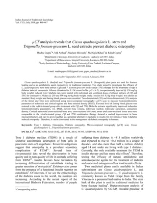
- 4. GUPTA et al.: µCT ANALYSIS OF CQ & TGF TREATED DIABETIC OSTEOPATHY 463 Blood glucose A 4.5 fold hike in blood glucose level was observed in diabetic-untreated rats when compared with vehicle-treated group (Fig. 2). This hike was sharply reduced by 3.2 fold, 3.4 fold, 3.2 fold, 3.9 fold and 4.7 fold in CQ, TFG, combined low dose, combined high dose and insulin-treated groups, respectively. Blood glucose levels were brought down to the normal physiological range in combined high dose (112.6 ± 9.45) and insulin-treated rats (93.5 ± 11.47) and this was similar to vehicle-treated rats (96.25 ± 11.09). µCT analysis of long bones (femur and tibia) We evaluated trabecular and cortical bone micro- architecture in all rats. 3D-µCT image revealed deterioration in bone quality with increased porosity in the trabecular region in all diabetic-untreated rats compared to the vehicle-treated group (Figs 3A&B). In diabetic-untreated rats (received vehicle), there was intense deficit in trabecular bone due to reduction in bone volume fraction (BV/TV), Conn.Dn, Degree of Anisotropy, Tb.No followed by an increase in Tb.Sp, Tb.Pf and SMI. When, the trabecular response in the proximal tibial metaphysis and the femur epiphysis to different treatments was compared with the diabetic- untreated group, bone volume fraction, mean values of Conn.Dn, Degree of Anisotropy were increased, Fig. 2 — Effect of ethanol extract of C. quadrangularis stem and T. foenum-graecum seeds supplementation on fasting blood sugar level. Values are expressed as mean ± SD ***p < 0.001(Diabetic- untreated (received vehicle) vs other treatment groups). Fig. 3 — Three dimensional µCT images of trabeculae of rat femur (A), and tibia (B) reveal the anti-osteoporotic effect of ethanol extract of Cissus quadrangularis stem and Trigonella foenum-graecum seeds in diabetic rats. VT: vehicle-treated normal; D: diabetic-untreated (received vehicle); D-CQ: diabetic-C. quadrangularis-treated; D-TFG: diabetic-T. foenum-graecum-treated; D-LD: diabetic CQ + TFG low dose-treated; D-HD: diabetic CQ + TFG high dose-treated.
- 5. INDIAN J TRADIT KNOWLE, VOL 17, NO 3, JULY 2018 464 whereas SMI, Tb.Pf and Tb.Sp were reduced. Rats given combined high dose of CQ and TFG had materially reduced trabecular separation comparable to vehicle-treated rats. Trabecular thickness was moderately improved after TFG supplementation (p < 0.01), but the mean values were insignificant among rest of the groups. Trabecular numbers were substantially increased in CQ, combined high dose and combined low dose treated rats, but this increase was modest in TFG and insulin administration. Trabecular thickness was increased in D-TFG (Table 1). Cortical bone strength was found reduced in diabetic-untreated rats as there was a decrease in B.Ar, Cs.Th, T.Ar and MMI in the mid-diaphysis of both tibia and femur when compared with vehicle- treated rats (p < 0.001). B.Ar, Cs.Th, T.Ar and MMI were increased substantially in rats given combined high dose (500 mg CQ + 500 mg TFG). B.Ar, Cs.Th, T.Ar and MMI in combined high dose group were significantly greater than that of insulin. T.Ar-B.Ar difference was increased in the diabetic-untreated group when compared with vehicle-treated rats but a marked decrease was noted in diabetic rats administered with insulin. A very slight decrease in T.Ar-B.Ar was noted in rats supplemented with CQ, TFG, D-LD and D-HD, but their differences were not significant (Table 2). BMD assessment In general, bone mineral density of femur bones was found more than the tibia (cortical as well as trabecular). Alloxan induced diabetes was accompanied by a reduction in BMD of both femur and tibia bones after 30 days of diabetes induction (0.1555 ± 0.0137) when compared with vehicle- Table 1 — Static trabaecular bone histomorphometric measurements of the femur and tibia using µCT Parameters VT D D-CQ D-TFG D-LD D-HD D-INS Femur BMD (g/cm2 ) 0.2895 ±0.03 0.1555±0.01 0.2544±0.04*** 0.2541±0.03*** 0.2490±0.03** 0.2356±0.04* 0.2555±0.06** BV/TV (%) 32.99±3.31 14.40±0.69 24.50±2.58*** 25.27±1.84*** 22.61±1.30*** 24.08±2.58*** 21.82±3.01*** Conn.Dn (mm-3 ) 118.4±15.32 52.54±17.78 121.0±16.16*** 96.52±20.43* 133.4±21.66*** 134.8±43.51*** 97.83±21.29* Degree of Anisotropy 1.816±0.05 1.477±0.03 1.730±0.02*** 1.583±0.04 1.625±0.02* 1.655±0.05** 1.817±0.14*** SMI 1.004±0.12 1.820±0.08 1.335±0.10*** 1.152±0.10*** 1.175±0.13*** 1.415±0.13*** 1.322±0.16*** Tb.No (mm-1 ) 2.689±0.32 1.369±0.24 2.251±0.14*** 2.098±0.11** 2.327±0.62*** 2.251±0.33*** 2.050±0.34* Tb.Pf (mm-1 ) 0.9975±0.04 4.396±0.48 1.755±0.71*** 1.106±0.32*** 1.648±0.98*** 2.033±0.82*** 1.913±0.12*** Tb.Sp (mm) 0.4998±0.06 0.8680±0.15 0.6375±0.15** 0.6316±0.1** 0.6253±0.10** 0.4502±0.04*** 0.6325±0.01** Tb.Th (mm) 0.1159±0.01 0.1086±0.006 0.1120±0.01 0.1309±0.01** 0.1230±0.01 0.1081±0.008 0.1041±0.01 Tibia BMD (g/cm2 ) 0.2372±0.03 0.1239±0.02 0.1819±0.04 0.2164±0.07** 0.2152±0.04*** 0.1944±0.02* 0.2035±0.05** BV/TV (%) 25.55±3.49 11.71±1.61 17.95±3.59** 20.02±5.43*** 21.53±3.32*** 17.37±1.50** 17.49±1.64** Conn.Dn (mm-3 ) 118.8±13.67 63.92±12.04 99.58±17.34* 121.6±18.90*** 120.2±23.12*** 129.0±28.00*** 89.37±8.77 Degree of Anisotropy 2.416±0.17 1.545±0.10 2.200±0.18*** 2.188±0.09*** 2.010±0.15*** 2.315±0.13*** 2.279±0.18*** SMI 1.453±0.13 2.249±0.20 1.855±0.14** 1.730±0.29*** 1.705±0.27*** 1.798±0.19*** 1.763±0.15*** Tb.No (mm-1 ) 2.329±0.28 1.124±0.13 1.619±0.36* 1.636±0.65* 1.846±0.42*** 1.752±0.23** 1.625±0.20* Tb.Pf (mm-1 ) 4.079±1.19 11.12±1.65 7.050±0.87*** 7.080±0.70*** 6.445±0.86*** 5.853±0.98*** 5.855±2.04*** Tb.Sp (mm) 0.331±0.05 0.721±0.07 0.429±0.11*** 0.541±0.18* 0.444±0.07*** 0.430±0.08*** 0.492±0.12*** Tb.Th (mm) 0.1088±0.006 0.1054±0.003 0.1111±0.007 0.1216±0.007**a 0.1143±0.007 0.1062±0.01 0.1085±0.012 Values are expressed as mean ± standard deviation pooled from 7 rats/group. BMD: bone mineral density, BV/TV: Percent bone volume, Conn.Dn: connection density, SMI: structure model index, Tb.No: trabecular number, Tb.Pf: trabecular pattern factor, Tb.Sp: trabecular separation, Tb.Th: trabecular thickness. ***p < 0.001; **p < 0.01; *p < 0.05 (Diabetic-untreated (received vehicle) vs other treatment groups); a p < 0.05 (vehicle-treated normal vs other treatment groups).
- 6. GUPTA et al.: µCT ANALYSIS OF CQ & TGF TREATED DIABETIC OSTEOPATHY 465 treated group (0.2895 ± 0.03776). This diabetes induced decrease in BMD of the trabecular region was prevented by CQ (p < 0.001), TFG (p < 0.001), combined low dose (p < 0.01) and combined high dose (p < 0.05) supplementation. The effect of combined low dose administration was comparable to insulin (p < 0.01). Diabetes did not produce any significant difference in BMD of the cortical region among groups (Tables 1 & 2). Discussion We used alloxan induced type I diabetic rat model to study the impact of diabetes on bone and to explore the potential alternative medicine in the form of natural extracts from potent medicinal plants. We did a comprehensive µCT scan analysis of long bones (femur and tibia) for diabetes induced osteopathy. Results of this study clearly demonstrate the anti- osteoporotic and anti-hyperglycaemic effect of CQ and TFG as evidenced by prevention of alloxan induced type I diabetes led increase in the level of blood glucose. Morphometric analysis of isolated rat tibia and femur and their BMD values revealed that the decline in bone quality with increased osteoporosis was prevented by CQ and TFG supplementation. Both the individual as well as combination therapy of CQ and TFG were found promising in improving bone microarchitecture. Furthermore, CQ was proved better than TFG in improving bone quality in diabetic rats in this experiment. One of the manifestations of diabetes was a reduction in body weight, which was clearly visible in alloxan induced type 1 diabetic control rats (received vehicle)18 . An average 3-4 times increase in glucose in diabetic rats was the consequence of alloxan induced beta cell destruction followed by the absence of insulin from the blood16 . This increment in blood glucose was prevented after treatment. Blood glucose was maintained in the normal physiological range in all groups of diabetes-treated rats. This explains that both CQ and TFG possess blood glucose lowering properties. Trabecular micro-architecture and cortical histomorphometry get altered in type I diabetes, as the bone becomes more porous4 . A reduction in trabecular number, trabecular thickness and disturbed trabecular layout in 3D space in both tibia and femur as well as a reduction in cortical thickness and bone mineral density (BMD) were the signs of osteopathy in TIDM. Impaired values of trabecular BMD and 3D histomorphometric parameters, viz. BV/TV, Conn.Dn, Tb.No, and Tb.Sp were significantly corrected after plant extract supplementation. Type I diabetes induced bone loss was most significantly prevented after treatment with CQ alone and CQ + TFG combined doses. Among the nonmetric parameters Tb.Pf, SMI, and degree of anisotropy were the parameters of trabecular bone’s mechanical strength. Tb.Pf is an index of connectivity of trabecular bone represents the ratio of convex to concave surfaces with larger Tb.Pf values reveal Table 2 — Static cortical bone histomorphometric measurements of the femur and tibia using µCT Parameters VT D D-CQ D-TFG D-LD D-HD D-INS Femur BMD 1.264±0.01 1.241±0.02 1.267±0.02 1.252±0.01 1.269±0.02 1.261±0.01 1.271±0.01* B.Ar (mm2 ) 3.202±0.17 1.960±0.17 2.301±0.23 2.665±0.13*** 2.743±0.13*** 3.219±0.08*** 2.768±0.58*** Cs.Th (mm) 0.3304±0.00 0.2639±0.01 0.3534±0.02*** 0.3219±0.01*** 0.3306±0.01*** 0.4034±0.00*** 0.3559±0.02*** MMI (mm4 ) 5.533±0.26 2.075±0.15 2.692±0.46** 4.322±0.19*** 3.102±0.30*** 4.642±0.17*** 2.247±0.19 T.Ar (mm2 ) 6.533±0.22 4.367±0.25 6.451±0.15*** 6.101±0.16*** 5.753±0.63*** 6.296±0.40*** 5.847±1.18*** T.Ar-B.Ar (mm2 ) 1.980±0.06 2.714±0.27 2.545±0.27 2.677±0.42 2.315±0.17 2.405±0.35 1.755±0.16*** Tibia BMD 1.191±0.025 1.145±0.043 1.170±0.026 1.171±0.014 1.178±0.022 1.173±0.018 1.163±0.033 B.Ar (mm2 ) 2.630±0.17 1.512±0.08 2.077±0.19*** 2.161±0.11*** 2.160±0.10*** 2.634±0.16*** 2.051±0.33*** Cs.Th (mm) 0.3197±0.01 0.2331±0.01 0.2827±0.03** 0.2619±0.01 0.3200±0.03*** 0.3247±0.01*** 0.2996±0.01** MMI (mm4 ) 3.349±0.29 1.331±0.06 3.107±0.36*** 2.793±0.16*** 1.823±0.1* 3.573±0.40*** 1.383±0.18 T.Ar (mm2 ) 4.091±0.18 2.765±0.13 4.353±0.22*** 4.163±0.17*** 3.411±0.26*** 4.287±0.28*** 2.786±0.03 T.Ar-B.Ar (mm2 ) 1.025±0.04 1.792±0.30 1.093±0.32*** 1.785±0.27 1.315±0.12** 1.280±0.21** 0.8508±0.03*** Values are expressed as mean ± standard deviation pooled from 7 rats/group. BMD: bone mineral density, B.Ar: mean total cross- sectional bone area, Cs.Th: cross-sectional thickness, MMI: mean polar moment of inertia, T.Ar: mean total cross-sectional tissue area. ***p < 0.001; **p < 0.01; *p < 0.05 (Diabetic-untreated (received vehicle) vs other treatment groups
- 7. INDIAN J TRADIT KNOWLE, VOL 17, NO 3, JULY 2018 466 convexity with isolated disconnected structures, whereas SMI indicates the relative prevalence of rods and plates in trabecular bone; with larger SMI values indicate a more rod like architecture - a characteristic of osteoporotic bone. The increase in SMI and Tb.Pf was prevented after treatment. BMD of cortical bone was largely unaffected by hyperglycaemia. Cortical bone strength was importantly determined by B.Ar, T.Ar, (T.Ar-B.Ar), Cs.Th and MMI. All cortical parameters except T.Ar-B.Ar were corrected in the group treated with a combined high dose of CQ + TFG and values were comparable to vehicle-treated rats. Taken together, our data revealed that a combination therapy of CQ and TFG has marked anabolic effect on bone micro-architecture and prevented diabetes induced bone loss. When the effect of treatment of CQ and TFG was compared with their combination therapy, it was observed that though TFG was good at correcting fasting blood glucose but moderately improved trabecular microarchitecture in diabetic rats. The above mentioned effects of CQ and TFG can be explained on the basis of phytoconstituents present in their extracts. Gas chromatography - mass spectrophotometric analysis of ethanolic extract of CQ reveals the presence of saponins, alkaloids, flavonols and flavonoids, terpenes, phytosterols, tannins, ascorbic acid (vitamin C), tocopherols (vitamin E), etc.10,15,19 . Quantitative analysis of the stem of CQ reveals a higher proportion of phosphorus and calcium20 . Similarly, the ethanolic extract of fenugreek showed the presence of saponins (fenugreekine, diosgenine), alkaloids (trigonellin), coumarins, phenolic derivatives, amino acids (4-hydroxyisoleucine, arginine), flavonoids, flavonols, mucilaginous fibers (galactomannan), vitamins and minerals21,22 . TFG seeds are also a rich source of phosphorus, magnesium, calcium, iron, and zinc23 . The anti-osteoporotic activity is explained as both plants are rich in calcium and phosphorus: the two minerals needed by bone for strengthening benefits and involve in bone homeostasis. As ascorbic acid is involved in matrix mineralization of bone and is found in CQ extract which explains why it has greater impact on bone healing than TFG extract24 . Flavonols found in these plant extracts have an anabolic effect on bone due to their ability to bind estrogen receptor25 . Blood glucose lowering effect of the TFG seed extract is attributed to GII compounds. GII compounds are reported to improve blood glucose utilization in glucose tolerance test, reduced fasting blood glucose and glycated haemoglobin in alloxan induced diabetic rabbits26 . As evident from our data of 30 days study period, bone histomorphometric values were markedly improved, which shows bone healing in all treatment groups when compared with diabetic-untreated group. But when the comparisons were made between vehicle-treated control and plant extracts treatments (D-CQ; D-TFG; D-LD; D-HD), Conn.Dn, degree of anisotropy, SMI and Tb.Th were comparable to vehicle-treated, however BMD, BV/TV %, Tb.Pf, Tb.No, and Tb.Sp were not. This indicates that a longer duration of treatment is required to bring the histomorphometric values to non-diabetic healthy rats. When the plant extracts treatments were compared with insulin therapy, except for degree of anisotropy and T.Ar-B.Ar, all bone histomor- phometric values were either comparable to or greater than that of insulin therapy. Conclusion The present study provides scientific evidence for the treatment of diabetic osteopathy by Ayurvedic herbal medicine. We conclude that the individual and combination therapy of two plant extracts, viz. Cissus quadrangularis and Trigonella foenum-graecum have osteoprotective potential against deterioration of bone microarchitecture in diabetic rats. Further clinical investigation is needed for the implication of this study on human health. Acknowledgement Authors are thankful to Council of Scientific and Industrial Research for providing financial assistance (File no. 09/107(350)/2010-EMR-I). We thank Dr AM Saxena, Department of Zoology, University of Lucknow for all kind of support and guidance. We are very thankful to Dr Naibedya Chattopadhyay, CSIR-CDRI, Lucknow, for providing µCT facility. We have no conflict of interest to declare. References 1 Notkins AL, Immunologic and genetic factors in type 1 diabetes, J Biol Chem, 277 (46) (2002) 43545-43548. 2 Alikhani M, Alikhani Z, Boyd C, MacLellan CM, Markos R, et al., Advanced glycation end products stimulate osteoblast apoptosis via the MAP kinase and cytosolic apoptotic pathways, Bone, 40 (2) (2007) 345-353. 3 Nicodemus KK & Folsom AR, Type 1 and type 2 diabetes and incident hip fractures in postmenopausal women, Diabetes Care, 24 (7) (2001) 1192-1197. 4 Silva MJ, Brodt MD, Lynch MA, Mckenzie JA, Tanouye KM, et al., Type 1 diabetes in young rats leads to progressive
- 8. GUPTA et al.: µCT ANALYSIS OF CQ & TGF TREATED DIABETIC OSTEOPATHY 467 trabecular bone loss, cessation of cortical bone growth, and diminished whole bone strength and fatigue life, J Bone Min Res, 24 (9) (2009) 1618-1627. 5 Botolin S, Faugere MC, Malluche H, Orth M, Meyer R, et al., Increased bone adiposity and PPARγ2 expression in type I diabetic mice, Endocrinology, 146 (8) (2005) 3622-3631. 6 Fulzele K, Riddle RC, DiGirolamo DJ, Cao X, Wan C, et al., Insulin receptor signaling in osteoblasts regulates postnatal bone acquisition and body composition, Cell, 142 (2) (2010) 309-319. 7 Cho NH, Whiting D, Forouhi N, Guariguata L, Hambleton I, et al., IDF Diabetes Atlas, 7th edn, International Diabetes Federation (IDF), Brussels, Belgium, 2015. 8 Maggio ABR, Rizzoli RR, Marchand LM, Ferrari S, Beghetti M, et al., Physical activity increases bone mineral density in children with type 1 diabetes, Med Sci Sports Exerc, 44 (7) (2012) 1206-1211. 9 Potu BK, Rao MS, Nampurath GK, Chamallamudi MR, Prasad K, et al., Evidence-based assessment of antiosteoporotic activity of petroleum-ether extract of Cissus quadrangularis L. on ovariectomy-induced osteoporosis, Ups J Med Sci, 114 (3) (2009) 140-148. 10 Kumar TS, Anandan A, Jegadeesan M, Appl A & Res S, Identification of chemical compounds in Cissus quadrangularis L. Variant-I of different sample using GC-MS analysis, Sch Res Libr, 4 (4) (2012) 1782-1787. 11 Sanyal A, Ahmad A & Sastry M, Calcite growth in Cissus quadrangularis plant extract, a traditional Indian bone- healing aid, Curr Sci, 89 (10) (2005) 1742-1745. 12 Kumar P, Kale RK, Mukherjee S, Prakash K, McLean P, et al., Antidiabetic effects of Trigonella foenum-graecum seed powder in a rat model, Toxicol Env Chem, 93 (10) (2011) 2085-2097. 13 Xue WL, Li XS, Zhang J, Liu YH, Wang ZL, et al., Effect of Trigonella foenum-graecum (fenugreek) extract on blood glucose, blood lipid and hemorheological properties in streptozotocin-induced diabetic rats, Asia Pac J Clin Nutr, 16 (1) (2007) 422-426. 14 Sreeja S, Anju VS & Sreeja S, In vitro estrogenic activity of fenugreek (Trigonella foenum graecum) seeds, Indian J Med Res, 131 (2010) 814-819. 15 Chidambara MKN, Vanitha A, Mahadeva SM & Ravishankar GA, Antioxidant and antimicrobial activity of Cissus quadrangularis L., J Med Food, 6 (2) (2003) 99-105. 16 Federiuk IF, Casey HM, Quinn MJ, Wood MD & Ward WK, Induction of type-1 diabetes mellitus in laboratory rats by use of alloxan: route of administration, pitfalls, and insulin treatment, Comp Med, 54 (3) (2004) 252-257. 17 Sharan K, Mishra J, Swarnkar G, Siddiqui J, Khan K, et al., A novel quercetin analog from an indian medicinal plant promotes peak bone mass achievement, bone healing after injury and exerts anabolic effect on osteoporotic bone: evidence toward the role of aryl hydrocarbon receptor as a mediator of osteogenic action, J Bone Min Res, 26 (9) (2011) 2096-2111. 18 El-Ghaffar EAA & Shehata SM, Antioxidant and anti- inflammatory effects of Acrocarpus fraxinifolius on hyperglycemia, hyperlipidemia and liver/kidney dysfunctions against alloxan induced Type 1 diabetes in rats, Indian J Tradit Knowle, 17 (2) (2018) 223-232. 19 Eswaran R, Anandan A, Doss A, Sangeetha G & Anand SP, Analysis of chemical composition of Cissus quadrangularis Linn. by GC-MS, Asian J Pharm Clin Res, 5 (2) (2012) 139-140. 20 Udayakumar R, Sundaran M & Krishna R, Mineral and biochemical analysis of various parts of Cissus quadrangularis L., Ancient Sci Life, 24 (2) (2004) 1-4. 21 Priya V, Jananie RK & Vijayalakshmi K, GC/MS determination of bioactive components of Trigonella foenum grecum, J Chem Pharm Res, 3 (5) (2011) 35-40. 22 Sohrevardi N & Sohrevardi F, Essential oil composition and antioxidant activity of Trigonella foenum graecum L. plant, Int J Agric Crop Sci, 4 (12) (2012) 793-797. 23 Ziwar JB, Estimation of Lipid Composition in Fenugreek Seed by GC/MS, Iraq Acad Sc Journal, 15 (10) (2010) 15-20. 24 Franceschi RT, Iyer BS & Cui Y, Effects of ascorbic acid on collagen matrix formation and osteoblast differentiation in murine MC3T3-E1 cells, J Bone Min Res, 9 (6) (1994) 843-854. 25 Prouillet C, Mazière JC, Mazière C, Wattel A, Brazier M, et al., Stimulatory effect of naturally occurring flavonols quercetin and kaempferol on alkaline phosphatase activity in MG-63 human osteoblasts through ERK and estrogen receptor pathway, Biochem Pharmacol, 67 (7) (2004) 1307-1313. 26 Puri D, Prabhu KM & Murthy PS, Antidiabetic effect of GII compound purified from fenugreek (Trigonella foenum graecum Linn.) seeds in diabetic rabbits, Indian J Clin Biochem, 27 (1) (2012) 21-27.