The lost buccal plate
•Download as PPTX, PDF•
0 likes•261 views

“Perio-Implant Synergy”- Two lectures on “Lost Buccal Plate- Complications and Management” and “Failing to Plan is Planning to Fail”. Organized by the Society of Periodontists and Implantologists of Kerala” at PMS Dental College, Trivandrum, India on 17/9/2018.
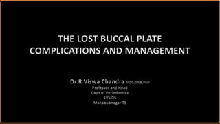
![Ganz SD. Thetriangle of bone—a formula for successful implant placement and restoration. Article in The Implant Society: [periodical]· January 1995.
Garcia JJ et al. A new protocol for immediate implants. The rule of the 5 triangles: A case report – EAO 2014.
BiotypePrimary
stability
Implant
design
Jumping Gap
Buccal Plate
Rule of five triangles](data:image/gif;base64,R0lGODlhAQABAIAAAAAAAP///yH5BAEAAAAALAAAAAABAAEAAAIBRAA7)
Recommended
Recommended
More Related Content
What's hot
What's hot (20)
Similar to The lost buccal plate
Similar to The lost buccal plate (20)
More from R Viswa Chandra
More from R Viswa Chandra (20)
Recently uploaded
Recently uploaded (20)
The lost buccal plate
- 1. THE LOST BUCCAL PLATE COMPLICATIONS AND MANAGEMENT
- 2. Ganz SD. Thetriangle of bone—a formula for successful implant placement and restoration. Article in The Implant Society: [periodical]· January 1995. Garcia JJ et al. A new protocol for immediate implants. The rule of the 5 triangles: A case report – EAO 2014. BiotypePrimary stability Implant design Jumping Gap Buccal Plate Rule of five triangles
- 3. “I found a way to see in the dark. Close your eyes.” ― J.R. Rim
- 4. Schropp L, et al. Int J Periodontics Restorative Dent. 2003 Aug;23(4):313-23. Hämmerle CH, et al. Clin Oral Implants Res. 2012 Feb;23 Suppl 5:80-2. Weng D, et al. Eur J Oral Implantol. 2011;4 Suppl:59-66. Two-thirds of resorption occurs within the first three months
- 5. The buccal plate is the weakest amongst the socket walls Buccal plate thinning, dehiscences or fenestrations are common reduction in almost 50% of cases post extraction *H. D. Barber and N. J. Betts. Implant Dentistry, Vol. 2, No. 3, 1993, pp. 191-193. Schropp L, Wenzel A, Kostopoulos L, Karring T. Int J Periodontics Restorative Dent. 2003;23:313–323. ThinningDehiscence Fenestration
- 6. What is a “lost buccal plate”? Esposito M et al. Eur J Oral Implantol 2009;2(3):167–184. Extraction Socket Dehiscence Defects Horizontal Defect Vertical Defect
- 7. What is a “lost buccal plate”? I II III Intact Socket Dehiscence- Fenestration Large Dehiscence
- 8. Fragility of the buccal plate Novaes Jr et al. J Periodontol 2011;82:872-877. Higher Density More marrow spaces Less thinner ̴ 1mm
- 9. Causes of buccal plate loss LOSS OF BUCCAL PLATE BIOLOGICAL BONE ›Bone Quality ›Bone Quantity MECHANICAL SOFT TISSUE ›Biotype ›Flap Design IMPLANT SIZE ›Diameter ›Length IMPLANT DESIGN ›Macrosurface ›Microsurface Hämmerle CH. et al., Int J Oral Maxillofac Implants. 2004;19 Suppl:26-8.
- 10. Immediate Implant Metal Show (IIM) Delayed Implant Shadow Show (DISS) Delayed Implant Actual Show (DIAS) Mazen Almasri. Surgical Science. 2013:4;110-113. Signs of a “losing or lost buccal plate”
- 11. The need for intervention *Vignoletti F, et al. Clin Oral Implants Res. 2012 Feb;23 Suppl 5:22-38. **Weng D, et al. Eur J Oral Implantol. 2011;4 Suppl:59-66. › Poorer maintenance of healthy periimplant soft tissues › Poorer aesthetic outcomes › 10 times greater need for hard tissue augmentation at implant placement without previous Ridge Preservation
- 12. Intervention Prevent volume loss Improve the aesthetic outcomes Cardaropoli D, et al. Int J Periodontics Restorative Dent. 2014 Mar–Apr;34(2):211-7. Morjaria KR, et al. Clin Implant Dent Relat Res. 2014 Feb;16(1):1-20.
- 13. PROBLEM OUTCOME IMMEDIATE IMPLANT DELAYED IMPLANT TECHNIQUE SP ESR ISD SP- Socket preservation ESR- Extraction Site Reconstruction ISD-Implant Site Development
- 14. Clinical decision tree for alveolar ridge-preservation procedures *RONALD E. JUNG,ALEXIS IOANNIDIS,CHRISTOPH H. F. H€AMMERLE & DANIEL S. THOMA. Periodontology 2000, Vol. 0, 2018, 1–11.
- 15. Selection criteria for regeneration in lost buccal plate It is important that the regenerative material used to fill defects correspond the number of walls of host bone remaining in contact with the graft. Misch & Misch (2010) and Ogunsalu (2011) gave a standard criteria in this regard. *Christopher Ogunsalu (2011). Bone Substitutes and Validation, Implant Dentistry - The Most Promising Discipline of Dentistry, Prof. Ilser Turkyilmaz (Ed.), ISBN: 978-953-307-481-8.
- 16. Additional active elements are beneficial in this graft since bone does not surround the defect. . Four wall defect ~ Labial bone loss
- 17. . Alloplasts are advantageous Two/Three wall defect ~ Labial bone loss
- 18. IMPLANT SITE DEVELOPMENT Bartee (2011) 1-2 Missing walls Autogenous bone Osteoinductive Materials Rigid membranes >2 Missing walls Block Grafts Rigid fixation No membranes Bartee BK. Implant Site Development and Extraction Site Grafting. 2011 by Osteogenics Biomedical, Inc
- 19. Managing the Plate of Bone Greenstein and Cavallaro (2013) NO ADDITIVE TREATMENT BONE GRAFT +/- GROWTH FACTORS BARRIER ONLY BARRIER + BONE GRAFT ›Flap positioned over defect ›Flap Placed at crest ›Flap positioned over defect ›Flap Placed at crest ›With Flap Advancement ›Without Flap Advancement ›With Flap Advancement ›Without Flap Advancement Managing the Buccal Gap and Plate of Bone: Immediate Dental Implant Placement. Continuing Education . Course Number: 159 2013
- 20. No additive treatment Flap placed over the defect Small defects with respect to height and width could be eliminated without the use of a membrane and/or a bone graft. Chen ST, Darby IB, Adams GG, et al. A prospective clinical study of bone augmentation techniques at immediate implants. Clin Oral Implants Res. 2005;16:176-184.
- 21. Objectives › One-stage treatment of hard and soft tissues › Preservation of the alveolar bone volume › Long-term good aesthetic outcome in front teeth with short treatment time.
- 22. No additive treatment Flap positioned at bone crest Gaps < 2 mm usually heal without allografts, xenografts, and barriers when implants are submerged Juodzbalys G, Wang HL. Soft and hard tissue assessment of immediate implant placement: a case series. Clin Oral Implants Res. 2007;18:237-243.
- 23. Compensating for osteogenic jumping distance LARGER DIAMETER IMPLANT BUCCAL POSITIONING GRAFTING ON BUCCAL BONE X Bone response? X Loss of prosthetic centre X More Bone loss M. G. Araujo, F. Sukekava, J. L. Wennstrom, and J. Lindhe, Clinical Oral Implants Research, vol. 17, no. 6, pp. 615 –624, 2006.
- 24. Objectives › The technique minimizes the treatment time › The treatment maintains the archetype of the soft and hard tissues
- 25. BONE GRAFT +/- GROWTH FACTORS ›Flap positioned over defect ›Flap Placed at crest Managing the Plate of Bone Greenstein and Cavallaro (2013)
- 26. Graft with or without Growth Factors With Flap over the defect
- 27. *RONALD E. JUNG,ALEXIS IOANNIDIS,CHRISTOPH H. F. H€AMMERLE & DANIEL S. THOMA. Periodontology 2000, Vol. 0, 2018, 1–11. Turchi JL. Dent Today. 2008 Jun;27(6):112, 114. If the endpoint is high quality bone, use Autografts and Bioactive glasses
- 28. Graft with or without Growth Factors With Flap at the crest Markus Glocker, , Thomas Attin and Patrick R. Schmidlin. Ridge Preservation with Modified “Socket-Shield” Technique: A Methodological Case Series. Dent. J. 2014, 2(1), 11-21; https://doi.org/10.3390/dj2010011 GLOCK’sTechnique
- 29. Growth factors combined with Socket- shield preserves up to 88 % of the ridge width and promote more new bone formation vs no membrane Perelman-Karmon et al. Int J Periodontics Restorative Dent. 2012 Aug;32(4):459-65. Jung RE, et al. J Clin Periodontol. 2013 Jan;40(1):90-8.
- 30. BARRIER ONLY ›With Flap Advancement ›Without Flap Advancement Managing the Plate of Bone Greenstein and Cavallaro (2013)
- 31. De Stavola L, Tunkel J. Int J Oral Maxillofac Implants. 2014 Jul-Aug;29(4):921-6. doi: 10.11607/jomi.3370 "Obtained Primary Closure” 87.6% “Compromised closure” 44.6% of complications attributable to improper closure Barrier only placed over defect With Flap advancement
- 33. It may be beneficial to use a barrier, and this would necessitate elevating a flap in order to achieve wound closure. Pearce AI, Richards RG, Milz S, et al. Animal models for implant biomaterial research in bone: a review. Eur Cell Mater. 2007 ;13:1-10.
- 34. Barrier only placed over defect No flap advancement *Rosen PS, Rosen AD. Compend Contin Educ Dent. 2013 Jan;34(1):34-8, 40. OPEN GBR CONCEPT Altering the amount of keratinized tissue Altering soft-tissue landmarks Increased pain, swelling or paraesthesia ProTiss®
- 35. ProTiss® With a flapless approach, it is suggested that overfill of the gap with bone helps support the soft tissue and reduces recession and bone loss. Tarnow D. Immediate vs. delayed socket placement: what we know, what we think we know and what we don’t know. American Academy of Periodontology Annual Meeting; November 14, 2011; Miami Beach, FL
- 36. TISSUE GRAFTS- PEDICULATED/ NON-PEDICULATED 1. El Chaar E, Oshman S, Cicero G, Castano A, Dinoi C, Soltani L, Lee YN.Soft Tissue Closure of Grafted Extraction Sockets in th e Anterior Maxilla: 2. A Modified Palatal Pedicle Connective Tissue Flap Technique. Int J Periodontics Restorative Dent. 2017;37(1):99 -107.
- 37. Objectives › Almost complete maintenance of the ridge volume is achieved › After 8–10 weeks, the soft tissue has a quality and maturity that is adequate for early implant restoration.
- 38. BARRIER + BONE GRAFT ›With Flap Advancement ›Without Flap Advancement Managing the Plate of Bone Greenstein and Cavallaro (2013)
- 39. Stevens MR, Emam HA, El Alaily M, Shar-awy M. Implant bone rings. One-stage three-dimensional bone transplant technique: a case report. J Oral Implantol 2010;1:69–74. 21. Barrier placed over graft With Flap advancement
- 40. Bone-ring techniques offer multiple advantages of a 1-stage procedure for immediate implant placement and 3-D bone augmentation. Proper treatment planning and careful surgical execution are essential to ensure predictability. Kaufman E, Wang PD. Localized vertical maxillary ridge augmentation using symphyseal bone cores: a technique and case report. Int J Oral Maxillofac Implants 2003;18:293–8.
- 41. Barrier placed over graft Without Flap advancement
- 42. Several recent articles have indicated that if a flap is not raised, there is better increase in bone dimensions when a graft is used. Vera C, De Kok IJ, Chen W, et al. Int J Oral Maxillofac Implants. 2012;27:1249-1257. Degidi M, Daprile G, Nardi D, Piattelli A. Clin Oral Implants Res. August 13, 2012. doi: 10.1111/j.16000501.2012.02561.x. Brownfield LA, Weltman RL.. J Periodontol. 2012;83:581-58
- 43. Objectives › Fast and scar-free soft-tissue regeneration › Optimal clinical and aesthetic result for the patient
- 44. A partially missing buccal plate is not a critical factor for primary stability Even complete loss of buccal plate is no issue if primary stability can be obtained Delayed implant placement approach is recommended in extreme buccal plate loss Biomaterials can be placed without a barrier Degidi M, Daprile G, Nardi D, Piattelli A. Clin Oral Implants Res. doi: 10.1111/j.16000501.2012.02561.x.
- 45. “Learn to do common things uncommonly well.” ― George Washington Carver
Editor's Notes
- 2mm buccal plate is crucial to avoid soft tissue recession. An approximate 2-4 mm of bone apical to the alveolus is necessary in order to have a greater possibility of obtaining a stable anchor, and thus obtain stability. To enhance primary stability self-tapping implants were developed, which compress the alveolar bone,
- Alveolar bone deficiency pre implant placement is one of the most common challenges that surgeons encounter on their daily practice. Bartee reported that following dental extraction, bone loss in the extraction socket takes place significantly during the first 6 months, with as much as 40% of the alveolar height and 60% of the width is lost. This magnitude of post extraction alveolar bone loss is sufficient to compromise implants placement that can extremely compromise implants overall success. Labial bone plate thinning, dehiscence, or fenestrations are other examples of such compromise. This can be of deleterious effect if occurred at the anterior maxillary region (the esthetic zone or the smile zone). In order to avoid such complications, the practitioner should not underestimate the need of proper alveolar ridge preparation for future implant placement. In order to optimize the socket width for future implant placement, the first step to undergo is atraumatic dental extraction which is often more difficult to accomplish in endodontically treated teeth, ankylosed, and previously traumatized teeth. The use of a thin periotome elevator will help luxating the roots, however, care should be taken to maintain an intact buccal plate, the weakest amongst the socket wall Extraction sites heal in a highly predictable fashion, with little intervention required for clinically acceptable wound healing to occur. The initial step involves the formation of a blood clot in the socket. At the apical aspect of the socket, the clot is rapidly replaced by a highly vascular granulation tissue, accompanied by ingrowth of blood vessels from the periodontal plexus. By about 14 days, this granulation tissue is replaced by an organized connective tissue matrix which is eventually mineralized to form bone. Socket healing progresses in an apical to coronal direction, so that by 21 days approximately 2/3 of the socket is filled with the connective tissue required to form bone (osteoid). Bone formation begins in the apex, progressing coronally to partially fill the socket with immature bone by 6 weeks. At the coronal aspect however, within hours of extraction migrating epithelium invades the clot, resulting in incomplete bone regeneration in the upper 1/3 to 1/4 of the socket. As a result, the extraction site heals in a concave fashion. Impaction of debris and bacteria into the healing socket further prevents the formation of bone.
- It is reasonable to conclude that the higher bone density, represented by the lower number of marrow spaces, in association with the thinner aspect of the buccal bone plates made them more fragile to absorb compared to the lingual bone plates, especially during mucoperiosteal procedures. Note that the buccal bone plate (BBP) appears almost without marrow spaces, differently from the lingual bone plate. Thelingualboneplate(A)issignificantlythickerandwithbigger marrowspacescomparedtothebuccal bone plate.
- A lot of articles discussed variable techniques in dealing with implant surface exposures at the time of implant placement (Immediate Implant Metal Show; IIMS). This problem can be treated by immediate grafting of the site using autogenous or non autogenous grafts. Moreover, implant metal show can be witnessed in few months after implant placement as a delayed implant shadow show (DISS) when the labial bone plate becomes thin or dehisced but is still covered with a relatively thin gingival flap. On the other hand, delayed implant actual show (DIAS) is witnessed when tissue loss occurs at both the bone and gingival envelop. DISS and DIAS management is critical and the methods of treatment are beyond the scope of this article. This technique was found to provide favorable long term esthetic results as no case of implant immediate metal show, delayed metal shadow show (DMSS) or delayed metal actual show (DMAS) were observed.
- We found that alveolar ridge preservation is effective in limiting physiologic ridge reduction as compared with tooth extraction alone. While immediate implant placement does not prevent bone resorption, Biomaterials can largely compensate for bone loss and preserve the contour of the alveolar ridge. Ridge Preservation can: › prevent volume loss and lead to an optimised hard and soft tissue situation irrespective of the chosen time for implantation › improve the aesthetic outcome by preserving the alveolar ridge volume and contour, when the objective of treatment is to place a bridge
- A partially missing buccal plate was not a critical factor for the stability and successful osseointegration of immediate implants, and these defects could heal clinically using GBR