
Recommended
Recommended
More Related Content
Similar to anti-VIII antibody.pdf
Similar to anti-VIII antibody.pdf (20)
Recently uploaded
Recently uploaded (20)
anti-VIII antibody.pdf
- 1. Clin. exp. Immunol. (1980) 39, 315-320. Circulating immune complexes containing anti-VIII antibodies in multi-transfused patients with haemophilia A M. D. KAZATCHKINE,* Y. SULTAN, E. J. BURTON-KEE & J. F. MOWBRAY Department of Experimental Pathology, St Mary's Hospital Medical School, London, and Department ofHaematology, Hopital Cochin, Paris, France (Acceptedfor publication 2July 1979) SUMMARY Evidence for the presence ofcirculating immune complexes was found in thirty-four out offifty- five samples from forty-seven patients with haemophilia A. In eleven patients the complexes, precipitated from the blood with polyethylene glycol, were digested with pepsin. The F(ab')2 antibody was tested, and found to have neutralizing activity against coagulant Factor VIII in two patients. In one ofthese no free antibody had ever been found in the plasma, while in the other the antibody was concentrated tenfold in the complex. In two other samples free without complexed antibody was found. In comparison, IgG-containing complexes were found in nine out ofnineteen patients with von Willebrand's disease and no complexes were found in the sera from twelve multi-transfused thalassaemics. PEG precipitation is a useful technique for the preparation of concentrated immune complexes for further study such as antigen identification. INTRODUCTION Acquired inhibitors ofFactor VIII develop in 8-14% ofpatients with severe haemophilia A treated with plasma or anti-haemophilic Factor concentrates (Shapiro & Hultin, 1975). These inhibitors can be recognized by their specific ability to inactivate procoagulant activity ofhuman Factor VIII (VIIIc) and have been identified in most series as oligoclonal IgG antibodies. Recent in vitro studies using anti- Factor VIII plasma, or semi-purified anti-VIIIc immunoglobulins have provided evidence for immune complex formation between anti-VIIIc and purified Factor VIII or Factor VIII from cryoprecipitates (Allain & Frommel, 1973; Lavergne, Meyer & Reisner, 1976; Lazarchick & Hoyer, 1977). Using a simple semi-quantitative test we have studied the occurrence ofcirculating immune complexes in vivo in multi-transfused patients with haemophilia A, and we have demonstrated the specificity of some of the complexed antibodies. MATERIALS AND METHODS Forty-five citrated plasmas and ten sera were obtained from forty-seven patients with haemophilia A. The ten sera were obtained from patients with the severe form ofthe disease (VIIIc below 0-01 iu/ml); ofthese, three (patients BP, BS and CP, Table 1) had a circulating inhibitor of Factor VIII, and one had a previously documented inhibitor but had no VIIIc- neutralizing activity in the serum at the time of the study (patient LA). With the exception of two patients with circulating inhibitor (BS and CP), all patients received either therapeutically or prophylactically 1500 to 100,000 iu of Factor VIII, as cryoprecipitate, within twelve weeks of the study. For comparison, plasma samples were obtained from nineteen patients with severe von Willebrand's disease who were regularly transfused with Factor VIII concentrate, sera were obtained from twelve multi-transfused patients with thalassaemia (sera kindly provided by Dr E. Letsky, Institute ofChild Health, London), * Present address: Department of Medicine, Harvard Medical School and Robert B. Brigham, Hospital, Boston, MA, USA. Correspondence: Dr M. Kazatchkine, The Seeley G. Mudd Building, 250 Longwood Avenue, Boston MA 02115, USA. 0099-9104/80/0200-0315 $02-00 () 1980 Blackwell Scientific Publications 315
- 2. 316 M. D. Kazatchkine et al. and thirty-eight plasmas and thirty-four sera were obtained from twenty-four healthy individuals, together with eight pools of serum from normal healthy subjects. Plasmas were stored at - 20'C and - 70'C; sera were separated within 2 hr aftdr clotting and stored at - 70'C in glass bottles until analysis. Circulating immune complexes. These were measured by a semi-quantitative assay using the precipitation ofcomplexes from serum or plasma with 2% polyethylene glycol (PEG) followed by measurement ofthe amount of IgG and Clq in the precipi- tate. For the test, 0-1 ml ofa 12% solution ofPEG 6000 (BDH Ltd., Poole) in veronal buffer containing 60 mM EDTA, pH 7-6, was added to 0 5 ml of the test sample in a polystyrene tube. After incubation for 18 hr at 4VC, the samples were centrifuged at 1000 g for 20 min at 40C. The supernatants were decanted and kept at 40C. The precipitates were washed once in 2 ml veronal buffer containing 2% PEG, 10 mm EDTA, pH 7-6, and redissolved in 0-5 ml of veronal buffer pH 7-6. The amount of IgG and Clq in the precipitate was determined by single radial immunodiffusion (SRID) using 1% agarose gels in veronal buffer containing 40 mm EDTA, pH 8-6, and appropriate dilutions of specific antiserum. The values in eighty normal samples were 0 39+ 0-15% of serum IgG concentration (mean+ s.d.) for the precipitated IgG from 1 ml serum and 44 ug± 20 (mean+ s.d.) for the precipitated Clq. Values of precipitated IgG and Clq from the test samples which were more than 2 standard deviations above the mean values obtained in the normal samples were regarded as positive. The measurement ofClq in the starting serum and in the supernatant by SRID served as an internal control for Clq recovery. The sensitivity of this assay allows the detection of 10 ,ug/ml of heat aggregated IgG. IgA in the precipitates was detected by double diffusion in agarose plates and measured by rocket electrophoresis at 5 v/cm overnight in 1% agarose gels containing 40 mm EDTA buffer, pH 8-6, and 2-5% v/v rabbit anti-human IgA antiserum. F(ab')2 antibodies. These were prepared from the PEG precipitates by pepsin digestion The precipitates obtained from 0 5 ml of starting serum were adjusted to pH 3 with 3 N and 0-2 N acetic acid, and 5% pepsin by weight in acetic acid was added to give a final substrate/enzyme ratio of 20: 1. The digestion was performed for 6 hr at 37°C; the pH was raised to 5 with 3 N NaOH and then to 8 with 0 5 M phosphate buffer pH 8-0. After addition to rabbit hyperimmune serum used as a carrier, the F(ab')2 fragments were precipitated with 20% Na2SO4 at 37°C for 30 min, and tested for antibody activity after overnight dialysis against phosphate buffered saline (Lachmann, 1971; Dambuyant, Burton-Kee & Mowbray, 1978). Complement components and immunoglobulins. Serum Clq, C3, C4, C5, IgG, IgA and IgM concentrations were measured by SRID in agarose using monospecific antisera raised in rabbits. C2 functional activity was measured in an assay using the serum ofa patient totally deficient in haemolytic and antigenic C2 as a substrate. Factor B activity was measured as described (Martin et al., 1976). Factor VIII inhibitor activity. This was measured as described in 1975 at a Bethesda conference (Kasper et al., 1975). Hepatitis B surface antigen (HBs) and antibody to HBs. These were detected and titrated by inhibition ofhaenmagglutination and passive haemagglutination tests. RESULTS Results obtained in patients with haemophilia A, von Willebrand's disease and transfusion-dependent thalassaemia are shown in Fig. 1. The amounts of 2% PEG precipitable IgG were significantly raised more than 2 standard deviations above the normal mean in thirty-four offifty-five samples from patients with haemophilia (thirty of forty-seven patients) (P<0.001 using Student's t-test). Two percent PEG precipitable Clq was raised in nine of fifty-five haemophilia samples (nine of forty-seven patients) (P<0.001). The test was positive for both IgG and Clq in seven patients. Nine of nineteen samples from patients with von Willebrand's disease were positive for IgG (P<0 001); none was positive for Clq. No statistically significant differences were found between the transfused thalassaemic group and the normal controls. Complement and immunoglobulin levels in ten sera and one plasma from eleven patients selected because of their severe haemophilia are shown in Fig. 2: six had CH50 and functional C2 values below the normal laboratory range; of these, five were positive for IgG and Clq in the immune complex assay. The immunochemical level of C4 was in the lower normal range; immunochemical concentra- tions of Clq, C3, C5, IgA, IgM and functional activity of Factor B in the serum were normal. Three sera had increased concentrations of IgG. IgA was detected in twenty-one PEG precipitates from fifty-five haemophilia samples (nineteen of forty-seven patients) including seventeen samples positive for complexes and four samples negative for both IgG and Clq in the pellets. Two per cent PEG precipitable IgA was present in measurable amounts in only nineteen samples (of which seventeen were positive for IgG and Clq), and represented 0 4 to 2.8% of the total serum IgA. IgA could be detected in three of eighty PEG precipitates from normal samples, but was too low to measure by rocket electrophoresis. Pepsin digestion and preparation of F(ab')2 fragments were carried out in the PEG precipitates from
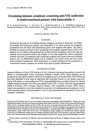
- 3. Immune Complexes in Haemophilia 317 4 300 0~~~~~~~~~~~~~~~~~~~ o ~~~~~~~~~~200 ~ 0~~~~~~~~~~~~~ a~~~~~~~~~~~~~~~~~~~a w (9 0 ~~~~00 Haemophlloa Thalassoemia| Haemophil ia ThalassaemiaI von Willebrand's Normols von Willebrand's Normals d isease disease FIG. 1. 2% PEG precipitable IgG and Clq in sera and plasma from patients with treated haemophilia A, repeatedly transfused patients with severe von Willebrand's disease or thalassaemia. Precipitated material is expressed for IgG, as percentage ofserum/plasma IgG concentration and for Clq as ug precipitated from one ml ofstarting serum or undiluted plasma. The horizontal line represents the upper limit ofnormal (mean+2 s.d.). 160 20 0 o IZ v 80 0 c a) 40 a- C, l . 0 CH50 Clq C2 C4 C3 C5 Bf IgA IgG IgM (funct.) (fund.) (funct ) FIG. 2. Complement and immunoglobulin levels in ten sera (-) and one plasma (0) from multi-transfused patients with severe haemophilia. The horizontal lines enclose the normal range. The bars represent the mean values for the eleven samples. the ten selected sera and from one plasma sample (Table 1). The F(ab')2 fragments were tested for Factor VIII neutralizing activity, anti-HBs, anti-CMV, and anti-Herpes simplex activities. F(ab')2 antibodies derived from two digested PEG precipitates had Factor VIII neutralizing activity, 2 iu in the F(ab')2 preparation from the PEG pellet of one ml of starting serum; both PEG precipitates containing anti-VIIIc antibodies were positive for IgG and Clq in the immune complex assay. Whole serum from one ofthe patients had an inhibitory activity to Factor VIII (patient CP, 32 iu), whereas serum from the second patient (patient LA) did not have detectable anti-VIIIc activity. Two other patients with anti-VIIIc activity (patients BP, 6 iu and BS, 32 iu) did not have neutralizing activity in their digested complex. No pepsin-digested PEG precipitates showed anti-HBs antigen activity, anti-CMV or anti- Herpes activity; however, in one serum (patient MJ), HBs antigen and anti-HBs antibody could be simultaneously detected. Electron microscopic examination of the eleven samples and PEG pellets (Dr Julian Hodgson) only revealed Australia antigen particles (spherical and tubular forms) after in vitro agglutination with anti-HBs antibody (Almeida & Waterson, 1969) in the two serologically HBs antigen positive samples; no evidence was found for Australia antigen particles in HBs antigen negative-immune complex positive sera or their PEG precipitates. I i -r so 0-0 : -j- Ao 0 0. 0 0 0 r. .
- 4. M. D. Kazatchkine et al. 4444d E -. -; -m -d E E E -, - 0 CC C CD D tv Cd 0 00 0 0E 00. = c = r. r . = I- I1 I1 1I 1I o I ++ +++ en n en en Ao r I n 4 -4 C1 eq v v A ¢ - 1 C m - - C en 318 0 bO ) 0- 00 04D - 44) cd 0. 0 E- .0 0) .. 0 r. 4.4 Cd 0. r. Cd 441 3 0 C) u ~0 t0. ce *-0) 0 _ 0. O .- 4) o 0. 0 - .Y cD 0 X H E Z o ° 0:1 * .I-+ en en + + + +
- 5. Immune Complexes in Haemophilia The latex agglutination test (Rheuma Wellcotest, Wellcome Reagents Ltd.), also performed on these eleven samples, was positive in four including the two HBs antigen-positive sera. Five samples tested were negative for the presence of cryoglobulins. In addition, ten of eleven patients were specifically examined to evaluate clinical evidence for immune complex disease (Table 1): none of the patients had immune complex disease-related signs or symptoms; none had proteinuria or abnormalities of renal function and urine sediment. However, four had abnor- malities of liver function including elevated transaminases and/or serum alkaline phosphatases. DISCUSSION Addition of low concentrations of PEG to serum results in the precipitation of high molecular weight proteins or macromolecular complexes, including antigen-antibody complexes (Zubler et al., 1977). As illustrated in this study of multi-transfused patients with haemophilia, the respective amounts of IgG and Clq in the precipitates may be indicative of the in vivo ability of circulating complexes to activate complement; furthermore, concentration of the complexes by precipitation also allows investigation of their specificity by direct electron microscopic examination, dissociation of the antigen and antibody, or isolation ofthe F(ab')2 fragments from the complexed antibodies (Dambuyant, Burton-Kee & Mowbray, 1979). Contamination of the PEG pellet with uncomplexed IgG in the conditions of the test is of the order of 0-39% and does not exceed 0.700, so that if an antibody specificity is found in the pepsin- digested complexes, it is unlikely to have been due to contamination with monomeric IgG. Evidence for the presence of circulating immune complexes has been found in thirty-four of fifty- five samples from forty-seven patients with haemophilia A (Kazatchkine et al., 1978) (Fig. 1). These results agree with those recently obtained by a different method for immune complex detection (N.K. Day, personal communication). IgG complexes were also found in some sera from patients with severe von Willebrand's disease repeatedly transfused with Factor VIII concentrates, but no complexes were found in the sera from transfused thalassaemics, suggesting that the hazards of transfusion are indeed greater with plasma-derived concentrates than with whole blood or washed red cells. Most of the complexes detected in the sera from patients with haemophilia did not contain increased amounts of2% precipitable Clq, suggesting that they could be large antigen excess complexes, IgG anti- IgG rheumatoid factor complexes, or complexes containing non-complement fixing or low affinity anti- bodies. Although only a small number have been studied, the inhibitors of Factor VIII have not been found to fix complement (Robboy et al., 1970). Clq-containing complexes were found only within a selected group of multi-transfused severe haemophiliacs in those patients in whom complement studies indicate the presence of moderate classical pathway complement consumption (Table 1 and Fig. 2). The poorly or non-complement activating properties of the immune complexes detected may account for the absence of immune complex disease-related clinical manifestations in haemophiliacs; it could be specu- lated, however, that circulating immune complexes play a role in the pathogenesis of the inflammatory joint episodes which are sometimes seen in these patients in the absence of bleeding. Anti-VIIIc specificity ofsome ofthe complexed antibodies was found in two ofnine immune complex- positive and pepsin-digested PEG precipitates from patients with severe haemophilia. Contamination by uncomplexed anti-Factor VIII antibody is unlikely, since in one patient Factor VIII neutralizing activity was recovered from the PEG precipitate but was not found in the starting serum, and since two patients with circulating inhibitor had no detectable anti-VIIIc activity in the digested PEG pellets. However, the incidence of VIlIc-anti-VIIIc complexes might have been underestimated in these samples, as the activity ofFactor VIII inhibitors may be damaged at the low pH used for pepsin digestion (Allain & Frommel, 1973). Inhibitors to Factor VIII interact specifically with the low molecular weight component of the Factor VIII molecule which is associated with the pro-coagulant activity (Rick & Hoyer, 1973) and which is absent or functionally and antigenically impaired (Zimmerman, De la Pointe & Edgington, 1977) in severe haemophilia A. The ability ofthe human antibody to form stable immune complexes with Factor VIII has recently been documented in vitro (Allain & Frommel, 1973; Lavergne et al., 1976; Lazarchick & Hoyer, 1977); elution patterns of immune complexes on gel filtration suggest 319
- 6. 320 M. D. Kazatchkine et al. that the low molecular weight component that interacts with antibody is univalent (Lazarchick & Hoyer, 1977), thus generating small non-complement fixing complexes. Such in vitro data is compatible with the observed in vivo persistence ofVIlIc-anti-IIIc complexes in one patient (patient CP) long after the last exposure to transfused Factor VIII (Table 1). Of the nine sera positive for immune complexes and studied in more detail (Table 1), in only one was there evidence suggesting the presence of HBs-anti-HBs complexes, as both antigen and antibody were simultaneously present in the serum; in this patient, the haemagglutination test may not have been sensitive enough to detect small amounts of specific anti-HBs F(ab')2 antibody. It has been suggested that immune complexes may be involved in the pathogenesis of chronic hepatitis in HBs antigen carriers (Almeida & Waterson, 1969; Nydegger et al., 1974) and of the asymptomatic liver disease present in patients with haemophilia (Spero et al., 1978; Mannucci et a!., 1975), but none of our four patients with abnormal liver functions had either anti-HBs activity or evidence of Australia antigen particles in the PEG precipitates. The higher than normal incidence of a positive latex test (Table 1) may be related to the chronic presence of HBs antigen (Markenson et al., 1975; Tedder & Briggs, 1977) and liver disease (Dudley, O'Shea & Sherlock, 1973); alternatively it could be due to the presence of altered IgG in the transfused material. This work was presented in part at the Annual Meeting of the American Federation for Clinical Research, San Francisco, CA, April, 1978. The work was carried out with grants from INSERM and DGRST (M .D. Kazatchkine) and the Medical Research Council (J. F. Mowbray). REFERENCES ALLAIN, J.P. & FROMMEL, D. (1973) Antibodies to Factor VIII. I. Variations in stability of antigen-antibody com- plexes in hemophilia A. Blood, 42, 437. ALMEIDA, J.D. & WATERSON, A.P. (1969) Immune com- plexes in hepatitis. Lancet, i, 983. DAMBUYANT, C., BURTON-KEE, J. & MOWBRAY, J.F. (1978) The use of preparation of F(ab')2 antibody from soluble immune complexes to determine the complexed antigens. J. Immunol. Methods, 24, 31. DAMBUYANT, C., BURTON-KEE, J. & MOWBRAY, J.F. (1979) Demonstration of two disease specific antigens in circu- lating immune complexes. Clint. exp. Immuniol. 37, 424. DUDLEY, F.J., O'SHEA, M.J. & SHERLOCK, S. (1973) Serum autoantibodies in hepatitis associated antigen (HAA) positive patients. A possible index of cell mediated immunity to the associated-infective agent. Clini. exp. Immunol. 13, 367. KASPER, C.K., ALEDORT, L.M., COUNTS, R.B., EDSON, J.R., FRATANTONI, J., GREEN, D., HAMPTON, J.W., HILL-. GARTNER, M.W., LAZERSON, J., LEVINE, P.H., MCMILLAN, C.W., POOL, J.G., SHAPIRO, S.S., SHULMAN, N.R. & VAN Eys, J. (1975) A more uniform measurement of Factor VIII inhibitors. Thromb. Diath. Haemorrh. 34, 869. KAZATCHKINE, M.D., MOWBRAY, J.F., BURTON-KEE, E.J. & SULTAN, Y. (1978) Circulating immune complexes in hemophilia. Clinical Research, 26, 350a. LACHMANN, P.J. (1971) The purification of specific antibody as F(ab')2 by the pepsin digestion of antigen-antibody precipitates and its application to immunoglobulin and complement antigens. Immnnochemistry, 8, 81. LAVERGNE, J.M. MEYER D. & REISNER, H. (1976) Charac- terization of human anti-Factor VIII antibodies purified by immune complex formation. Blood, 48, 931. LAZARCHICK, J. & HOYER, L.W. (1977) The properties of immune complexes formed by human antibodies to Factor VIII. 7. clin. Invest. 60, 1070. MIANNUCCI, P.M., CAPITANIO, A., DEL NINO, E., COLOMBO, M. PARETI, F. & RUGGERI, Z.M. (1975) Asymptomatic liver disease in hemophiliacs. ]. chin. Pathol. 28, 620. MARKENSON, J.A., DANIELS, C.A., NOTKINS, A.L., NOOF- NAGLE, J.H., GERETY, J. & BARKER, L.F. (1975) The interaction of rheumatoid factor with hepatitis B surface antigen-antibody complexes. Clin. exp. Immunol. 19, 209. MARTIN, A., LACHMANN, P.J., HALBWACHS, L. & HOBART, M.J. (1976) Haemolytic diffusion plate assays for factors B and D of the alternative pathway of complement activation. Immunochemistry, 13, 317. NYDEGGER, U.E., LAMBERT, P.H., GERBER, H. & MIESCHER, P.A. (1974) Circulating immune complexes in the serum in systemic lupus erythematosus and in carriers of hepa- titis B antigen. ]. c/in. Invest. 54, 297. RICK, M.E. & HOYER, L.W. (1973) Immunologic studies of antihemophilic factor (AHF, Factor VIII). V. Immuno- logic properties of AHF subunits produced by salt dissociation. Blood, 42, 737. ROBBOY, S.J., LEWIS, E.J., SCHUR, P.H. & COLMAN, R.W. (1970) Circulating anti-coagulants to Factor VIII. Am. 7. Med. 49, 742. SHAPIRO, S.S. & HULTIN, M. (1975) Acquired inhibitors to the blood coagulation factors. Semin. Thromb. Haemo- stasis, 1, 336. SPERO, J.A., LEWIS, J.H., VAN THIEL, D.H., HASIBA, U. & RABIN, B.S. (1978) Asymptomatic structural liver disease in hemophilia. N. Engl. jr. Med. 298, 1373. TEDDER, R.S. & BRIGGS, M. (1977) Anti-e and rheumatoid factor activity in hepatitis B. Lancet, i, 1262. ZIMMERMAN, T.S., DE LA POINTE, L. & EDGINGTON, T.S. (1977) Interaction of Factor VIII antigen in hemophilic plasmas with human antibodies to Factor VIII. ]. clin. Invest. 59. 984. ZUBLER, R.H., PERRIN, L.H., CREIGHTON, W.D. & LAMBERT, P.H. (1977) Use of polyethylene glycol (PEG) to con- centrate immune complexes from serum or plasma samples. Ann. Rheum. Dis. 36, suppl. 1, 23.