
Recommended
More Related Content
What's hot
What's hot (20)
Similar to Parotid gland and its anatomy;blood supply;nerve supply; anomalies
Similar to Parotid gland and its anatomy;blood supply;nerve supply; anomalies (20)
More from Priyanka Pai
More from Priyanka Pai (14)
Recently uploaded
Recently uploaded (20)
Parotid gland and its anatomy;blood supply;nerve supply; anomalies
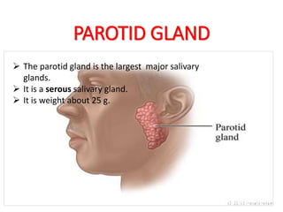
- 1. PAROTID GLAND The parotid gland is the largest of the three pairs of salivary glands. It is a serous salivary gland. It is weight about 25 g. The parotid gland is the largest major salivary glands. It is a serous salivary gland. It is weight about 25 g. HHHHHHHHHHHH
- 3. LOCATION • The parotid gland lies in the pyramidal fossa , which has following boundaries : Anteriorly - posterior border of the ramus of mandible. Posteriorly - mastoid process and sternocleidomastoid Superiorly - external acoustic meatus.
- 4. PAROTID CAPSULE • The investing layer of the deep cervical fascia forms the parotid capsule. • The fascia splits to enclose the gland. Attachments : • Superficial lamina - zygomatic arch • Deep lamina –styloid process and tympanic plate of the temporal bone • The deep lamina, extending between the styloid process and the mandible, is thickened to form the stylomandibular ligament. • The stylomandibular separates the parotid gland from the submandibular salivary gland.
- 5. EXTERNAL FEATURES The gland resembles a three-sided inverted pyramid. • It has - An apex - Base or Superior surface - 3 surfaces: - Superficial surface. - Anteromedial surface. - Posteromedial surface. - 3 borders: - Anterior. -Posterior. -Medial.
- 6. Apex: • It projects downwards overlapping the posterior belly of digastric and reaches the carotid triangle. Structures passing: 1. Cervical branch of the facial nerve. 2. Anterior and posterior divisions of retromandibular vein.
- 7. Base : • It is concave and related to the external acoustic meatus and posterior aspect of temporomandibular joint. Structures passing : • Superficial temporal vessels • Auriculotemporal nerve.
- 8. Superficial Surface: • It lies between anterior and posterior border. • It is covered from superficial to deep by : 1. Skin. 2. Superficial fascia containing greater auricular nerve, superficial parotid lymph nodes, and platysma. 3. Parotid fascia.
- 9. Anteromedial Surface: • It lies between anterior and medial border. • It is deeply grooved by the posterior border of the ramus of the mandible. • It is related to: 1. Masseter. 2. Medial pterygoid. 3. Posterior border of the ramus of the mandible. 4. Lateral aspect of the temporomandibular joint. Structures passing : - Maxillary vessels
- 10. Posteromedial Surface: • It lies between posterior and medial border. • It is related to : 1. Mastoid process, sternocleidomastoid, and posterior belly of digastric. 2. Styloid process and styloid group of muscles. Structures passing : 1. Facial nerve trunk . 2. External carotid artery.
- 11. Anterior Border: • It separates the superficial surface from the anteromedial surface. • It rest on the masseter muscle. Structures passing (from above downward): 1. Temporal branch of the facial nerve 2. Zygomatic branch of the facial nerve. 3. Transverse facial vessels. 4. Upper buccal branch of the facial nerve. 5. Parotid duct. 6. Lower buccal branch of the facial nerve. 7. Marginal mandibular branch of the facial nerve.
- 12. Posterior Border: • It separates the superficial surface from the posteromedial surface. • It rest on the sternocleidomastoid Structures passing: • 1. Posterior auricular vessels. • 2. Posterior auricular branch of the facial nerve.
- 13. Medial Border: • It separates the anteromedial surface from the posteromedial surface. • It is related to the lateral wall of the pharynx
- 14. STRUCTURES PRESENT WITHIN THE PAROTID GLAND • From superficial to deep these are: 1. Facial nerve. 2. Retromandibular vein. 3. External carotid artery. 4. Deep parotid lymph nodes.
- 15. Facial nerve : • It enters the gland through the posteromedial surface and divides into five terminal branches . • Temporal , Zygomatic, Buccal and Marginal mandibular branch leave the gland through its anterior border. • The Cervical branch leave the gland through its apex. • The five terminal branches of the facial nerve radiate like a goose-foot . • Such branching pattern of the facial nerve is termed pes anserinus.
- 16. Retromandibular vein : • The retromandibular vein is formed within the gland by the union of the superficial temporal and maxillary veins. • In the lower part of the gland, the vein divides into anterior and posterior divisions which emerge at the apex of the gland.
- 17. External carotid artery: • The external carotid artery enters the gland through the posteromedial surface. • Within the gland it divides into superficial temporal and maxillary arteries. • The superficial temporal leave the gland through its base and maxillary artery through its anteromedial surface. • Posterior auricular artery leave the gland through its posterior border. • The transverse facial artery, branch of superficial temporal artery emerges through the anterior border of the gland.
- 18. Parotid Duct (Stenson’s Duct) • Parotid duct is about 5 cm long and emerges from the middle of the anterior border of the gland. Course: • After emerging from the gland, it runs forward over the masseter muscles. Relations on masseter muscle: Superiorly: - Accessory parotid gland - Upper buccal branch of the facial nerve - Transverse facial vessels Interiorly: - The lower buccal branch of the facia nerve.
- 19. • At the anterior border of masseter, it turns inwards, and piercing the following structures - buccal pad of fat - buccopharyngeal fascia - buccinator muscle. • After piercing the buccinator muscle, the parotid duct runs forwards between buccinator and the buccal mucosa. Opening of the duct: • Finally, the duct opens into the vestibule of mouth opposite the crown of upper second molar teeth.
- 20. NERVE SUPPLY: • Parasympathetic • Sympathetic • Sensory 1. Parasympathetic (secretomotor) supply : Inferior salivatory nucleus - glossopharyngeal nerve - tympanic branch of glossopharyngeal (Jacobson’s nerve)- tympanic plexus - lesser petrosal nerve - relay into otic ganglion - Postganglionic fibres - auriculotemporal nerve to supply the parotid gland. 2. Sympathetic supply: • It is derived from sympathetic plexus around external carotid artery formed by postganglionic fibres derived from superior cervical ganglion. • The sympathetic fibres are vasomotor. 3.Sensory supply: It is derived from: (a) Auriculotemporal nerve. (b) Great auricular nerve
- 21. BLOOD SUPPLY: • Arterial supply - External Carotid Artery • Venous drainage - External jugular veins. LYMPHATIC DRAINAGE: • Afferent - superficial and deep parotid lymph nodes. • Efferent - jugulodigastric lymphnode.
- 22. APPLIED ANATOMY 1.Parotid swellings are very painful due to the unyielding nature of the parotid fascia. 2.Mumps: • It is an infectious disease of parotid caused by paramyxovirus. • Mumps can cause complications in the adults like orchitis, pancreatitis and oophoritis. 3. A parotid abscess is best drained by horizontal incisions known as Hilton's method.
- 23. 4. Frey’s syndrome: • Penetrating wounds of the parotid gland may damage auriculotemporal and great auricular nerves. • During regeneration the secretomotor fibres of auriculotempora joints with great auricular nerves. • Thus when a person eats beads of perspiration appears on parotid region. 5. parotid sialogram: • The parotid duct can be demonstrated radiologically by injecting radio-opaque dye.