
Recommended
More Related Content
Similar to INFLAMMATION @.docx
Similar to INFLAMMATION @.docx (20)
Recently uploaded
Recently uploaded (20)
INFLAMMATION @.docx
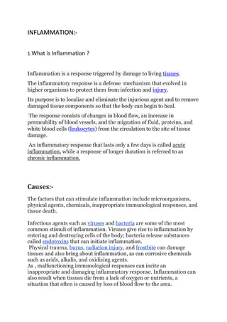
- 1. INFLAMMATION:- 1.What is Inflammation ? Inflammation is a response triggered by damage to living tissues. The inflammatory response is a defense mechanism that evolved in higher organisms to protect them from infection and injury. Its purpose is to localize and eliminate the injurious agent and to remove damaged tissue components so that the body can begin to heal. The response consists of changes in blood flow, an increase in permeability of blood vessels, and the migration of fluid, proteins, and white blood cells (leukocytes) from the circulation to the site of tissue damage. An inflammatory response that lasts only a few days is called acute inflammation, while a response of longer duration is referred to as chronic inflammation. Causes:- The factors that can stimulate inflammation include microorganisms, physical agents, chemicals, inappropriate immunological responses, and tissue death. Infectious agents such as viruses and bacteria are some of the most common stimuli of inflammation. Viruses give rise to inflammation by entering and destroying cells of the body; bacteria release substances called endotoxins that can initiate inflammation. Physical trauma, burns, radiation injury, and frostbite can damage tissues and also bring about inflammation, as can corrosive chemicals such as acids, alkalis, and oxidizing agents. As , malfunctioning immunological responses can incite an inappropriate and damaging inflammatory response. Inflammation can also result when tissues die from a lack of oxygen or nutrients, a situation that often is caused by loss of blood flow to the area.
- 2. Signs The four cardinal signs of inflammation—redness (Latin rubor), heat (calor), swelling (tumor), and pain (dolor)—were described in the 1st century AD by the Roman medical writer Aulus Cornelius Celsus. Redness is caused by the dilation of small blood vessels in the area of injury. Heat results from increased blood flow through the area and is experienced only in peripheral parts of the body such as the skin. Fever is brought about by chemical mediators of inflammation and contributes to the rise in temperature at the injury. Swelling, called edema, is caused primarily by the accumulation of fluid outside the blood vessels. The pain associated with inflammation results in part from the distortion of tissues caused by edema, and it also is induced by certain chemical mediators of inflammation, such as bradykinin, serotonin, and the prostaglandins. A fifth consequence of inflammation is the loss of function of the inflamed area, a feature noted by German pathologist Rudolf Virchow in the 19th century. Loss of function may result from pain that inhibits mobility or from severe swelling that prevents movement in the area. Chemical mediators of inflammation Although injury starts the inflammatory response, chemical factors released upon this stimulation bring about the vascular and cellular changes outlined above. The chemicals originate primarily from blood plasma, white blood cells (basophils, neutrophils, monocytes, and macrophages), platelets, mast cells, endothelial cells lining the blood vessels, and damaged tissue cells. One of the best-known chemical mediators released from cells during inflammation is histamine, which triggers vasodilation and increases vascular permeability. Stored in granules of circulating basophils and mast cells, histamine is released immediately when these cells are injured.
- 3. Other substances involved in increasing vascular permeability are lysosomal compounds, which are released from neutrophils, and certain small proteins in the complement system, namely C3a and C5a. Many cytokines secreted by cells involved in inflammation also have vasoactive and chemotactic properties. The prostaglandins are a group of fatty acids produced by many types of cells. Some prostaglandins increase the effects of other substances that promote vascular permeability. Others affect the aggregation of platelets, which is part of the clotting process. Prostaglandins are associated with the pain and fever of inflammation. Prostaglandins are synthesized from arachidonic acid, as are the leukotrienes, another group of chemical mediators that have vasoactive properties. The plasma contains four interrelated systems of proteins—complement, the kinins, coagulation factors, and the fibrinolytic system—that generate various mediators of inflammation. Activated complement proteins serve as chemotactic factors for neutrophils, increase vascular permeability, and stimulate the release of histamine from mast cells. They also adhere to the surface of bacteria, making them easier targets for phagocytes. The kinin system, which is activated by coagulation factor XII, produces substances that increase vascular permeability. The most important of the kinins is bradykinin, which is responsible for much of the pain and itching experienced with inflammation. The coagulation system converts the plasma protein fibrinogen into fibrin, which is a major component of the fluid exudate. The fibrinolytic system contributes to inflammation primarily through the formation of plasmin, which breaks down fibrin into products that affect vascular permeability. Process:- In response to any harmful or foreign molecule, inflammation generally starts within few minutes by activation of immune system . Innate system comprises immune cells that include lymphocytes, dendritic cells (DC’s), neutrophils, macrophages and mast cells that play significant functions in inflammatory reactions.
- 4. 1. Firstly, the pathogens get adhere to particular receptors i.e. G-protein attached receptors ,Pattern realization receptors and Chemokine receptors . 2. The fabrication of the inflammatory cytokines including IL-1, IL-6, chemokines and TNF is initiated by these receptors. These inducers quickly change the vascular endothelial permissibility and releases antibodies, complement factors and neutrophils in the site of septicity. [The inflammatory cytokines increases the excretion of coagulation factors and C-reactive protein by means of the liver cell. They invoke brain endothelium and smooth the secretion of prostaglandins. They are important for the key symptoms of pain and fever via their detrimental effects on CNS (central nervous system) . Alternatively the viral contamination follows one kind of signaling pathway via producing different type of cytokines known as type1 interferons (IFN’s). Furthermore, parasitic infections and allergens invoke the assembly of other inflammatory cytokines IL-13, IL-5, and histamine where the rest of the pathway is nearly the same . Inflammation is a complex biochemical mechanism which leads to the stimulation of infectious agents and promotes injury. It causes tissue damage and pain monitored through treatment .Chemical agents are secreted by immune cells like cytokines, chemokine and reactive oxygen species at injury site to eliminate pathogens. A major constituent of inflammatory process is arachidonic acid which is a by product of fast acting cell membrane. Arachidonic acid is transformed into prostaglandins and thromboxane enzyme by cyclooxygenase (COX).] 3. Neutrophil:- 1. Leukocyte margination and endothelial adhesion:- Neutrophils begin to attach strongly to the endothelium by using carbohydrate ligands to show symptoms of inflammation. Endothelial cells in their stimulated form are responsible for the production of surface bonded and soluble particles. They produce a strong adhesion between neutrophils and endothelium.
- 5. 2.Migration across the endothelium, known as transmigration, via the process of diapedesis: Chemokine gradients stimulate the adhered leukocytes to move between adjacent endothelial cells. The endothelial cells retract and the leukocytes pass through the basement membrane into the surrounding tissue .(Neutrophils leave the bloodstream and travels across endothelium . ) 3.Movement of leukocytes within the tissue via chemotaxis: Leukocytes reaching the tissue interstitium bind to extracellular matrix proteins via expressed integrins and CD44 to prevent them from leaving the site. A variety of molecules behave as chemoattractants, for example, C3a or C5, and cause the leukocytes to move along a chemotactic gradient towards the source of inflammation. Production of particular cells like cell adhesion molecules (CAMs), their activators and chemical stimulus is responsible for the neutrophils emigration . 4.Phagocytosis:- Extravasated neutrophils in the cellular phase come into contact with microbes at the inflamed tissue. Phagocytes express cell-surface endocytic pattern recognition receptors (PRRs) that have affinity and efficacy against non-specific microbe-associated molecular patterns (PAMPs). Most PAMPs that bind to endocytic PRRs and initiate phagocytosis . Upon endocytic PRR binding, that endocytoses the plasma membrane containing the PRR-PAMP complex, and the microbe. pathways have been implicated to traffic the endocytosed phagosome to intracellular lysosomes, where fusion of the phagosome and the lysosome produces a phagolysosome. The reactive oxygen species, superoxides and hypochlorite bleach within the phagolysosomes then kill microbes inside the phagocyte. Phagocytic efficacy can be enhanced by opsonization. Plasma derived complement C3b and antibodies that exude into the inflamed tissue during the vascular phase bind to and coat the microbial antigens. As well as endocytic PRRs, phagocytes also express opsonin receptors Fc receptor and complement receptor 1 (CR1), which bind to antibodies
- 6. and C3b, respectively. The co-stimulation of endocytic PRR and opsonin receptor increases the efficacy of the phagocytic process, enhancing the lysosomal elimination of the infective agent. Molecular or cellular actions of infectious response tend to increase blood movement, capillary damage, leukocytes access and the creation of chemical agents . Stimulation of these chemical agents initiates the formation of inflammatory cytokines including TNF, IL-1, chemokines and IL-6 that causes tissue damage. 5.Due to phagocytic activity of cells, migratory neutrophils are eventually removed from inflammatory site through apoptosis and produce anti- inflammatory cytokines .