20140824 Abnormalities in human pluripotent cells due to reprogramming mechan...
Gray et al. 2004 Science copy
1. 8. C. Toyoshima, H. Nomura, T. Tsuda, Nature 432, 361
(2004).
9. T. L.-M. Sørensen et al., J. Biol. Chem. 279, 46355
(2004).
10. J. V. Møller, G. Lenoir, M. Le Maire, B. S. Juul, P. Champeil,
Ann. N. Y. Acad. Sci. 986, 82 (2003).
11. J. D. Clausen, B. Vilsen, D. B. McIntosh, A. P. Einholm,
J. P. Andersen, Proc. Natl. Acad. Sci. U.S.A. 101,
2776 (2004).
12. Materials and methods are available as supporting
material on Science Online.
13. S. Danko, K. Yamasaki, T. Daiho, H. Suzuki, J. Biol.
Chem. 279, 14991 (2004).
14. C. Toyoshima, G. Inesi, Annu. Rev. Biochem. 73, 269
(2004).
15. A. Barth, W. Kreutz, W. Mantele, J. Biol. Chem. 272,
25507 (1997).
16. I. M. Glynn, J. Physiol. 462, 1 (1993).
17. T. Sørensen, B. Vilsen, J. P. Andersen, J. Biol. Chem.
272, 30244 (1997).
18. B. Forbush III, I. Klodos, in The Sodium Pump:
Structure, Mechanism, and Regulation, J. H. Kaplan,
P. de Weer, Eds. (Rockefeller Univ. Press, New York,
1991), pp. 211.
19. F. Cornelius, J. V. Møller, FEBS Lett. 284, 46 (1991).
20. X. Yu, S. Carroll, J. L. Rigaud, G. Inesi, Biophys. J. 64,
1232 (1993).
21. D. Levy, M. Seigneuret, A. Bluzat, J. L. Rigaud, J. Biol.
Chem. 265, 19524 (1990).
22. X. Yu, L. Hao, G. Inesi, J. Biol. Chem. 269, 16656 (1994).
23. T. Seekoe, S. Peall, D. B. McIntosh, J. Biol. Chem. 276,
46737 (2001).
24. M. Toustrup-Jensen, B. Vilsen, J. Biol. Chem. 278, 11402
(2003).
25. M. Shigekawa, L. J. Pearl, J. Biol. Chem. 251, 6947 (1976).
26. W. P. Jencks, J. Biol. Chem. 264, 18855 (1989).
27. W. L. DeLano, PyMOL Molecular Graphics System (2002);
available online at http://www.pymol.sourceforge.net.
28. C. R. Lancaster, Nature 432, 286 (2004).
29. We thank B. Holm, B. Nielsen, and C. Lauridsen for
expert technical assistance; the staff at the Protein
Structure Factory beamline BL14.1 of Free University
Berlin at Berliner Elektronenspeicherring-Gesellschaft
fu¨r Synchrotronstrahlung m. b. H., the staff at European
Synchrotron Radiation Facility (ESRF) beamline ID29;
J. L. Karlsen and R. Jørgensen for extensive computer
support; and J. Nyborg for fruitful scientific discussions.
This work was supported by an Ole Rømer Research
grant; the Center for Structural Biology; the Dansync
program under the Danish Natural Science Research
Council; a grant from the Lundbeck Foundation (P.N.),
and grants from the Danish Medical Research Council,
Aarhus University Research Foundation, and Novo
Nordic Foundation (J.V.M.). Coordinates and structure-
factor amplitudes have been deposited to the Protein
Data Bank under code 1XP5.
Supporting Online Material
www.sciencemag.org/cgi/content/full/306/5705/2251/
DC1
Materials and Methods
Figs. S1 to S4
Table S1
Movie S1
12 October 2004; accepted 23 November 2004
10.1126/science.1106289
Mouse Brain Organization Revealed
Through Direct Genome-Scale
TF Expression Analysis
Paul A. Gray,1,3
*. Hui Fu,2,3
* Ping Luo,1,3
* Qing Zhao,4
Jing Yu,5
Annette Ferrari,3
Toyoaki Tenzen,5
Dong-in Yuk,4
Eric F. Tsung,6
-
Zhaohui Cai,6
John A. Alberta,3
Le-ping Cheng,1,3
Yang Liu,1,3
Jan M. Stenman,5
M. Todd Valerius,5
Nathan Billings,4
Haesun A. Kim,2,3
` Michael E. Greenberg,1,8
Andrew P. McMahon,5
David H. Rowitch,4,7
Charles D. Stiles,2,3
¬ Qiufu Ma1,3
¬
In the developing brain, transcription factors (TFs) direct the formation of a
diverse array of neurons and glia. We identifed 1445 putative TFs in the
mouse genome. We used in situ hybridization to map the expression of over
1000 of these TFs and TF-coregulator genes in the brains of developing mice.
We found that 349 of these genes showed restricted expression patterns that
were adequate to describe the anatomical organization of the brain. We
provide a comprehensive inventory of murine TFs and their expression
patterns in a searchable brain atlas database.
The mammalian nervous system contains
thousands of distinct neuronal and glial cell
types that are distinguished on the basis of
morphology, projection, and marker gene ex-
pression (1). Transcription factors (TFs) play a
pivotal role in brain development by directing
the formation of neurons and glia from uncom-
mitted progenitor cells (2). To determine the
extent to which TFs describe the diversity of
the mammalian central nervous system (CNS),
we visualized the spatial and temporal expres-
sions of TF-encoding genes on a genome-wide
scale in the developing mouse CNS.
To identify unique genomic loci that encode
putative TFs in the mouse genome, we ana-
lyzed and annotated existing public and private
databases (3–6). Candidates were classified as
TFs only if their predicted protein sequence
included a Protein Families Database (Pfam)–
defined TF-DNA binding domain (3). We iden-
tified 1445 unique transcriptional units in the
mouse genome with putative TF-DNA bind-
ing domains. The nonoverlapping genes for
each DNA binding family (7, 8) are summa-
rized in Table 1 and tables S1 and S2. Recent
protein prediction databases have estimated that
there are over 2300 different TF proteins in the
mouse genome (6, 9). This higher number is
primarily due to the separate counting of each
possible protein variant as a unique transcrip-
tional unit, as well as the duplicate counting of
genes with multiple DNA binding domains.
The largest single class of TF proteins (È678
members, not including nuclear hormone re-
ceptors) was the zinc-finger family. Homeo-
box TFs had 227 members, and there were 50
nuclear hormone receptors and 116 basic helix-
loop-helix (bHLH) TFs (Table 1 and table S1).
The human genome encodes 20,000 to 25,000
genes (10). If a similar number of genes are
encoded in the mouse genome, then TF genes
make up more than 7% of the total.
We designed gene-specific polymerase
chain reaction (PCR) primer pairs to produce
in situ hybridization probes for 1174 TF-
encoding genes. This probe set covers 91% of
genes that belong to 32 out of the 33 major TF
families (table S1). For the remaining (also
the largest) gene family, which encodes zinc-
finger proteins (divided into 12 subgroups),
71% of the genes were covered by the probe
set (table S1). These primers were used to
perform PCR on cDNA templates prepared
from embryonic day 13.5 (E13.5) and newborn
Epostnatal day zero (P0)^ mouse brains. A small
number of additional probes were acquired
from embryonic mouse kidney or testis cDNA.
Among the 1174 TF-encoding genes screened,
972 (83%) showed positive PCR products. We
monitored the recovery of nuclear hormone
receptors as a metric for sensitivity. We cloned
46 of the 50 nuclear hormone receptors that are
encoded in the mouse genome. Only one nu-
clear hormone receptor known to be expressed
in the brain, the androgen receptor (11), was
missed by our procedure. In total, 983 TF-
encoding genes were subsequently cloned or
acquired (Table 1 and table S1). We also cloned
104 transcription cofactor genes (Table 1 and
table S1), yielding a total number of 1087
genes, which are collectively referred to as TF-
encoding genes. These cloned in situ plasmids
1
Department of Neurobiology, 2
Department of Mi-
crobiology and Molecular Genetics, Harvard Medical
School, Boston, MA 02115, USA. 3
Department of
Cancer Biology, 4
Department of Pediatric Oncology,
Dana-Farber Cancer Institute, 1 Jimmy Fund Way,
Boston, MA 02115, USA. 5
Department of Molecular
and Cellular Biology, Harvard University, 16 Divinity
Avenue, Cambridge, MA 02138, USA. 6
Informatics
Program, 7
Division of Newborn Medicine, 8
Division of
Neuroscience, Children’s Hospital, Boston, MA 02115,
USA.
*These authors contributed equally to this work.
.Present address: Molecular Neurobiology Laborato-
ry, The Salk Institute, 10010 North Torrey Pines Road,
La Jolla, CA 92037, USA.
-Present address: Department of Biology, Boston
College, 140 Commonwealth Avenue, Chestnut Hill,
MA 02467, USA.
`Present address: Department of Biological Sciences,
Rutgers University, 101 Warren Street, Newark, NJ
07102 USA.
¬To whom correspondence should be addressed. E-mail:
Qiufu_Ma@dfci.harvard.edu (Q.M.); Charles_Stiles@dfci.
harvard.edu (C.D.S.)
R E P O R T S
www.sciencemag.org SCIENCE VOL 306 24 DECEMBER 2004 2255
2. have been deposited at the American Type Cul-
ture Collection (ATCC) for open distribution.
We synthesized digoxigenin-labeled probes
for the TF-encoding genes. To visualize TF ex-
pression at an early developmental stage, we
analyzed the expression of 1013 TF-encoding
genes using whole-mount in situ hybridization
on E10.5 mouse embryos. Of these 1013, at
least 142 were clearly expressed in a spatially
restricted manner (table S6 and fig. S7). We
also performed in situ hybridization for 1034
TF genes on transverse sections through the
E13.5 and P0 head and trunk, as well as on
sections through the postnatal cerebellum at P7,
P15, and P21. Of 1034 genes examined in the
E13.5 and/or P0 nervous system, 349 showed
spatially restricted expression patterns (table
S3). For TFs that belong to non–zinc-finger
families, È38% of them showed restricted ex-
pression patterns in the CNS. However, only
È10% of zinc-finger genes showed restricted
patterns. Collectively, È27% of all the TFs ex-
hibited spatially restricted patterns (table S3).
We divided the CNS into seven general
areas for annotation: the cortex, striatum (and
other basal ganglia), thalamus, hypothalamus,
midbrain, hindbrain, and spinal cord. Very
few of the 349 TFs with spatially restricted
expression patterns were expressed in only
one region of the brain (table S4). Nearly all
TF-encoding genes expressed in the neonatal
brain were also detected in postmitotic
neurons at E13.5. Digital images of the entire
in situ hybridization set have been deposited
in the Mahoney Transcription Factor Atlas
(12), as well as in the Jackson Laboratory_s
Gene Expression Database (13).
The in situ hybridization data show that
postmitotic anatomical diversity within the
CNS can be described by TF expression. For
example, the neocortex is a highly laminated
and regionally organized anatomical struc-
ture (14). We found that several dozen TF-
encoding genes occupy different dorsoventral
positions or different laminae of the neocortex
(Fig. 1). In the striatum, a major basal ganglia
component crucial for movement control is
organized in a somatotopic fashion (15). This
somatotopic organization is echoed by the
gradient expression of many TF-encoding
genes in this area (fig. S1). The thalamus and
hypothalamus are organized into discrete ana-
tomical and functional nuclei that are marked
by the overlapping expression pattern of spe-
cific TFs within these regions (figs. S2 and
S3). In the retina, a large number of TF genes
are expressed with distinct density within the
retinal ganglion and amacrine cell layers (figs.
S4 and S5), which may correlate with the
morphological diversity of these cell types (1).
In the postnatal cerebellum, laminar-specific
TF expression marks the immature and mature
granule cells and Purkinje cells (fig. S6).
The genome-scale whole-mount and sec-
tion in situ hybridizations also identified over
100 TF genes expressed in spatially restricted
patterns within non-neuronal tissues such as
the nose, oral cavity, teeth, salivary gland,
inner ear bone, mandibular bone, eye muscle,
facial muscle, skin, and others (fig. S8 and
table S4). Although not the explicit focus of
this study, the open-source TF data set gener-
ated in this work provides detailed information,
enabling comprehensive study of developing
cranial facial tissues. The in situ plasmid set
provided here can be used to define TF expres-
sion patterns in other tissues and at other de-
velopmental times in neural tissues.
Our atlas of TF expression provides a
visual filter for functional analysis of the TF
gene set in brain development. Over 7% of the
murine genome may encode TFs. However,
our studies suggest that at a given developmen-
tal stage, fewer than 27% of these TF-encoding
genes are expressed in spatially restricted pat-
terns consistent with a direct role in the for-
mation of specific neural types. The regulatory
elements for these spatially restricted TFs might
be useful reagents for driving the expression of
reporter genes that will mark specific neural cell
types, as described recently by Gong et al. (16).
The TF expression profile in postmitotic neu-
rons will be useful to study the expression of
neural-specific genes encoding ion channels,
neurotransmitter receptors, and synaptic pro-
teins, whose molecular control remains largely
unknown. On a clinical front, TF mutations
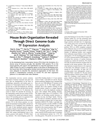
Fig. 1. Diversity of transcription factor expression in the P0 mouse cerebral cortex. Nonradioactive
in situ hybridization patterns for 20 representative TFs or TF cofactors on sections through the
forebrain of P0 mice are shown. Labels at the bottoms of individual panels indicate Locuslink gene
names. (A) Over a dozen TF-encoding genes occupy different dorsoventral positions of the
hippocampus (top row) and neocortex (middle and bottom rows). (B) A dozen genes show laminar
specific expression in the neocortex. HC, hippocampus; BG, basal ganglia; SC, superior colliculus;
TH, thalamus; HY, hypothalamus; DG, hippocampal dentate gyrus; CA1 to 3, hippocampal CA1,
CA2, and CA3 regions. Scale bar in (A), 1 mm; all images in this section show the same
magnification. Scale bar in (B), 0.2 mm; all images in this section show the same magnification.
R E P O R T S
24 DECEMBER 2004 VOL 306 SCIENCE www.sciencemag.org2256
3. have already been shown to underlie certain
disorders in speech (17), appetite control (18),
breathing patterns (19), and autism (20) in hu-
mans. The TF atlas presented here may have
practical overtones for understanding addi-
tional neurological or behavioral disorders in
children and adults.
References and Notes
1. R. H. Masland, Curr. Biol. 14, R497 (2004).
2. R. Shirasaki, S. L. Pfaff, Annu. Rev. Neurosci. 25, 251
(2002).
3. E. L. Sonnhammer, S. R. Eddy, E. Birney, A. Bateman,
Nucleic Acids Res. 26, 320 (1998).
4. V. Matys et al., Nucleic Acids Res. 31, 374 (2003).
5. D. L. Wheeler et al., Nucleic Acids Res. 32, D35 (2004).
6. P. D. Thomas et al., Genome Res. 13, 2129 (2003).
7. D. Stryke et al., Nucleic Acids Res. 31, 278 (2003).
8. J. Hansen et al., Proc. Natl. Acad. Sci. U.S.A. 100,
9918 (2003).
9. R. H. Waterston et al., Nature 420, 520 (2002).
10. International Human Genome Sequencing Consorti-
um, Nature 431, 931 (2004).
11. N. M. Shah et al., Neuron 43, 313 (2004).
12. The Mahoney Transcription Factor Atlas is available
at http://mahoney.chip.org/mahoney/.
13. See the Jackson Laboratory’s Gene Expression Database,
accession no. J:91257, available at www.informatics.
jax.org/.
14. D. D. O’Leary, Y. Nakagawa, Curr. Opin. Neurobiol.
12, 14 (2002).
15. E. R. Kandel, J. H. Schwartz, T. M. Jessell, Principles of
Neural Science (McGraw-Hill, New York, 2000).
16. S. Gong et al., Nature 425, 917 (2003).
17. C. S. Lai, S. E. Fisher, J. A. Hurst, F. Vargha-Khadem,
A. P. Monaco, Nature 413, 519 (2001).
18. J. L. J. Holder, N. F. Butte, A. R. Zinn, Hum. Mol. Genet.
9, 101 (2000).
19. J. Amiel et al., Nature Genet. 33, 459 (2003).
20. N. Gharani, R. Benayed, V. Mancuso, L. M. Brzustowicz,
J. H. Millonig, Mol. Psychiatry 9, 474 (2004).
21. We thank J. Olsen, J. Chan, and P. Santos for their
technical assistance and R. DePinho for providing the
National Institute on Aging 15K cDNA set. We also
thank R. Segal and S. Pfaff for critical reading of this
manuscript. The work was supported by the Charles
Dana Foundation, the National Institute of Neuro-
logical Disorders and Stroke (Q.M., C.D.S., D.H.R., and
A.P.M.), the National Institute of Dental and Cranio-
factial Research (Q.M.), the National Institute of Dia-
betes and Digestive and Kidney Dieseases (A.P.M.), a
Ford Foundation Postdoctoral Fellowship for Minorities
(P.A.G.), a Parker B. Francis Fellowship in Pulmonary
Medicine (P.A.G.), and the Pew Trust (Q.M.).
Supporting Online Material
www.sciencemag.org/cgi/content/full/306/5705/2255/
DC1
Materials and Methods
Figs. S1 to S8
Tables S1 to S7
References
7 September 2004; accepted 15 November 2004
10.1126/science.1104935
Activity-Dependent Internalization
of Smoothened Mediated by
b-Arrestin 2 and GRK2
Wei Chen,1
* Xiu-Rong Ren,2
Christopher D. Nelson,2
Larry S. Barak,3
James K. Chen,4
. Philip A. Beachy,4
Frederic de Sauvage,5
Robert J. Lefkowitz2
*
Binding of Sonic Hedgehog (Shh) to Patched (Ptc) relieves the latter’s tonic
inhibition of Smoothened (Smo), a receptor that spans the cell membrane
seven times. This initiates signaling which, by unknown mechanisms,
regulates vertebrate developmental processes. We find that two molecules
interact with mammalian Smo in an activation-dependent manner: G protein–
coupled receptor kinase 2 (GRK2) leads to phosphorylation of Smo, and bbb-
arrestin 2 fused to green fluorescent protein interacts with Smo. These two
processes promote endocytosis of Smo in clathrin-coated pits. Ptc inhibits
association of bbb-arrestin 2 with Smo, and this inhibition is relieved in cells
treated with Shh. A Smo agonist stimulated and a Smo antagonist (cyclopamine)
inhibited both phosphorylation of Smo by GRK2 and interaction of bbb-arrestin 2
with Smo. bbb-Arrestin 2 and GRK2 are thus potential mediators of signaling by
activated Smo.
Hedgehog (Hh) signaling is mediated by
regulation of a protein called Smoothened
(Smo) that spans the cell membrane seven
times (7MS), activation of which sets in
motion transcriptional events that control
growth and patterning in vertebrate develop-
ment (1, 2). Dysregulated Smo activity also
leads to several forms of cancer (3–7). Hh
binds to a receptor that spans the cell mem-
brane 12 times, Patched (Ptc), and relieves
inhibitory control of Smo by Ptc. However,
almost nothing is known of the mechanisms
operating just downstream of Smo to medi-
ate and modulate its actions. b-Arrestins are
cytosolic proteins that bind to most acti-
vated 7MS receptors after the receptors
have been phosphorylated by GRKs, which
promotes internalization of the receptors
and some forms of signaling (8, 9). Elements
that regulate receptor functions often show
Table 1. Nonredundant numbers of putative TFs in the mouse genome. Columns describe the total
number of unique genomic loci encoding predicted DNA binding TFs by domain and the numbers and
relative percentages analyzed. The second to last column describes the number of family members
available as Genetrap cell lines in the Baygenomics and/or German Gene Trap Consortium libraries. The
last column describes the percentage of family members with available enhancer trap cell lines. Genes
that encode multiple DNA binding domains are listed and counted in one family for clarity. Nuclear
hormone receptors are not included in zinc-finger genes. Transcription cofactors and these non-TF genes
we analyzed are also included. Asterisks indicate the cofactor and non-TF gene numbers we analyzed
rather than the total number in the genome. HMG, high-mobility group; bZIP, basic helix-loop-helix and
leucine zipper proteins; non-TFs, genes that do not encode TFs; nuclear rec, nuclear hormone receptors;
ZF, zinc finger; ETS, erythroblast transformation–specific; PHD, plant homeodomain; btb/poz, broad-
complex, tramtrack, and bric-a-brac/poxvirus and zinc-finger proteins.
Domain No. of genes No. cloned % cloned Genetrap available % trapped
Homeobox 227 170 74.9 12 5.3
bHLH 116 100 86.2 22 19.0
HMG 58 41 70.7 14 24.1
bZIP 57 41 71.9 16 28.1
Nuclear Rec 50 46 92.0 10 20.0
Forkhead 40 29 72.5 12 30.0
ETS 28 26 92.9 8 28.6
ZF C2H2 490 287 58.6 171 34.9
ZF PHD 60 44 73.3 42 70.0
ZF C2CH 39 18 46.2 11 28.2
ZF btb/poz 28 18 64.3 8 28.6
Other 252 163 64.7 124 49.2
TF Total 1445 983 68.0 450 31.1
Cofactors* 133 104 78.2 48 36.1
Non-TFs* 336 261 77.7 95 28.6
Total genes 1914 1348 70.4 493 25.8
1
Department of Medicine, 2
Howard Hughes Medical
Institute, Departments of Medicine and Biochemistry,
3
Department of Cell Biology, Duke University Medical
Center, Durham, NC 27710, USA. 4
Howard Hughes
Medical Institute, Department of Molecular Biology
and Genetics, Johns Hopkins University School of Med-
icine, Baltimore, MD 21205, USA. 5
Department of Mo-
lecular Oncology, Genentech, South San Francisco, CA
94080, USA.
*To whom correspondence should be addressed.
E-mail: lefko001@receptor-biol.duke.edu (R.J.L.) and
w.chen@duke.edu (W.C.).
.Present address: Department of Molecular Pharma-
cology, Stanford University School of Medicine, Stan-
ford, CA 94305, USA
R E P O R T S
www.sciencemag.org SCIENCE VOL 306 24 DECEMBER 2004 2257