
Recommended
More Related Content
Similar to REtinal detachment.pptx
Similar to REtinal detachment.pptx (20)
Recently uploaded
Recently uploaded (20)
REtinal detachment.pptx
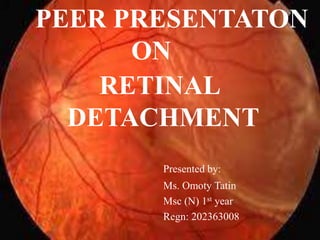
- 1. PEER PRESENTATON ON RETINAL DETACHMENT Presented by: Ms. Omoty Tatin Msc (N) 1st year Regn: 202363008
- 2. OBJECTIVES: At the end of the class the students will be able to : 1. Define Retinal detachment 2. List the causes and risk factors 3. Explain the pathology and Pathophysiology 4. List the symptoms 5. Enumerate the types
- 3. 6. List the diagnostic evaluation and principles of treatment 7. Discuss the Medical, Pharmacologic and surgical management 8. List the complications 9. Discuss the nursing responsibilities and coping and supportive measures
- 5. INTRODUCTION: The role of vision in our lives is difficult to define, because it is so deeply personal and intimate. Loss of vision means loss of independence. Among the various cause of blindness, retinal detachment is one which is an ocular emergency.
- 6. DEFINITION: Retinal detachment is a disorder of the eye in which the retina peels away from its underlying layer of support tissue.
- 8. INCIDENCE: The average incidence of RRDs among males was 7.99 cases per 100 000 and among females was 7.56 cases per 100 000 in China. The incidence of retinal detachment has been reported between 0.04% and 0.36% by previous studies.
- 9. CAUSES: Spontaneous due to degenerative changes in the retina or vitreous Trauma, inflammation or other problems Older than 40 Common cause is the tear or hole in the retina,
- 10. Diabetic retinopathy Previous retinal detachment in the other eye Family history of retinal detachment Metabolic and vascular diseases Severe myopia
- 11. RISK FACTORS: Aging Previous retinal detachment in one eye Family history of retinal detachment Extreme nearsightedness (myopia)
- 12. Previous eye surgery Previous severe eye injury Previous other eye disease or disorder
- 13. PATHOPHYSIOLOGY
- 14. Due to etiological factors (a torn or break in retina) Vitreous fluid or serous fluid leaks in between the layers of retina or behind the retinal layers Detachment of retinal layer
- 15. Retina can peel away from the underlying layer of blood vessels Lack of oxygenation in tissues of retina Vision disturbances
- 16. SYMPTOMS: The sudden appearance of many floaters Flashes of light in one or both eyes (photopsia) Blurred vision Gradually reduced side (peripheral) vision A curtain-like shadow over the field of vision
- 17. FLOATERS
- 18. PHOTOPSIA
- 20. TYPES: 1. Rhegmatogenous 2. Tractional 3. Exudative
- 21. RHEGMATOGENOUS It occurs due to break in the retina (called a retinal tear)
- 22. TRACTIONAL It occurs when scar tissue grows on the retina's surface, causing the retina to pull away from the back of the eye
- 23. EXUDATIVE It occurs due to inflammation, injury or vascular abnormalities. Fluid accumulating underneath the retina without the presence of a hole, tear or break.
- 26. DIAGNOSIS: History Physical Examination Checking of visual acuity External examination for signs of trauma and checking of the visual field. Assessment of pupil reaction
- 27. Measurement of intraocular pressure in both eyes. Slit lamp examination Ultrasound imaging Visual acuity Measurement
- 29. VISUAL ACUITY MEASUREMENT: SNELLEN CHART
- 30. TREATMENT: General principles of treatment: Find all retinal beaks Seal all retinal breaks Relieve vitreo retinal traction
- 31. MANAGEMENT: Sedation ( local anesthesia) Bed rest for 2 to 4 weeks and eye patch Restricted eye movement
- 32. PHARMACOLOGIC INTERVENTION Drops as prescribed of Cyclopentolate hydrochloride (Cyclogyl) Antibiotic drops such as Gentamicin; prednisolone acetate to prevent eye infections Other Drugs: Antiemetics and analgesics
- 33. SURGICAL MANAGEMENT Cryotherapy or Laser surger(photocoagulation) Freezing (cryopexy) Electrodiathermy Pneumatic retinopexy Scleral buckling Vitrectomy
- 34. CRYOTHERAPY OR LASER SURGERY
- 36. ELECTRODIATHERMY A tiny hole is made in the sclera to drain subretinal fluid, allowing the pigment epithelium to adhere to the retina.
- 38. SCLERAL BUCKLING
- 39. VITRECTOMY
- 40. COMPLICATIONS
- 41. Early complication- Glaucoma, infections, failure of retina to reattach Late complications- Infection, diplopia, refractive errors Re-enactment of the retina may occur any time.
- 42. Due to postoperative swelling of tissue in anterior chamber leads to increased intraocular pressure in glaucoma. Due to the retina separated from blood supply for a long time even after reattachment patients vision does not improve
- 43. NURSES RESPONSIBILITIES Pre-operative: Provide emotional support to the patient Instruct the patient to remain quiet in prescribed position Allow the patient and family to discuss their concerns.
- 44. Prepare the patient by cleaning his face and administer preoperative medications as ordered. Post-operative: Assess the status of eye dressing and presence of bleeding or drainage.
- 45. Place the patient in fowler or semi-fowler’s position to reduce the intraocular pressure. Take measures to prevent postoperative complications Encourage ambulation and independence as tolerated. Administer medication for pain, nausea, and vomiting as directed
- 46. NURSING DIAGNOSIS: Acute pain related to trauma to the incision site and increased intraocular pressure Impaired sensory perception vision related to impaired sensory reception
- 47. Anxiety related to lack of knowledge about the disease and its treatment High risk of injury related to loss of vitreous, intraocular hemorrhage and IOP Risk for infection related to trauma to the incision.
- 48. NURSING INTERVENTIONS Assess visual status and functional vision in the unaffected eye to determine self care needs. Discourage straining during defecation, bending down and hard coughing, sneezing or vomiting to avoid activities that increase intraocular pressure.
- 49. Assist with ambulation, as needed, to help the client remain independent Provide assistance with activities of daily living to minimize frustration and strain. Orient the client to his environment to reduce the risk of injury.
- 51. Have the patient or significant others demonstrate the correct technique for instilling eye drops. Instruct the patient to wash her or his hands before and after removing the dressing.
- 52. Do not touch any part of the eye with the dropper. Teach the patient to use warm or cold compresses for comfort several times a day. Teach the patient to avoid vigorous activities and heavy lifting for the immediate postoperative period.
- 53. COPING AND SUPPORT Get glasses. Brighten the home Make the home safer Enlist the help of others Get help from technology Talk to others with impaired vision
- 56. CONCLUSION Retinal detachment is a vision threatening condition that requires early surgery. It can be diagnosed best by retinal examination usually indirect opthalmoscopy. Treatment outcome has improved with modern surgical techniques, but the key to successful reattachment is early detection and prompt referral by primary eye care workers
- 57. BIBLIOGRAPHY
- 58. BOOKS: Correia C. Medical surgical Nursing- Nursing Speciality as per INC syllabus. First Edition Vol 2; Jaypee Brothers Medical Publishers (P) Ltd, New Delhi, 2017; Page no: 227-29 Chintamani, Lewis’s Medical- Surgical Nursing- Assessment and Management of clinical Problems. 7th Ed; Reed Elsevier India Private Limited, New Delhi, 2011; Page no: 424-26 Reeves J. C, Roux G, Lockhart R. Medical-Surgical Nursing. International Edition; McGraw Hill Nursing Core Series, page no: 18 Waugh A, Grant A. Ross and Wilson Anatomy and physiology in illness and health, 12th Edition, Reed Elsevier India Private Limited, New Delhi, 2014; Page no: 196- 205
- 59. INTERNET: Mayo Clinic (7 Sept 2022), Retinal Detachment; cited on 13/ 10/ 23; Available from: https://www.mayoclinic.org Gabby E.A ( 12 April 2023), Retinal Detachment, Edited by Amy Boshnack; Cited on 13/10/13; Available from: https://www.healthline.com
- 60. JOURNAL Paolo Chelazzi, Claudia Azzolini, Claudia Bellina, Francesca Cappelli, Ilaria Del Genovese, Laura Caraffa, Francesco Scullica, "Efficacy and Safety of Vitrectomy without Using Perfluorocarbon Liquids and Drainage Retinotomy Associated with Postoperative Positioning Based on Residual Subretinal Fluid for Rhegmatogenous Retinal Detachment", Journal of Ophthalmology, vol. 2021, Article ID 5588479, 9 pages, 2021. Available from: https://doi.org/10.1155/2021/5588479