2. Objectives :
- To know the definition & classification of
Dermatitis/Eczema
- To recognize the primary presentation of different
types of eczema
- To understand the possible pathogenesis of each
type of eczema
-To know the scheme of managements lines
4. Hypersensitivity Reaction
Type 1: Immediate Hypersensitivity Reaction
Mediated by IgE to specific antigens
Mast cells stimulated and release histamine
Reaction within 15-30 minutes of exposure
Examples: Anaphylaxis (e.g.penicillin) ,Urticaria , Angioedema .
Type 2: Cytotoxic Antibody mediated Reaction
Mediated by IgG and IgM to specific antigens
Examples: Transfusion Reaction ,Rhesus Incompatibility (Rh
Incompatibility), Hashimoto‘ thyroiditis.
5. Cont…
Type 3: Immune Complex Reaction
Antigen-Antibody complexes deposit in tissue
Reaction within 1-3 weeks after exposure
SLE, serum sickness , vasculitis:Examples
Type 4: Delayed-Type Hypersensitivity
Mediated by T-Lymphocytes to specific antigens
Reaction within 2-7 days after exposure
Examples: Allergic contact dermatitis (e.g. Nickel allergy)
6. Eczema (Dermatitis)
Definition:
is an inflammatory skin disease.
Skin inflammation characterized by:
-itchy, scaly, patches of erythema
Pathogenesis:
It is an epidermal reaction to specific Antigens; these
antigens may be internal or external, acting
singularly or in combination
7. Clinical picture:
Most eczemas share certain general features, and each
different type of eczema will have some distinguishing
markers of their own. Eczema can be broadly classified as
acute, subacute, and chronic
8. Clinical picture (cont….)
- Acute:
-eryhema
- papules & vesicles
- oozing
-Subacute:
- scales
- Excoriation
- -Chronic:
-lichenificaion & hyperkeratosis
Clinically an eczematous disease may start at any stage and
evolve into another
16. Atopic Dermatitis
chronic relapsing itchy skin disease in
genetically predisposed patients
associated with personal or family history of
asthma, allergic rhinitis, conjunctivitis
or atopic eczema
17. Affects 15-30% of children, 2-10% of adult
60% begin during the first year
85% begin before 5 years
Up to 70%: spontaneous remission before
adolescence
18. Pathogenesis:
-Genetic pedisposition
-immune mediated (increase IgE), T-helper cell2 activation
-Impaired skin barrier.
defective epidermal differentiation (filaggrin
mutations) and resultant impaired barrier function of
the skin
- Allergy, increased tendency to certain allergens
- Infection : skin of pts with AD is colonized by S aureus.
infection with S aureus often causes a flare of AD
- AD and Food! minor role
19. AD associated with local infiltration of Th 2
that secrete IL-4, IL-5, IL-13, IL-31
More than 50% develop asthma
75% develop Allergic Rhinitis
Complex interrelationship of genetic,
environmental, and immunologic.
22. Allergens are taken up by dendritic cells and presented to T cells. In the absence of childhood microbial
exposure, the balance between T helper 1 (TH1) and TH2 cells is altered. TH2 cells encourage the
production of immunoglobulin E (IgE) by B cells. Allergen-specific IgE then binds to the high-affinity
receptor for IgE (FcepsilonRI) on mast cells. Allergen exposure induces crosslinking of receptor-bound IgE
with subsequent mast-cell degranulation and the release of pro-inflammatory molecules.
IL, interleukin; TCR, T-cell receptor.
32. Childhood atopic dermatitis
Children develop lesions at antecubital and
popliteal fossae, neck, wrists, and ankles.
Lichenification, excoriations, and dry skin are
common as well as post-inflammatory
hyperpigmentation
35. In adults
• most common manifestation: hand dermatitis.
• chronic severe form of generalized and
lichenified atopic eczema.
Adult atopic dermatitis
64. Clinical Picture:
Seborrheic dermatitis is defined by clinical
parameters which include:
1-erythematous red-yellow , poorly circumscribed
patches & thin plaques with bran-like to flaky
(greasy) scales.
2-Limitation to those periods of life when
sebaceous gland are active i.e. the 1st few
months of life & post puberty (infantile & adult
forms).
65. Cont….
3- A predilection for areas rich in sebaceous
glands
e.g: scalp , face, ears , presternal region &
flexural areas (axillae, inguinal &
inframammary folds , umbilicus).
4-A mild course with moderate discomfort.
66.
67. Cradle cap: is coherent scaly & crusty mass covering most of
the scalp & can be seen in infanile seborreic dermatitis.
71. Treatment:
-Medicated shampoo (e.g. containing coal tar,
selenium sulfide or ketoconazole)
-Topical antifungal.
-low potency topical steroid.
- Topical immunomodulators (tacrolimus & pimecrolimus)
- A preparation of salicylic acid (2–5%, depending upon
the scaling) can be used for the scalp.
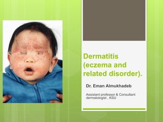
72. Infantile atopic dermatitis Infantile Seborrheic dermatitis
-Markedly pruritic
-Presents as erythema, papules and
vesicles
-Prominent on the cheeks and extensor
surface of the limbs.
-Asymptomatic
-Present as greasy scales over an
erythematous base
-Prominent on the
scalp , nasolabial fold and
body folds
75. Allergic contact dermatitis (ACD)
Definition:
Dermatitis resulting from type 4 reaction
following exposure to topical substances in
sensitized individuals (requires induction and
elicitation phase (lag time to reaction).
Common allergens eliciting contact dermatitis:
nickel (affects 10% of women and 1% of men),
perfumes, fragrances , preservatives.
hair dyes,
rubber latex
76. Clinical picture:
-Acute form present with
crusted erythematous
papules, vesicles &
bullae that is well
demarcated &
localized to the site of
contact with the
allergen.
-ACD can be more
diffuse in
distribution.
91. Irritant contact dermatitis (ICD)
-Is localized non immunologically mediated
inflammatory reaction.
-ICD results from direct cytotoxic effect d.t single
or repeated application of a chemical
substance to the skin.
Most common irritants are:
Water
Abrasives
Chemicals, e.g. acids and alkalis
Solvents and detergents
92. Clinical picture:
-Similar to ACD but ICD never extend beyond
the area of contact.
-tend to be painful rather than pruritic .
-can occur from the 1st exposure to the irritant
unlike ACD which only occur in previously
sensitized individual.
99. Nummular (discoid) dermatitis
-Sharply circumscribed eczema , nummular
means ( coin -shaped)
-Pathogenesis: Probably microbial in origin
i.e. 2ry to bacterial colonization or
disseminaion of bacterial toxins.
102. Dyshidrotic dermatitis (pompholyx)
Acute dermatitis which is often vesicular with tiny deep
seated vesicles along the sides of the fingers associated
with pruritus
103. Cont..
-Not considered as a separate disease
-Can be associated with atopy , of patients with
dyshidrosis, 50% have atopic dermatitis.
-Exogenous factors (eg, contact dermatitis to
nickel,chemicals) also play a role.
-Affect hands & feet.
108. Stasis dermatitis
-seen in patient with
signs of venous
hypertension like
chronic lower limb
edema, varicose vein.
-can be complicated by
superimposed allergic
contact dermatitis.
116. Neurodermatitis
-Include dermatitis which results from repeated
rubbing & scratching of the skin .
-Chronic itching and scratching can cause the skin
to thicken and have a leather texture with
exaggeration of skin marking.
-A scratch-itch cycle occurs which is difficult to
break .
118. lichen simplex chronicus
Present as thick hyperkeratotic plaque with
accentuation of skin marking that occurs on any
site that the patient can reach, including the
following:
-Scalp
-Nape of neck
-Extensor forearms and elbows
-Vulva and scrotum
-Upper medial thighs, knees, lower legs, and
ankles
124. Distinctive morphological features of
different forms of dermatitis
type Features of dermatitis Other skin
findings
Atopic Symmetry, changes with age Xerosis
Seborrheic Greasy scale, face and scalp affected Oiliness
Nummular Coin-shaped or discoid macules and
patches
Xerosis
Stasis Affects lower legs, ankles Edema,
Xerotic Mild, widespread; typically fall &
winter
Xerosis, hyper-
pigmentation
Allergic contact sites of contact,
may have geometric patterns
Irritant contact typically affects hands, face Xerosis, fissuring.
125. Summary
Describe the cutaneous features of dermatitis.
-Differentiate acute from chronic dermatitis
Contrast irritant versus allergic contact
dermatitis
Describe the presentation of atopic dermatitis
at different ages
Indicate cutaneous findings that are unique
for each type of dermatitis