2. -INTRODUCTION
-A CHEST TUBE
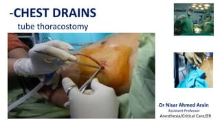
--Chest drain
--Thoracic catheter
--Tube thoracostomy
--Intercostal Drain
--It is a flexible plastic tube that is
inserted through the chest wall
between the two layers of the
pleura.
-- The other end of the tube is attached
to a drainage device placed below
chest level, allowing the air or fluid
to drain from the pleural space
5. -INDICATIONS OF
CHEST DRAIN
-PNEUMOTHORAX
-When there is Air or Gas in the pleural space.
--In any ventilated patient
--Tension pneumothorax after initial
needle relief
--Persistent or recurrent pneumothorax
after simple aspiration
--Large simple spontaneous
pneumothorax in patients over 50
years age
8. -PLEURAL EFFUSION
CHYLOTHORAX
--Lymphatic fluid in the pleural space
EMPYEMA
--Pyogenic infection of pleural space
HEMOTHORAX
--Accumulation blood in the pleural space
HYDROTHORAX
--Accumulation of serous fluid in the pleural
space
POST OPERATIVE
--For Example
a-Thoracotomy
b-Esophagectomy
c-Cardiac surgery
10. -EQUIPMENT FOR THORACOSTOMY
-All the equipment required to insert
a chest tube should be available
--Sterile gloves and gown
--Skin antiseptic solution e.g Iodine
--Sterile drapes
--Gauze swabs
--A selection of syringes and needles
(21 – 25 gauge)
--Local Anesthetic e.g Lignocaine 1%
--Scalpel and Blade
--Suture (e.g “1” silk)
--Instrument for blunt dissection
12. -INSERTION TECHNIQUE
--British Thoracic Society recommends the tube is inserted in an
area described as the “Safe Zone” a region bordered by: the
lateral border of Pectoralis Major and the Anterior border of
Latissimus dorsi and a Horizontal line superior to the Nipple.
More specially the tube is inserted Into the
5th intercostal space slightly anterior to the mid axillary line
14. --1-Inject a local Anesthetic solution into the location of the
target 4rth or 5th intercostal space
--2-On an obese patient it can be difficult to palpate the ribs.
However the 4rth or 5th rib will be approximately at a level
just above the nipple.
--3-Make a small incision through the skin
--4-Using Hemostats (Forceps) in a spreading motion to part the
skin and subcutaneous tissue widening the opening.
-INSERTION TECHNIQUE
15. -INSERTION TECHNIQUE --5-Force your finger into the opening and spread subcutaneous
tissue sufficiently to feel the ribs
--6-Using hemostats, push the closed end through the intercostal
muscles and spread the Hemostats forcefully and open to form
a hole in the intercostal muscles
--7-Push your finger back into the space, and twisting your finger
press any tissue back from the Hole
--8-Grasp the chest tube end with forceps and feed it into the
opening.
--9- Tube insertion should be made just above and parallel
to a Rib
16. -INSERTION TECHNIQUE
--10-After insertion, the tube is typically secured by
a suture and the entry area covered by dressings
“Purse string” sutures must not be used.
In the case of Acute Hemothorax, however large
bore tubes (28 – 30 F Minimum) continue to be
recommended for their dual role of Drainage of
the thoracic cavity and assesment of continuing
blood loss
17. -INSERTION TECHNIQUE
--11-The chest tube is then attached to a
drainage system which only allows one
direction of flow
-The respiratory swing in the fluid in the
chest tube is useful for assessing tube
patency and confirms the position of
the tube in the pleural cavity
-Once tube is in place, a x ray is performed
to verify the location of the drain.
19. - MANAGEMENT
OF DRAINAGE SYSTEM
-CLAMPING OF DRAIN
--A bubbling chest tube should never be
clamped.
--Drainage of a large pleural effusion should
be controlled to prevent the potential
complications of re expansion
pulmonary edema.
--***In case of Pneumothorax, clamping of
the chest tube should usually be
avoided.
20. -REMOVAL OF CHEST TUBE
--The timing of removal is dependent on the
original reason for insertion and patients
clinical progress
--In case of Pneumothorax, the chest tube
should not be clamped at the time of its
removal there is no evidence that clamping
a chest drain at the time of its removal is
beneficial
--The chest tube should be removed during
expiration with a brisk firm movement
while an assistant ties the previously placed
closure suture