1. COLLEGE OF MEDICALLABORATORYSCIENCE
CENTRALPHILIPPINE UNIVERSITY
MLS 1102a
HUMANANATOMYAND PHYSIOLOGYWITH PATHOPHYSIOLOGY
Exercise No. 5 Score:
INTEGUMENTARYSYSTEM
Group No. 2
Names: Eureka Reign R. Guihama
Edzel T. Gustilo
Natasha Lilliane D. Loot
Date Performed: November 13, 2021
Learning outcome: At the end of this exercise, the student should be able to
1.Describe the major structures of the skin and identify their functions
2.Identify important skin structures in a diagram
3.Compare and contrast the feature of thick skin and thin skin
4.Identify and describe the accessory structures of the skin (hair and nails)
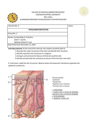
A. Instruction: Label the skin structures. Observe where the basement membrane separates the
epidermis and dermis.
*dermal papillae
*epidermis
*hypodermis
*papillary layer of dermis
*reticular layer of dermis
1. EPIDERMIS
2. PAPILLARY LAYER OF DERMIS
3.RETICULAR LAYER OF DERMIS
4. HYPODERMIS
5. DERMAL PAPILLAE
3. B. Describe the structure and function of the following parts of the skin:
1. Dermis The dermis is a fibrous structure composed of collagen, elastic tissue, and other
extracellular components that includes vasculature, nerve endings, hair follicles,
and glands. Also, dermis is a connective tissue layer sandwiched between the
epidermis and subcutaneous tissue. Its role is to support and protect the skin and
deeper layers, assist in thermoregulation, and aid in sensation.
2. Epidermis An epidermis is composed mainly of keratinocytes and it is the outermost layer
of the skin. Its thickness varies depending on which part of the body it is located.
Example on the eyelids it is thinner and is thickest on the palms and soles. Its
function is to provide a waterproof barrier and create our skin tone. Also, it
protects our body by keeping things that might be harmful to us and keeping the
things our body needs to function properly.
3. Hypodermis The hypodermis is the subcutaneous layer lying below the dermis and it consists
largely of fat. It contains the cells known as fibroblasts, adipose cells, connective
tissue and larger nerves and blood cells. It provides the main structural support
for the skin, as well as insulating the body from cold and aiding shock absorption.
4. Hair Root The hair root is in the skin and extends down to the deeper layers of the skin. It is
surrounded by the hair follicle which is also connected to a sebaceous gland. Each
hair follicle is attached to a tiny muscle that can make the hair stand up. Its main
function is the uptake of water and nutrients from the rhizosphere.
5. Hair Shaft It is formed of three layers: The medulla – the deepest layer of the hair shaft, only
seen in large and thick hairs. The cortex – the middle layer of the hair shaft which
provides the strength, color and texture of a hair fiber. The cuticle – the outer
layer of the hair shaft is thin and colorless. The primary purpose for this is to trap
a layer of air to add insulation.
6. Hair Follicle It is a stocking-like structure that contains cells and connective tissue and
surrounds the root of a hair. It exists within the dermis and the epidermis, the
two top layers of the skin. The hair follicle serves as an anchor for the hair shaft.
7. Sweat Gland Sweat glands are coiled tubular structures vital for regulating human body
temperature. It consists of a secretory unit consisting of a base rolled into a
glomerulum, and a duct that carries the sweat away.
8. Sebaceous
Gland
Sebaceous glands are usually attached to hair follicles and release a fatty
substance, sebum, into the follicular duct and then to the surface of the skin.
4. 9.Arrector Pili This is a tiny muscle that attaches to the base of a hair follicle at one end and to
dermal tissue on the other end. In order to generate heat when the body is cold,
the arrector pili muscles contract all at once, causing the hair to "stand up
straight" on the skin. It lies flat and prevents heat from being trapped by the layer
of still air between the hairs.
C. Discussion:
1. How does thin skin differ from thick skin? Explain briefly.
In terms of people, one obvious difference between a thin and a thick skinned person
is that people with thicker skin contains more adipose tissue which means there is more body
fat making a person look bigger and its skin thicker that is why fat people could tolerate an
environment with a lower temperature compared to those people who are thin-skinned.
Talking about the parts of the body, the thinnest skin covers the eyelids and most of the body,
except on the soles of the feet and palms of the hands where thick skin is present. Thick skin
has no hair follicles or sebaceous glands, whereas thin skin does. Thick skin actually has a
thinner dermis layer than thin skin, but is still thicker due to the stratum lucidum layer present
in the epidermis which protects the area most common to damages. Stratum lucidum can be
found only in thick skin not in thin skin.
5. D. Identify the nail structures on the diagram
(a)
*eponychium
*free edge
*lunula
*nail body
(b)
*eponychium
*free edge
*hyponychium
*lunula
*nail root
*nail body
*nail matrix
1. FREE EDGE
2. NAIL BODY
3. LUNULA
4. EPONYCHIUM
5. NAIL MATRIX
6. NAIL ROOT
7. EPONYCHIUM
8. LUNULA
9. NAIL BODY
10. FREE EDGE
11. HYPONYCHIUM
6. E. Describe the structure of the following parts of the nail(s):
1. Free edge The end of the nail plate that is shaped during Manicure & Pedicure. It is made
up of tightly packed, hard, keratinized epidermal cells. The nail plate leaves the
end of the finger and forms a projection that is called the free edge. This is
attached to the nail bed and appears as white. The function of the free edge is
to protect the fingertip and the hyponychium.
2. Nail body It is also made up of tightly packed, hard, keratinized epidermal cells. It is
found between the free edge and the lunula, just above the nail bed. It forms
a back-support for picking up small objects with the fingers and protects the
layer beneath it from germs to avoid infections.
3. Nail groove These are the grooves on the skin at the sides of the free edge, and the nail
follows them as a guideline when it grows.
4. Lunula The white, half-moon shaped point where the matrix and nail bed meet.
5. Eponychium
(Cuticle)
The overlapping skin surrounding the nail. It is situated between the skin of
the finger and the nail plate. It fuses these structures together and provides a
waterproof barrier. Its job is to protect the matrix from being invaded by
bacteria and physical damage.
6. Nail root This is also known as the germinal matrix. Its edge appears as a white crescent,
known as the lunula. The root portion of this nail lies below the skin,
underneath the nail, and extends several millimeters into the finger. It produces
most of the volume of the nail and the nail bed.
7. Nail bed Referred also as the sterile matrix. It extends from the edge of the nail root, or
lunula, to the hyponychium. The nail bed contains blood vessels, nerves, and
melanocytes that produce melanin. As the root grows the nail, the nail streams
down along the nail bed and adds material to the underside of the nail to make
it thicker. When the nail grows properly, the nail bed is smooth, but if the nail
doesn't grow correctly, the nail may split or develop ridges that aren't
cosmetically attractive.
8. Hyponychium The hyponychium is the area between the free edge of the nail plate and the
skin of the fingertip. It also provides a waterproof barrier.
7. 9. Nail fold The lateral nail fold overlaps the nail on the sides, helping to anchor the nail
body and to protect the nail plate edges. The nail fold that meets the proximal
end of the nail body forms the nail cuticle, also called the eponychium.