Expression profile of BRCA1 and BRCA2 genes in premenopausal Mexican women wi...
2014 Scope poster
1. Hormonal Effect on Occludin Expression in the Endometrial Adenocarcinoma HEC-1A and HEC-1B cells.
Morgan R. Gallo, Kelsey A. Rice, Taylor D. Vickers and Maria E. Cuevas
Biology Department, Southwestern University
Methods
Objectives
Results
According to the American Cancer Society1, endometrial cancer is the most
common female reproductive cancer in the United States with an incidence of 1
in 37 women. Alterations of tight junction (TJs) proteins have been reported in a
number of human cancers. Occludin is one of several TJ proteins responsible
for the proper structure and functions of TJs, including restriction of paracellular
transport and maintenance of cell polarity2. While disruption of TJs has been
associated with tumorigenesis, very few studies have investigated the role of
occludin in the development and progression of endometrial cancer. In this
study we evaluated the possible effects of estradiol and 4-hydroxytamoxifen (4-
OHT) on occludin expression and the invasive capability of HEC-1A and
HEC-1B adenocarcinoma cell lines. Results show that HEC-1A cells
overexpressed two low molecular weight (46, 58 kDa) and the expected 65 kDa
isoforms of occludin, whereas HEC-1B only expressed the 46 kDa isoform.
After treatment with 0-100 nM estradiol (E2), we observed a biphasic effect on
occludin expression on both cell lines. In contrast, when cells were treated with
4-OHT a dose-dependant inhibition on occludin expression was observed. In
addition, we observed a decrease on the invasive capability of HEC-1A and
HEC-1B with increased E2 concentration. Our data suggest that at low
concentrations, E2 promotes invasion of the HEC-1A and HEC-1B cells by
increasing the expression levels of the two low molecular weight isoforms of
occludin.
Cell Lines and Tissue Culture Conditions: Cells were cultured in their
respective medium (HEC-1A in McCoys 5A and HEC-1B in MEM)
supplemented with 10% fetal bovine serum (FBS),1% penicillin and
streptomycin and 2mM L-glutamine (PSG). Cells were maintained in a 5%CO2
atmosphere at 37°C.
Western Blot Analysis. Protein extracts were run on a pre-cast 10% SDS-PAGE gel
for occludin protein analysis. Polyacrylamide gels were then transferred to Immobilon-P
PVDF membranes. The membranes were probed for 1 h at RT with 1 µg/ml mouse-anti
occludin primary antibody (Life Technologies) in 5% milk/PBS solution. Membranes
were then probed at RT for 1 h with a 1:3000 dilution of goat-anti mouse HRP-
conjugated secondary antibody (BioRad Laboratories). For signal detection the
enhanced chemiluminescence (ECL) kit (Amersham) was used according to
manufacturer’s instructions.
The specific objectives of the present study were to determine:
1) Occludin expression levels in a panel of female cancer cell lines.
2) The possible effect of estradiol (E2) and 4-hydroxytamoxifen (4-OHT) on
occludin expression in the endometrial adenocarcinoma HEC-1A and HEC-1B
cell lines.
3) The possible effect of E2 and 4-OHT on the invasive capability of HEC-1A and
HEC-1B.
HEC-1A
46
Actin
46
58
65
kDa
A. E2 (nM)
B. 4-OHT (nM)
0 10 50 100 FBS
0 10 50 100 FBS
Actin
46
58
65
kDa
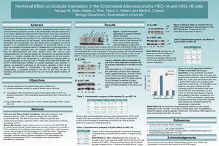
Figure 1: Levels of occludin
expression in a panel of female
cancer cell lines.
Extracts were prepared from log
phase cultures of the following cell
lines: human normal mammary
epithelial (HMEC); breast (MCF-7,
MDA-MB-231), cervical (HeLa); ovarian (SK-OV-3); and endometrial (RL95-2, HEC-1A
and HEC-1B). Equal amounts of each protein extract were subjected to SDS-PAGE
and transferred onto PVDF membrane. Following incubation with occludin antibody
we observed strong signals corresponding to 65, 58 and 46 kDa in HEC-1A and
MCF-7. Notably, HEC-1B only expressed the lower molecular weight isoform.
Actin
46
Actin
0 10 50 100 FBS
Figure 2: Biphasic effect of estradiol (A)
and 4-OHT dose-dependent inhibition (B)
on occludin expression in the HEC-1A cell
line.
Cells were plated (2 x 10⁵ cells/well) onto 6-
well plates and cultured in their respective
media, supplemented with 10%FBS/1%PSG
for 48 h. Prior to hormone exposure cells
were washed twice with PBS, serum starved
for 24 h followed by addition of media
containing charcoal stripped fetal bovine
serum (CSFBS) containing 0-100 nM E2 or 4-
OHT.
Conc (nM) 65 kDa 58 kDa 46 kDa 58 /65 kDa 46/65 kDa
0 1.0 1.0 1.0 1.0 1.0
10 1.60 ± 1.0 2.8 ± 1.85 1.20 ± 0.14 1.70 0.70
50 0.98 ± 0.3 0.44 ± 0.18 0.80 ± 0.22 0.45 0.80
100 2.44 ± 1.9 1.40 ± 0.35 1.30 ± 0.9 0.60 0.54
FBS 13.6 ± 15.6 2.00 ± 0.23 0.70 ± 0.3 0.14 0.50
Table 1: Densitometric analysis of the biphasic E2 on HEC-1A
A. E2 (nM)
46
Actin
0 10 50 100 FBS
B. 4-OHT (nM)
Figure 3: Biphasic effect of estradiol (A) and
4-OHT dose-dependent (B) expression in the
HEC-1B cell line. The experiment was
conducted as described in figure 2.
Table 3: Densitometric analysis the effects of
E2 and 4-OHT on HEC-1B
Conc (nM) 46 kDa
0 1.0
10 2.0 ± 0.6
50 1.54 ± 0.7
100 1.63 ± 0.5
FBS 2.4 ± 1.5
Table 2: Densitometric analysis of 4-OHT effect on HEC-1A
Conc (nM) E2 4-OHT
46 kDa
0 1.0 1.0
10 1.0 1.0
50 0.96 0.80
100 1.190 0.74
FBS 1.39 1.1
MAIN RESULTS: HEC-1A Experiment: We observed an
increase in the lower molecular weight (58 and 46 kDa) occludin
isoforms at 10 nM E2. The same concentration also increased
the ratio of 58/65 kDa by 11-fold and the 46/65 kDa by 14-fold.
MAIN RESULTS: Notably, we only
observed the 46 kDa isoform of
occludin. However, we again
observed the estrogen biphasic and
4-OHT dose-dependent inhibition of
occludin expression.
kDa
0 10 50 100
Estradiol concentration (nM)
ND
0.1
0.2
0.3
0.4
0.5
Absorbance(560nm)
HEC-1A
HEC-1B
Figure 4: E2 dose-dependent inhibition
on HEC-1A and HEC-1B invasion
capabilities. Invasive potential was tested
using a Boyden Chamber with 8 µm pore size
polycarbonate membrane coated with ECMatrix
(EMD, Inc.). Cells were serum starved for 48 h
in phenol free medium prior to plating. Cell
suspension containing 3 x 10⁵ cells/300 µl was
loaded on the upper chamber and FBS
containing medium was added to the lower
chamber as the chemoattractant. E2 and
4-OHT (0-100 nM) were diluted in medium
before plating. Cells were incubated for 24 h
at 37oC in 5%CO2 atmosphere. Inserts were
removed and the cells on the lower chamber
were stained and collected following the
manufacturer’s instructions.
MAIN RESULTS: We observed a dose-dependent inhibition on the invasive
capabilities in both cell lines . At 10 nM E2 concentration, HEC-1B appeared to
exhibit the highest invasive potential. Furthermore, the inhibitory effect seems to be
more pronounced in the HEC-1B cell line.
Abstract
Western blots were quantified by computer-aided determination of OD using
ImageJ (NIH) and normalized to actin. Means (± SD) of three experiments
in each point of changes in densitometry of occludin 65, 58 and 46 kDa
isoforms in response to treatments with E2.
Means (± SD) of two experiments in each point of changes
in densitometry of occludin 46 kDa isoform in response to 4-
OHT.
References
1. American Cancer Society (online). Available:
http://www.cancer.org/cancer/endometrialcancer/detailedguide/endometrial-uterine-
cancer-key-statistics (last updated 02/03/2014)
2. Schneeberger EE and Lynch RD. The tight junction: a multifunctional complex. Am J
Physiol Cell Phsysiol 2004; 286:1213-1228.
Acknowledgements
We would like to thank the Howard Hughes Medical Institute Grant (HHMI) and
Southwestern University for their support of the SCOPE program.