Recommended

Recommended
More Related Content
Similar to MonicaGiraldoChica_Atlanta2016_ABIDE_v4
Similar to MonicaGiraldoChica_Atlanta2016_ABIDE_v4 (20)
MonicaGiraldoChica_Atlanta2016_ABIDE_v4
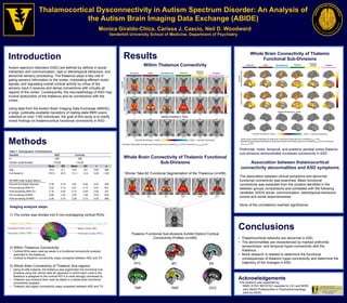
- 1. Introduction Autism spectrum disorders (ASD) are defined by deficits in social interaction and communication, rigid or stereotypical behaviors, and abnormal sensory processing. The thalamus plays a key role in gating sensory information to the cortex, modulating efferent motor signals, and regulating overall cortical activity by virtue of the sensory input it receives and dense connections with virtually all regions of the cortex. Consequently, the neuropathology of ASD may involve dysfunction of the thalamus and its connections with the cortex. Using data from the Autism Brain Imaging Data Exchange (ABIDE), a large, publically-available repository of resting-state fMRI scans collected on over 1100 individuals, the goal of this study is to clarify mixed findings on thalamocortical functional connectivity in ASD. Thalamocortical Dysconnectivity in Autism Spectrum Disorder: An Analysis of the Autism Brain Imaging Data Exchange (ABIDE) Monica Giraldo-Chica, Carissa J. Cascio, Neil D. Woodward Vanderbilt University School of Medicine, Department of Psychiatry Conclusions • Thalamocortical networks are abnormal in ASD. • The abnormalities are characterized by marked prefrontal, sensorimotor, and temporal hyper-connectivity with the thalamus. • More research is needed to determine the functional consequences of thalamic hyper-connectivity and determine the trajectory of the changes in ASD. Acknowledgements Methods Table 1. Demographic Characteristics Variable ASD Controls N 228 228 Gender (male:female) 119:29 119:29 Mean SD Mean SD t p Age 16.6 6.1 16.6 6.0 0.65 .948 Full-Scale IQ 101.9 22.0 111.3 13.4 5.45 <.001 RS-fMRI Data Quality Metrics Percent Scrubbed Volumes 5.34 5.19 4.91 5.00 0.91 .362 Pre-scrubbing RMS FD 0.22 0.13 0.21 0.12 0.47 .641 Post-scrubbing RMS FD 0.16 0.05 0.15 0.04 0.93 .351 Pre-scrubbing DVARS 2.95 1.03 2.97 1.02 0.23 .816 Post-scrubbing DVARS 2.25 0.74 2.25 0.73 0.05 .960 Imaging analysis steps: 1) The cortex was divided into 6 non-overlapping cortical ROIs: 2) Within Thalamus Connectivity: • Cortical ROIs were used as seeds in a functional connectivity analysis restricted to the thalamus; • Cortical-to-thalamic connectivity maps compared between ASD and TD 3) Whole Brain Connectivity of Thalamic Sub-regions: • Using all 456 subjects, the thalamus was segmented into functional sub- divisions using the ‘winner take all’ approach in which each voxel in the thalamus is assigned to the cortical ROI it is most strongly connected to; • Thalamic sub-divisions then used as seeds in a whole-brain functional connectivity analysis; • Thalamic sub-region connectivity maps compared between ASD and TD Temporal Cortex (TMP) Prefrontal Cortex (PFC) Motor Cortex (MT) Somatosensory Cortex (SS)Parietal Cortex (PAR) Occipital Cortex (OCC) Results PFC MT SS TMP PAR OCC Whole Brain Connectivity of Thalamic Functional Sub-Divisions Prefrontal, motor, temporal, and posterior parietal cortex thalamic sub-divisions demonstrated increased connectivity in ASD. Association between thalamocortical connectivity abnormalities and ASD symptoms The association between clinical symptoms and abnormal functional connectivity was examined. Mean functional connectivity was extracted from the clusters identified in the between groups comparisons and correlated with the following variables: ADOS social, communication, stereotypical behaviors scores and social responsiveness. None of the correlations reached significance. This research was supported by: - NIMH (5 R21 MH101321 awarded to CJC and NDW) - Jack Martin Professorship in Psychopharmacology (held by NDW). Within Thalamus Connectivity All results thresholded at cluster-level Family-wise error corrected p(FWE)=.05 for voxel-wise p(uncorrected)=.001. Whole Brain Connectivity of Thalamic Functional Sub-Divisions ‘Winner Take All’ Functional Segmentation of the Thalamus (n=456) Thalamic Functional Sub-divisions Exhibit Distinct Cortical Connectivity Profiles (n=456) Within group results thresholded at whole-brain voxel-level Family-wise error corrected p(FWE)=.001. Between group results thresholded at whole-brain cluster-level Family-wise error corrected p(FWE)=.05 for voxel-wise p(uncorrected)=.001.