AACR 2015 Poster 04-16-2015 2005 hrs
•Download as PPTX, PDF•
0 likes•204 views

Recommended

Recommended
More Related Content
What's hot
What's hot (20)
Similar to AACR 2015 Poster 04-16-2015 2005 hrs
Similar to AACR 2015 Poster 04-16-2015 2005 hrs (20)
More from Mohammed Talha Shekhani, MD
More from Mohammed Talha Shekhani, MD (8)
AACR 2015 Poster 04-16-2015 2005 hrs
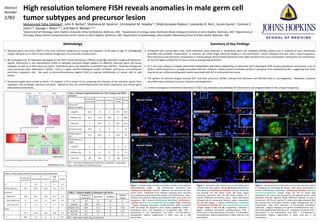
- 1. High resolution telomere FISH reveals anomalies in male germ cell tumor subtypes and precursor lesion Mohammed Talha Shekhani1, John R. Barber4, Stephania M. Bezerra1, Christopher M. Heaphy1,3, Nilda Gonzalez Roibon1, Leonardo O. Reis1, Gunes Guner1, Corinne E. Joshu3,4, George J. Netto1,2,3, and Alan K. Meeker1,2,3 1Department of Pathology, Johns Hopkins University School of Medicine, Baltimore, MD; 2Department of Urology, James Buchanan Brady Urological Institute at Johns Hopkins, Baltimore, MD; 3Department of Oncology, Sidney Kimmel Comprehensive Cancer Center at Johns Hopkins, Baltimore, MD; 4Department of Epidemiology, Johns Hopkins Bloomberg School of Public Health, Baltimore, MD Methodolgy Testicular germ cell tumor (TGCT) is the most common malignancy of young men between 15-40 years of age. It is biologically unique, because it is in cells of non-somatic lineage and is an unusually curable cancer. We evaluated over 70 specimens (arranged on two TGCT tissue microarrays (TMAs)) using high resolution (single-cell) telomere- specific fluorescent in situ hybridization (FISH) to delineate telomere length patterns in different testicular germ cell tumor subtypes, as well as in their precursor lesion, intratubular germ cell neoplasia unclassified type (IGCNU). Telomere biology has never previously been addressed in IGCNU. Oct4 is a highly specific histological marker of IGCNU, seminoma, and embryonal carcinoma neoplastic cells. We used co-immunofluorescence against Oct4 to improve identification of cancer cells in said lesions. Telomere lengths were scored as short (=1), medium (=2) or long (=3) by comparing the intensity of the telomere signals from neoplastic cells to benign reference cell types. Reference cells are interstitial/stromal cells (short telomeres) and normal germ cells (medium telomeres). Abstract Number: 3783 Summary of Key Findings Compared with normal germ cells, short telomeres were observed in intratubular germ cell neoplasia (IGCNU) lesions and in majority of pure seminomas (p=0.006 and p=0.0005, respectively). In contrast, we noted longer telomere lengths in non-seminomas—cancer subtypes that also carry a worse prognosis. Non-seminoma and seminoma components in mixed subtype demonstrated telomeres with slight variation from pure counterparts. Among the non-seminomas, EC has the highest proportion of cases scored as having long telomeres. EC is the only subtype to display telomerase-independent alternative lengthening of telomeres (ALT) phenotype (43% of pure embryonal carcinomas). Loss of ATRX or DAXX expression is strongly associated with ALT. However, neither protein anomalies existed in sub-group of EC evaluated by IHC—suggesting that there may be (as yet undiscovered) genetic events associated with ALT in embryonal carcinoma. The pattern of telomere lengths among TGCT and their precursor, IGCNU, indicate that telomeres are affected early in carcinogenesis. Moreover, telomere anomalies may contribute to tumor initiation and progression. Further investigation of telomeric aberration in TGCT may delineate new pathways for intervention and prognostication in this unique malignancy. Table 2. Telomere length distributions for TGCT subtypes and IGCNU precursor Embryonal Carcinoma Seminoma Teratoma Yolk Sac Tumor Mixed Cancer cell telomere lengths Long (%) 42.9 14 0 33.3 N/A Medium (%) 14.3 27.9 100 0 N/A Short (%) 0 58.1 0 66.7 N/A ALT Present (%) 42.9 0 0 0 15 IGCNU Present (%) 71.4 58.1 0 33.3 70 IGCNU telomere length Long (%) 0 8 0 0 0 Medium (%) 0 8 0 0 7.1 Short (%) 100 84 0 100 92.9 ALT = alternative lengthening of telomeres; and IGCNU = intratubular germ cell neoplasia of unclassified type Table 3. Telomere lengths in mixed germ cell tumors Embryonal Carcinoma Seminoma Teratoma Yolk Sac Tumor N 18 9 12 9 Long (%) 38.9 66.7 0 0 Medium (%) 38.9 11.1 25 77.8 Short (%) 5.6 22.2 75 22.2 ALT (%) 16.7 0 0 0 ALT = alternative lengthening of telomeres Figure 1. Example image a healthy seminiferous tubule and an adjacent IGCNU/diseased tubule. (A) DAPI/Nuclear Stain/Blue and Cy3/Telomere-FISH/Red channels. Telomeres are punctate red dots within nuclei. Filled/solid arrow indicates neoplastic germ cell with short telomeres (nearly absent FISH signal) in the IGCNU tubule. Open- headed arrow indicates healthy spermatogonial stem cell’s nucleus for comparison. (B) 3 channel (DAPI/Nuclear Stain/Blue, Cy3/Telomere- FISH/Red and Oct4 immunostaining/Green) merged image. *(Asterisk) = Region displaying non-specific autofluorescence (often associated with red blood cell fragments from nearby capillaries); IGCNU = Intratubular Germ Cell Neoplasia of Unclassified type; FISH = fluorescence in situ hybridization; and DAPI = 4',6-diamidino-2- phenylindole. Original magnification is 200X, scale bar is 100 micrometers. Figure 2. Seminoma cells compared to normal healthy germ cells from the same patient. (A) and (B) Display DAPI/Nuclear Stain/Blue and Cy3/Telomere-FISH/Red channels. Telomeres are punctate red dots within nuclei. (A) Shows region containing seminoma cells and (B) shows contents of a healthy unaffected seminiferous tubule. Insets demonstrate enlarged cells for unequivocal telomere length comparisons. (C) and (D) Display 3 channel (DAPI/Nuclear Stain/Blue, Cy3/Telomere-FISH/Red and Oct4 immunostaining /Green) merged images of (A) and (B), respectively. *(Asterisk) = Region displaying non-specific autofluorescence; FISH = fluorescence in situ hybridization; and DAPI = 4',6-diamidino- 2-phenylindole. Original magnification is 400X, scale bar is 50 micrometers. Figure 3. Comparison of an embryonal carcinoma (A, top) with another EC displaying ALT phenotype (B, below). Both panels demonstrate 3 channel (DAPI/Nuclear Stain/Blue, Cy3/Telomere-FISH/Red and Oct4 immunostaining/Green) merged image. (A) This EC shows long telomeres in the cancer cells compared to the adjacent stroma. Inset delineates dramatic telomere length difference between EC and a stromal cell. (B) This ALT positive EC shows ultra-bright telomeric DNA foci (arrows) and intracellular telomere length heterogeneity that is characteristic. Note short telomeres in surrounding interstitium. *(Asterisk) = Region displaying non-specific autofluorescence; ALT = alternative lengthening of telomeres; EC = embryonal carcinoma; FISH = fluorescence in situ hybridization; and DAPI = 4',6-diamidino-2- phenylindole. Original magnification is 400X, scale bar is 50 micrometers.