1543 2165%282004%29128-1279%3 apcsasc-2%2e0%2eco%3b2
•
1 like•223 views

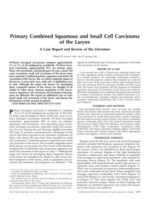
![A, Full-thickness squamous dysplasia consistent with squamous cell carcinoma in situ (hematoxylin-eosin, original magnification 3100). B and C,
A dense submucosal proliferation (B) of hyperchromatic neoplastic cells with minimal cytoplasm and nuclear molding (C) (hematoxylin-eosin,
original magnifications 3100 [B] and 3200 [C]). D, Strong cytokeratin (AE1/AE3) immunoreactivity in the small cell carcinoma component (original
magnification 3200). E, Strong CD56 immunoreactivity (original magnification 3400). F, Focal chromogranin immunoreactivity (original magni-fication
3400).
COMMENT
Combined small cell carcinoma, as defined by the In-ternational
Association for the Study of Lung Cancer, con-sists
of small cell carcinoma with a component of squa-mous
cell carcinoma or adenocarcinoma.11 Combined pri-mary
squamous and small cell carcinoma of the larynx is
rare, and only 13 cases have been reported in the literature
to date.3–10 The clinical features of these cases of combined
small cell carcinoma of the larynx, together with those of
the current case, are summarized in the Table. They have
clinical features similar to those of small cell carcinoma.
All patients were men within an age range of 41 to 83
years (median, 55 years), and all had a significant history
of smoking (Table). The presenting symptoms included
1280 Arch Pathol Lab Med—Vol 128, November 2004 Combined Small Cell Carcinoma of Larynx—Jaiswal & Hoang](data:image/gif;base64,R0lGODlhAQABAIAAAAAAAP///yH5BAEAAAAALAAAAAABAAEAAAIBRAA7)
Recommended
Recommended
More Related Content
What's hot
What's hot (19)
Viewers also liked
Viewers also liked (20)
Similar to 1543 2165%282004%29128-1279%3 apcsasc-2%2e0%2eco%3b2
Similar to 1543 2165%282004%29128-1279%3 apcsasc-2%2e0%2eco%3b2 (20)
1543 2165%282004%29128-1279%3 apcsasc-2%2e0%2eco%3b2
- 1. Primary Combined Squamous and Small Cell Carcinoma of the Larynx A Case Report and Review of the Literature Vilkesh R. Jaiswal, MD; Mai P. Hoang, MD c Primary laryngeal carcinomas comprise approximately 2% to 5% of all malignancies worldwide. Of these laryn-geal carcinomas, approximately 99% are primary squa-mous cell carcinomas. During the past 30 years, about 160 cases of primary small cell carcinoma of the larynx have been reported. Combined primary squamous and small cell carcinoma of the larynx, the so-called composite tumor of the larynx, is even more rare, with only 13 published cases to date. Although the major risk factors for developing these composite tumors of the larynx are thought to be similar to other more common neoplasms of the larynx, such as squamous cell carcinoma, the treatment and prog-nosis are different. We report an additional case of com-bined small cell carcinoma of the larynx and discuss the histogenesis of this unusual neoplasm. (Arch Pathol Lab Med. 2004;128:1279–1282) Primary laryngeal carcinoma is estimated to comprise 2% to 5% of all malignancies worldwide. In the Unit-ed States, this translates to about 12 500 new cases of pri-mary laryngeal carcinomas annually. Of these laryngeal carcinomas, approximately 99% or more are primary squamous cell carcinomas.1 The remaining types of pri-mary laryngeal carcinomas, especially small cell carcino-ma, are rare. During the past 30 years, slightly more than 500 cases of primary neuroendocrine carcinomas of the larynx have been reported in the literature worldwide. Of these, only about 160 cases have been classified as small cell carcinomas.2 Combined primary squamous and small cell carcinoma of the larynx, the so-called composite tumor of the larynx, is even more rare. Our review of the literature revealed only 13 published cases of primary squamous and small cell carcinoma of the larynx to date.3–10 Although the major risk factors for developing these composite tumors of the larynx are thought to be similar to other more com-mon neoplasms of the larynx, such as squamous cell car-cinoma, the treatment and prognosis are different.1–10 We Accepted for publication June 30, 2004. From the Department of Pathology, The University of Texas South-western Medical Center, Dallas. The authors have no relevant financial interest in the products or companies described in this article. Corresponding author: Mai P. Hoang, MD, Department of Pathology, The University of Texas Southwestern Medical Center, 5323 Harry Hines Blvd, Dallas, TX 75390-9073 (e-mail: mai.hoang@utsouthwestern.edu). report an additional case of primary squamous and small cell carcinoma of the larynx. REPORT OF A CASE A 41-year-old man with a 20-pack-year smoking history and no other significant medical history presented with hoarseness of 6 months’ duration. An endoscopic examination revealed a lesion in the left posterior ventricle that extended up to the left false vocal cord. At the same time, a bulky right laryngeal lesion was seen involving both the laryngeal ventricle and false vocal cord. The lesions were biopsied, and the diagnosis of combined squamous and small cell carcinoma of the larynx was rendered. Both chest radiography and computed tomography demonstrated no evidence of pulmonary disease. The patient underwent 2 cy-cles of chemotherapy, including cisplatin and etoposide, and is currently undergoing radiotherapy. He is alive 8 months status post diagnosis. MATERIALS AND METHODS Four-micrometer-thick sections were cut from the paraffin blocks and stained with hematoxylin-eosin. Additional paraffin sections were obtained for immunohistochemical studies, which were performed by avidin-biotin peroxidase complex technique, the heat-induced epitope retrieval buffer, and primary antibodies against cytokeratins (AE1/AE3, 1:50, Zymed Laboratories, South San Francisco, Calif), CD56 (1:10, Monosan, San Francisco, Calif), chromogranin (1:700, Dako Corporation, Carpinteria, Calif), CD20 (L26, 1:40, Signet Laboratory, Dedham, Mass), CD3 (1:200, Dako), and leukocyte common antigen (1:40, Dako). Appropriate positive and negative controls were included. PATHOLOGIC FINDINGS Biopsies from the left true vocal cord and left laryngeal ventricle revealed full-thickness squamous dysplasia con-sistent with squamous cell carcinoma in situ (Figure, A). No invasive tumor was identified on these small biopsy specimens. In contrast, biopsies of the right hemilarynx revealed a submucosal and dense infiltrate of neoplastic cells arranged in solid nests and sheets with minimal in-tervening stroma (Figure, B). The neoplastic cells were pleomorphic with hyperchromatic nuclei, high nuclear-cy-toplasmic ratio, and marked crushed artifact (Figure, C). There was no glandular or squamous differentiation pres-ent in this component of the tumor. These neoplastic cells strongly expressed cytokeratins (AE1/AE3) (Figure, D) and CD56 (Figure, E), and focally expressed chromogran-in (Figure, F). They were negative for CD20, CD3, and leukocyte common antigen. Based on these findings, the diagnosis of combined squamous and small cell carcino-ma of the larynx was rendered. Arch Pathol Lab Med—Vol 128, November 2004 Combined Small Cell Carcinoma of Larynx—Jaiswal & Hoang 1279
- 2. A, Full-thickness squamous dysplasia consistent with squamous cell carcinoma in situ (hematoxylin-eosin, original magnification 3100). B and C, A dense submucosal proliferation (B) of hyperchromatic neoplastic cells with minimal cytoplasm and nuclear molding (C) (hematoxylin-eosin, original magnifications 3100 [B] and 3200 [C]). D, Strong cytokeratin (AE1/AE3) immunoreactivity in the small cell carcinoma component (original magnification 3200). E, Strong CD56 immunoreactivity (original magnification 3400). F, Focal chromogranin immunoreactivity (original magni-fication 3400). COMMENT Combined small cell carcinoma, as defined by the In-ternational Association for the Study of Lung Cancer, con-sists of small cell carcinoma with a component of squa-mous cell carcinoma or adenocarcinoma.11 Combined pri-mary squamous and small cell carcinoma of the larynx is rare, and only 13 cases have been reported in the literature to date.3–10 The clinical features of these cases of combined small cell carcinoma of the larynx, together with those of the current case, are summarized in the Table. They have clinical features similar to those of small cell carcinoma. All patients were men within an age range of 41 to 83 years (median, 55 years), and all had a significant history of smoking (Table). The presenting symptoms included 1280 Arch Pathol Lab Med—Vol 128, November 2004 Combined Small Cell Carcinoma of Larynx—Jaiswal & Hoang
- 3. Clinical and Pathologic Data of 14 Cases of Combined Squamous and Small Cell Carcinoma of the Larynx Case No. Source, y Age, y/Sex Site Positive Lymph Nodes Distant Metastasis Therapy Follow-up 1 Yucel et al,3 2000 32/M Supraglottic 1 2 SL, MND, chemotherapy AWD, 1 y 2 Gianoli et al,4 1992 83/M Right supraglottic 2 2 TL, MND NED, 8 mo 3 Cosby and Babin,5 1988 56/M Left supraglottic 1 2 TL, RND, chemotherapy Lost to follow-up 4 Chen et al,6 1986 55/M Right hemilarynx, left supraglottic 1 2 TL, RND, chemotherapy, XRT DOD, 9 mo 5 Chen et al,6 1986 55/M Right supraglottic 2 2 SL, RND, chemotherapy AWD, 13 mo 6 Ferlito et al,7 1985 Gnepp et al,8 1983 57/M Right pyriform sinus 1 1 TL, RND, XRT DOD, 3.5 mo 7 Ferlito et al,7 1985 56/M Epiglottis 2 2 Chemotherapy, XRT NED, 5 y 8 Ferlito et al,7 1985 45/M Right aryepiglottic fold and pyriform sinus 1 2 Chemotherapy, XRT DOD, 21 mo 9 Ferlito et al,7 1985 47/M Right aryepiglottic fold and pyriform sinus 1 2 Chemotherapy, XRT AWD, 39 mo 10 Ferlito et al,7 1985 54/M Anterior commisure, vo-cal cords, subglottic 2 2 TL, RND NED, 2 y 11 Ferlito et al,7 1985 50/M Left hemilarynx 1 NA TL, RND, XRT DOD, 14 mo 12 Mills et al9, 1983 49/M Right pyriform sinus 1 2 TL, RND, XRT NED, 6 mo 13 Eubesi et al,10 1978 63/M Right hemilarynx 1 2 HL, RND AWD, 10 mo 14 Current case 41/M Left vocal cord, right hemilarynx NA 2 Chemotherapy, XRT NED, 8 mo * Plus sign indicates metastasis present; minus sign, metastasis absent; NA, information not available; SL, supraglottic laryngectomy; MND, modified neck dissection; TL, total laryngectomy; RND, radical neck dissection; XRT, radiation therapy; HL, hemilaryngectomy; AWD, alive with disease; NED, no evidence of disease; and DOD, died of disease. hoarseness, dysphagia, and in some cases, a neck mass. Although this composite tumor has been reported at var-ious locations in the larynx, the supraglottic region is the most frequently involved site—similar to primary laryn-geal small cell carcinoma.8 Paraneoplastic processes, such as Eaton-Lambert syndrome and syndrome of inappro-priate secretion of antidiuretic hormone, may occur in as-sociation with laryngeal small cell carcinoma, but these syndromes have not been reported with combined small cell carcinoma of the larynx.12 The clinical course of combined small cell carcinoma is similar to that of small cell carcinoma of the larynx; it displays aggressive biologic behavior with early distant metastasis as a rule.3–10 Most patients were given multi-modality therapy, which included a combination of sur-gery, radiation therapy, and chemotherapy (Table). Al-though survival for more than 3 years has been reported, most patients die within 2 years of the diagnosis, even with adjuvant radiation and chemotherapy (Table). In combined carcinoma of the larynx, small cell carci-noma was commonly associated with a synchronous and invasive squamous cell carcinoma. An in situ squamous cell carcinoma was seen in one case reported by Ferlito et al7 and in the current case. However, only small biopsies and no resections were performed on these patients, there-fore an invasive squamous cell carcinoma cannot be ex-cluded. Before rendering a diagnosis of a combined squa-mous and small cell carcinoma of the larynx, it is impor-tant to ascertain through clinical workup that the small cell carcinoma component is not a metastasis from either a gastrointestinal or bronchogenic origin. Other neuroen-docrine neoplasms, such as carcinoid tumor, atypical car-cinoid tumor, pure small cell carcinoma, and large cell neuroendocrine carcinoma, should be considered in the differential diagnosis. Nonkeratinizing squamous cell car-cinoma must be excluded. Basaloid squamous cell carci-noma is also a consideration; however, this tumor would show large epithelial lobules with comedo-like central ne-crosis, abundant stroma with hyalinization along the tu-mor nests, and lack of CD56 expression.13 In these settings, a panel of appropriate immunohistochemical stains would be helpful. It is believed that the neuroendocrine Kulchitsky cell (or argyrophilic cell) found in the mucosa of the larynx and other endodermally derived tissue is the histogenetic pre-cursor of small cell carcinoma of the larynx. Combined small cell carcinoma has been described in the lung, tra-chea, and esophagus.14–16 It has been speculated that these biphasic neoplasms are derived from a common neoplastic stem cell that can differentiate into the Kulchitsky cells (neuroendocrine cells) as well as squamous or glandular cells in the appropriate microenvironment.3 This hypoth-esis is not unlikely, since the neural crest cells have been shown to have broad differentiating capability.17 These findings lend support to the hypothesis that neoplastic cells of tumors such as ours, which display both squamous and neuroendocrine differentiation, are derived from common pluripotential stem cells. References 1. Fu Y-S, Wenig BM, Abemayor E, Wenig BL. Head and Neck Pathology. Phil-adelphia, Pa: Churchill-Livingstone; 2001:330–455. 2. Ferlito A, Barnes L, Rinaldo A, Gnepp DR, Milroy CM. A review of neuro-endocrine neoplasms of the larynx: update on diagnosis and treatment. J Laryngol Otol. 1998;112:827–834. 3. Yucel OT, Sokmensuer C, Gedikoglu G, Ayas K. Combined small cell and squamous cell carcinoma of the larynx: short communication. Tumori. 2000;86: 434–436. 4. Gianoli GJ, Butcher RB, Martin EJ. Composite tumor of the larynx. Ear Nose Throat J. 1992;71:81–82, 85–87. 5. Cosby WN, Babin RW. Simultaneous oat cell and squamous cell carcinoma of the larynx. Military Med. 1988;153:196–198. 6. Chen DA, Mandell-Brown M, Moore SF, Johnson JT. ‘‘Composite’’ tumor: mixed squamous cell and small cell anaplastic carcinoma of the larynx. Otolar-yngol Head Neck Surg. 1986;95:99–103. 7. Ferlito A, Recher G, Caruso G. Primary combined small cell carcinoma of the larynx. Am J Otolaryngol. 1985;6:302–308. 8. Gnepp DR, Ferlito A, Hyams V. Primary anaplastic small cell (oat cell) car-cinoma of the larynx: review of the literature and report of 18 cases. Cancer. 1983;51:1731–1745. 9. Mills SE, Cooper PH, Garland TA, Johns ME. Small cell undifferentiated Arch Pathol Lab Med—Vol 128, November 2004 Combined Small Cell Carcinoma of Larynx—Jaiswal & Hoang 1281
- 4. carcinoma of the larynx: report of two patients and review of 13 additional cases. Cancer. 1983;51:116–120. 10. Eusebi V, Betts CM, Giangaspero F. Primary oat cell carcinoma of the lar-ynx. Virchows Arch A Pathol Anat Histol. 1978;380:349–354. 11. Hirsch FR, Matthews MJ, Aisner S, et al. Histopathologic classification of small cell lung cancer: changing concepts and terminology. Cancer. 1988;62: 973–977. 12. Medina JE, Moran M, Geopfert H. Oat cell carcinoma of the larynx and Eaton-Lambert syndrome. Arch Otolaryngol. 1984;110:123–126. 13. Cho KJ, Jang JJ, Lee SS, Zo JI. Basaloid squamous carcinoma of the oe-sophagus: a distinct neoplasm with multipotential differentiation.Histopathology. 2000;36:331–340. 14. Mangum MD, Greco FA, Hainsworth JD, Hande KR, Johnson DH. Com-bined small-cell and non-small-cell lung cancer. J Clin Oncol. 1989;7:607–612. 15. Thornley PE, Howatson SR. Primary carcinoma of the trachea: mixed squa-mous and oat cell type. Thorax. 1980;35:72–73. 16. Fujiwara Y, Nakagawa K, Tanaka T, Utsunomiya J, Nishigami T, Uematsu K. Small cell carcinoma of the esophagus combined with superficial esophageal cancer. Hepatogastroenterology. 1996;43:1360–1369. 17. Clarke DL, Johansson CB, Wilbertz J, et al. Generalized potential of adult neural stem cells. Science. 2000;288:1660–1663. 1282 Arch Pathol Lab Med—Vol 128, November 2004 Combined Small Cell Carcinoma of Larynx—Jaiswal & Hoang