1. Cytocompatibility of Magnesium Alloys With Human Urothelial Cells
Michael Deo1, Qiaomu Tian1 and Huinan Liu1,2
1Department of Bioengineering, 2Materials Science and Engineering
University of California, Riverside, CA, 92521
Acknowledgments:
Discussion:
References:
Results: Cellular Characterization
Results: Substrate Characterization
Introduction
Method:
24hrs48hrs
AZ31_O AZ31_P Mg_PMg_OMg_Y_PMg_Y_O
0hrs
48hours
GlassAZ31_O AZ31_P Mg_PMg_OMg_Y_P CellsPUMg_Y_O
24hours
Magnesium Substrates
Polished, Sonicated,
and Sterilized
• Mg_Y_O
• M_Y_P
• AZ31_O
• AZ31_P
• Mg_O
• Mg_P
• Glass
• Polyurethane
Substrates (Oxidized and Polished):
Magnesium Substrates
and Controls Placed
into Porous Transwells,
as Prescribed
Cells and Transwell
with Substrate
Combined; culture
period (24, 48hrs)
Cell Characterization: Fluorescence Imaging
Substrate Characterization: SEM/EDS
Cellular Media Characterization: ICP-OES, pH meter
Collect Data/Images
and Conduct Statistical
Analysis
Two Study Periods:
24 and 48 hour period; Conduct each period in
triplicate
HUC Grown and
Cultured in vitro in
Flask
Cells Trypsinized and
Seeded into Well
Plates
Transwell
Single Well
Cellular Media
Substrate
Porous
Membrane
HUCs
Discussion:
Results: Cellular Media Characterization
M
g_Y_OM
g_Y_PAZ31_OAZ31_PM
g_O
M
g_P
PU
G
lass
C
ellsM
edia
7.0
7.5
8.0
8.5 24 hrs
48 hrs
#
#
pH
M
g_Y_OM
g_Y_PAZ31_OAZ31_P
M
g_O
M
g_P
PU
G
lass
CellsM
edia
0.0
2.0
4.0
6.0
24 hrs
48 hrs
*
*
#
#
Mg
2+
Concentration(100mg/L)
M
g_Y_O
M
g_Y_P
AZ31_O
AZ31_P
M
g_O
M
g_P PU
G
lass
C
ells
0.0
0.5
1.0
1.5
2.0
2.5
24 hrs
48 hrs
*
*#
#
CellDensity(Final/Initial)M
g_Y_OM
g_Y_PAZ31_OAZ31_P
M
g_O
M
g_P
PU
G
lass
0.75
1.00
1.25
24 hrs
48 hrs
#
#
MassRatio(Final/Initial)
Glass
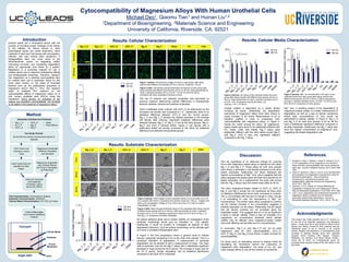
Figure 3 (above): SEM images of each substrate before (0 hrs) and after (24 or 48
hrs) cultured with HUCs in respective time periods. Scale bar: 100 µm. Images were
taken at an acceleration voltage of 10 kV and a spot size of 4.5 with an original
magnification of ×500.
Figure 1 (above): Fluorescence images of HUCs in each group after being
cultured with different time periods (24 hrs or 48 hrs). Scale bar: 100 µm
Figure 5 (above): pH values of the collected media from each
group after being cultured with different time periods (24 hrs or
48 hrs). Data represented as mean ± standard deviation (N=3).
p<0.05; # for comparisons among the alloys, not
controls;┌─#─┐ for 48 hrs.
Ureteral stents are a biomedical device with the
purpose of providing proper drainage of the kidney
to the bladder, as kidney stones or other
physiological issues can cause obstruction. Stent
materials of past have had issues with encrustation,
infection, and pain among other symptoms. A
biodegradable stent can solve some of the
aforementioned issues by degrading safely,
minimizing corrosion, and completing degradation
within an appropriate time frame for a patient.
Magnesium is a promising material for use in
ureteral stents due to observed antibacterial effects
and biodegradable properties. Therefore, research
into magnesium as a potential biocompatible alloy
for ureteral stent use is warranted. Alloys for this
study were chosen on the basis of favorable
features such as the antibacterial properties of
magnesium yttrium (Mg_Y). Thus, this research
seeks to establish firm research on the
cytocompatible effects of magnesium alloys on in
vitro human urothelial cells (HUCs) using the
hypothesis: HUCs density, an indication of cell
viability and therefore cytcompatibility, will increase
or be stable in the presence of magnesium alloys.
The null hypothesis of no observed change for culturing
HUCs with magnesium-based alloys is rejected for the alloys
Mg_Y_O and Mg_Y_P. These alloys for both time periods
have shown a decrease in cell density compared to the other
tested substrates. Additionally, the alloys displayed the
highest concentrations of Mg2+ ions, which suggests that the
alloys degraded the fastest given the fixed time periods for all
tested substrates. As a supplemental, the mass ratio shows
that the Mg_Y alloys have the lowest mass ratios at 48 hrs.
The other magnesium-based metals of AZ31_O, AZ31_P,
Mg_O, and Mg_P accept the null hypothesis as there were
no statistical significances found when compared to controls.
This is corroborated with the lack of change in mass, though
it is interesting to note the discrepancy in Mg2+ ion
concentrations. The similar mass ratios compared to controls
yet the marked increase in ion concentration suggests a
possible deposition on the alloys. Additionally, the pH values
and cell density numbers are not consistent given the
substrates tested. This suggests that pH is not as significant
a factor in cellular viability. There is also an indication of a
magnesium ion concentration threshold where cellular
viability may be affected since magnesium-based metals
have both differing cell densities and ion concentrations.
In conclusion, Mg_Y_O and Mg_Y_P are not an ideal
magnesium alloy for HUC cytocompatibility. AZ31_O,
AZ31_P, Mg_O, and Mg_P have shown a better promise for
cytocompatibility.
For future work, an interesting avenue to explore would be
elucidating the mechanism behind the cytotoxicity of
magnesium alloy degradation. The show of ion, pH, and
mass change seems to only be the surface of cytotoxicity.
1. Cipriano A, Zhao T, Johnson I, Guan R, Garcia S, Liu H.
In vitro degradation of four magnesium-zinc-strontium
alloys and their cytocompatibility with human embryonic
stem cells. Journal of Material Science. 2013; 24(4): 989-
1003.
2. Guan R, Cipriano A, Zhao Z, Lock H, et al. Development
and evaluation of a magnesium-zing-strontium alloy for
biomedical applications – Alloy processing,
microstructure, mechanical properties, and
biodegradation. Materials Science and Engineering.
2013; 33(7): 3661-69.
3. Johnson I, Liu H. A Study on Factors Affecting the
Degradation of Magnesium and a Magnesium-Yttrium
Alloy for Biomedical Applications. PLoS ONE. 2013; 8(6).
4. Lock J, et al. Degradation and antibacterial properties of
magnesium alloys in artificial urine for potential
resorbable ureteral stent applications. Journal of
Biomedical Materials Research. 2014; 102(3): 781-92.
This project was made possible due to Dr. Huinan Liu
for the use of all materials, equipment, and laboratory
space. I would also like to give thanks to Maria-Franco
Aguilar and Wendy Acosta for their patience and the
opportunity given to me to become a UC LEADS
scholar. Support and assistance in understanding basic
concepts to abstract thought given from graduate
students Aaron Cipriano, Nhu Nguyen, Cheyann
Wetteland, Ian Johnson, and Naiyin Zhang. Finally, I
would like to thank my undergraduate peers for
providing social support during all the late nights in the
laboratory.
Figure 2 (left): Cell density change (final/initial cell density) of each group after
being cultured with different time periods (24 hrs or 48 hrs). Data represented as
mean ± standard deviation (N=3). p<0.05; * for 24 hrs and # for 48 hrs
compared with other substrates including controls.
The use of oxidized and polished substrates was warranted by
previous research determining marked differences in characteristic
behavior between oxidized and polished substrates.
HUCs proliferated when cultured with AZ31_O as determined by the
increased cell density ratio compared to 1.0. Additionally, there is no
statistical difference between AZ31_O and the control groups.
Mg_Y_O and Mg_Y_P showed the highest decrease in the average
cell density after culturing. Significant statistical differences were
detected between Mg_Y_O, Mg_Y_P and all the other groups. Mg_O,
Mg_P and AZ31_P showed similar results in cell density with a
particularly stable cell density compared to the initial. No statistical
difference was detected among these groups.
Figure 4 (left): Mass change (final/initial mass) of the substrates after being cultured
at different time periods (24 hrs or 48 hrs). Data represented as mean ± standard
deviation (N=3). p<0.05; Statistical significance shown as # for 48 hrs of Mg_Y_O
and Mg_Y_P compared to all other substrates.
.
As cellular adhesion gives rise to cellular viability, an investigation of the
substrate morphology would give an indication of cell viability.
Additionally, it would be prudent to investigate all aspects of alloy
degradation behaviors, such as surface morphology, as the ultimate goal
is to have a complete biodegradable stent.
In Figure 3, the time progression shows a general trend of marked
fissures, especially comparing 0 hrs to the other time periods. This is a
visualized indication of degradation. A measurement of substrate
degradation can be alluded to with a measurement of mass. The mass
ratio of particular note are the Mg_Y alloys with a statistically significant
decrease in mass during the 48 hr period. The increase in mass ratio for
the 24 hr period, though interesting, has no statistical significance
compared to the other 24 hr substrates.
Figure 6 (above): Mg2+ ion concentration (100 mg/L) of the
collected media from each group after being cultured with
different time periods (24 hrs or 48 hrs). Data is represented
as mean ± standard deviation (N=3). p<0.05; * for 24 hrs and
# for 48 hrs in comparison to other groups.
Cellular viability is dependent on a variety factors
including pH levels. Additionally, the degradation
byproducts of magnesium alloys have been shown to
cause increase in pH levels. Measurement of pH is
therefore justified in order to understand HUC
cytocompatibility with magnesium alloys. In Figure 5,
results are varied among the alloys. All magnesium-
based metals were found to be statistically different with
PU, Glass, Cells, and Media. Mg_Y alloys were
statistically different with the other alloys except Mg_O
and Mg_P. AZ31_P was only significant different
compared to the Mg_Y alloys.
Mg2+ ions, a resulting product of the degradation of
magnesium-based metals, is a better measurement of
degradation as well as a sign of cellular cytotoxicity
where high concentrations of ions would be
detrimental to cellular viability. In Figure 6, Mg_Y_O
and Mg_Y_P for both time periods of 24 an 48 hrs
have a statistically significant difference compared to
all other substrates. Additionally, the Mg_Y alloys also
have the highest concentration of magnesium ions,
suggesting the fastest degradation rate.
Liu Research Group
www. Liugroup.org