
Recommended
Recommended
More Related Content
What's hot
What's hot (20)
Similar to Active self correction of back posture
Similar to Active self correction of back posture (20)
More from Meziat
Recently uploaded
Recently uploaded (20)
Active self correction of back posture
- 1. Manual Therapy 19 (2014) 392e398 Contents lists available at ScienceDirect Original article Active self-correction of back posture in children instructed with ‘straighten your back’ command Dariusz Czaprowski a,b,*, Paulina Paw1owska a, qukasz Stolinski b, Tomasz Kotwicki c a Department of Physiotherapy, Józef Rusiecki University College, Bydgoska 33, 10-243 Olsztyn, Poland b Rehasport Clinic, Poznan, Poland c Department of Pediatric Orthopedics and Traumatology, University of Medical Sciences in Poznan, Poland a r t i c l e i n f o Article history: Received 5 December 2012 Received in revised form 12 October 2013 Accepted 21 October 2013 Keywords: Self-correction Body posture Spine curvatures a b s t r a c t The ability to adopt the properly corrected body posture is one of the factors determining the effec-tiveness of therapeutic programmes. This study determined the active self-correction expressed by the change of sagittal spinal curvatures (in standing and sitting positions) in 249 children (136 females, 113 males, aged 10e14 years) instructed with ‘straighten your back’ command (SYB). Spinal curvatures (sacral slope-SS, lumbar lordosis-LL, global, lower and upper thoracic kyphosis-TK, LK, UK, respectively) were assessed using Saunders inclinometer. The assessment was done in spontaneous standing and sitting positions and in the positions adopted after the SYB. In a standing position SYB led to the significant (P 0.001) increase in SS, and the significant (P 0.01) decrease in LL, TK, LK, UK. In a sitting position SYB led to significant changes (P 0.001) from kyphotic to lordotic position of SS and LL and to the significant (P 0.001) reduction of TK (36.5 10.8 vs. 23.5 11) and the flattening of LK (15.2 8.7 vs. 1.0 8.4). There were gender-based discrepancy regarding active self-correction only for LL in a standing and UK in a sitting position. Females demon-strated a significant decrease in LL (P 0.001). UK significantly increased only in males (P 0.001). The ‘straighten your back’ command leads to moving the spine away from mid-range towards end range of motion. Therefore, the command should not be used to elicit the most optimal back posture. Further studies are needed to determine if the active self-correction is different in females and males. 2013 Elsevier Ltd. All rights reserved. 1. Introduction ‘Good’ posture is the complex interplay between biomechanical and neuromuscular functions which safely loads spinal segments and conserves energy (Claus et al., 2009a). Although it is widely accepted that a ‘good’ posture is vital for proper functioning of the body, it proves to be difficult to define by means of quantitative factors (Claus et al., 2009a). One of the basic features determining the quality of body posture is spinal curvatures in sagittal plane (Kendall et al., 2005). It is suggested that a correct standing position should involve slight lumbar lordosis and slight thoracic kyphosis (Kendall et al., 2005). Kyphotic shape of lower thoracic kyphosis is of importance as well since it serves an important role in maintaining rotational stabili-sation of the spine (Kotwicki, 2002). However, it seems to be more difficult to define the optimal sitting position. Some authors claim that spinal curves in sitting should be similar to “ideal” standing position (Lee, 2003; O`Sullivan, 2004; Claus et al., 2009a). Currently, a number of children and youth are being diagnosed with postural faults as well as back and neck pain (Jones and Macfarlane, 2005; Kendall et al., 2005; Geldhof et al., 2007). One reason, among other factors, may be prolonged poor sitting (Murphy et al., 2004; Geldhof et al., 2007). Prolonged sitting has also been reported to be a common aggravating factor for subjects with low back pain (LBP) (Williams et al.,1991). Commonly adopted relaxed postures (sway standing, slump sitting) has been also re-ported to frequently exacerbate LBP (O`Sullivan, 2000; O`Sullivan et al., 2002). Therefore, youths can be referred to various thera-peutic programmes aimed at improving the quality of body posture along with fostering the awareness of the importance of correct posture when sitting and standing (Geldhof et al., 2007). Teaching the appropriate active self-correction might be one of the elements of such programmes (Weiss et al., 2006; Romano et al., 2008). Ac-cording toWeiss et al. (2006) the ability to adopt and maintain the properly corrected body posture whilst completing activities of daily living is one of the factors determining the effectiveness of corrective programmes concerning the improvement of body * Corresponding author. Department of Physiotherapy, Józef Rusiecki University College, Bydgoska 33, 10-243 Olsztyn, Poland. Tel./fax: þ48 89 5260400. E-mail address: dariusz.czaprowski@interia.pl (D. Czaprowski). Manual Therapy journal homepage: www.elsevier.com/math 1356-689X/$ e see front matter 2013 Elsevier Ltd. All rights reserved. http://dx.doi.org/10.1016/j.math.2013.10.005
- 2. D. Czaprowski et al. / Manual Therapy 19 (2014) 392e398 393 posture. Active self-correction is also an essential part of the pro-gramme of conservative treatment for idiopathic scoliosis (Romano et al., 2008; Zaina et al., 2009) which may prove that the quality of performing active self-correction is important. Giving different commands such as ‘straighten your back’ might be one of the ways of improving one’s body posture. The command is used during therapeutic sessions as well as included in guidance provided by a physiotherapist (Bulinska, 2005). Our experience and observations show that it is also commonly given by parents and teachers. However, it has not been yet determined whether sub-jects following the aforementioned command adopt an optimal position of the spine and hence whether the instructions prove useful in improving the quality of body posture in youths. The aim of this study was to determine the active self-correction expressed by the change in the magnitude of spinal curvatures in the sagittal plane both in standing and sitting positions in children aged between 10 and 14 years instructed with ‘straighten your back’ command. As yet there have been no studies examining whether females and males perform active self-correction differ-ently, we have additionally conducted the assessment of changes in sagittal curvatures of the spine in individuals instructed with ‘straighten your back’ command for females and males separately. 2. Material and methods 2.1. Subjects The recruitment of the subjects to the study took place during the presentations for parents and their children. The presentations were given in 5 randomly selected primary schools. The information about the study was placed on notice boards and school websites with the school master’s consent. 450 parents and their children participated in the meetings. Finally, the study included 249 chil-dren (136 females and 113 males) aged 10e14 years (11.80.8),who met the following criteria: written informed consent of parents who allowed their children to participate in the study, no participation in corrective gymnastics classes, no previous guidance on how to ac-quire the correct posture, no neurological disorders, injuries or musculoskeletal pain in the preceding 12 months. The basic de-mographics of the study group are given in Table 1. 2.2. Measurement protocol 2.2.1. Evaluation of sagittal curvatures of the spine All of the children underwent the evaluation of spinal curvatures in sagittal plane. The assessment was carried out with Saunders inclinometer (Baseline Digital Inclinometer, The Saunders Group Inc, Chaska, MN, USA). The measurements were conducted ac-cording to the producer’s instructions following the American Medical Association guidelines (Saunders, 1998; Andersson and Cocchiarella, 2004). Prior to measurements, a non-toxic skin marker was used to mark the following measurement points found by palpation (Muscolino, 2008; O`Sullivan et al., 2010): lumbosacral junction e L5/S1 (LS point), thoracolumbar junction e T12/L5 (TL point), cervicothoracic junction e C7/T1 (CT point) and T6/T7 junction (T6 point) (Fig. 1). In order to assess the angle of sacral slope, the inclinometer was reset in the horizontal position and placed on the LS point. The angle of lumbar lordosiswas determined after the inclinometer was reset at the LS point and the readingwas taken at the TL point. The measurement of global thoracic kyphosis angle started with resetting the inclinometer at the TL point and then itwas applied to CT point. Additionally, the magnitude of lower (T6/T7eT12/L1) and upper thoracic kyphosis (C7/T1eT6/T7) was determined. The inclinometer was placed on the TL point, after which it was reset and applied to T6 point to determine the magnitude of lower thoracic kyphosis. In order to assess the upper kyphosis, the inclinometer was reset at the T6 point and placed at the CT point. Each measurement was carried out three times. The average values of the three measurements were used for the analysis (Saunders, 1998; Andersson and Cocchiarella, 2004). The assessment of sagittal curvatures of the spine was carried out with subjects in spontaneous standing and sitting positions and the position adopted after the ‘straighten your back’ command. The first measurement was carried out in a standing position. The subjects were neither provided with any guidance nor received any feedback on their posture. Kyphotic curves were represented as positive angles, whereas lordotic curves were recorded as negative (Claus et al., 2009a). All the measurements were performed by one investigator. 2.2.2. Measurement of sagittal curvatures of the spine in a standing position The assessment was conducted with subjects in a spontaneous standing position, shoeless (O`Sullivan et al., 2002). Their lower limbs were extended at the knee joint, with feet hip-width apart. The upper limbs were relaxed at the side of the body. Subjects were requested to view a designated point ahead at eye level. First, the magnitude of sagittal curvatures of the spine was measured with subjects standing in a habitual, spontaneous posi-tion, in line with the above mentioned methodology. Immediately afterwards, every subject was given the ‘straighten your back’ command and after 5 s the measurement was taken (Fig. 2). 2.2.3. Measurement of sagittal curvatures of the spine in a sitting position The examination was conducted on a therapeutic table with a subject in a sitting position, with no back support. The height of the tablewas adjusted to every subject individually to achieve the most natural and comfortable position. The height of the seat was adjusted to the posterior knee crease level to achieve the flexion of hip and knee joints at 90 (Claus et al., 2009a). The positions of hip and knee joints were verified with a goniometer. The subject’s hands rested on laps and their feet rested on 20-cm high box. Every subject was requested to adopt a relaxed, spontaneous position after being instructed with ‘sit as you usually do’ command (O`Sullivan et al., 2010). Subjects were also requested to view a designated point ahead at eye level (Caneiro et al., 2010; O`Sullivan et al., 2010). After 5 s, spinal curvatures were measured following the aforementioned measurement guidelines. Afterwards, the subjects were instructed with ‘straighten your back’ command and, after 5 s, the measurement was repeated (Fig. 3). 2.2.4. Active self-correction evaluation In order to determine the effect of active self-correction, the angular values of each spinal curvature were compared in different positions: spontaneous standing and sitting positions as well as positions adopted after ‘straighten your back’ command. The re-sults obtained during the examinations were compared for the whole group as well as for females and males separately. The local Ethical Commission granted permission for this research (permission number: 2/2012). Table 1 Demographics of the study group (n ¼ 249). Mean Minimum Maximum SD Age (years) 11.8 10.0 14.0 0.8 Height (m) 1.51 1.3 1.74 0.1 Weight (kg) 44.4 21.0 72.0 10.2 BMI (kg m2) 19.2 11.0 35.1 4.0
- 3. 394 D. Czaprowski et al. / Manual Therapy 19 (2014) 392e398 2.2.5. Pilot reliability study The reliability of the measurements performed in a spontaneous standing position expressed by Cronbach’s alpha coefficient was as follows (Czaprowski et al., 2012): (1) 0.85 for sacral slope; (2) 0.87 for lumbar lordosis; (3) 0.83 for thoracic kyphosis; (4) 0.82 for lower thoracic kyphosis; and (5) 0.86 for upper thoracic kyphosis. That indicates good reliability of the measurement (Bland and Altman, 1997; Czaprowski et al., 2012). The measurement error was calculated at (1) 3.3; (2) 3.2; (3) 3.8; (4) 3.3; and (5) 2.8, respectively (Czaprowski et al., 2012). Additionally, prior to the study, reliability of measurements and measurement error for spontaneous sitting position were assessed. The reliability level and measurement error were as follows: (1) 0.89 and 2.3; (2) 0.99 and 2.5; (3) 0.91 and 1.9; (4) 0.97 and 2.5; (5) 0.97 and 1.7 for sacral slope, lumbar lordosis, thoracic kyphosis, lower thoracic kyphosis, and upper thoracic kyphosis, respectively. That indicates excellent and good reliability of these measurements (Bland and Altman, 1997). 2.3. Statistical analysis Statistical analysis was performed with Statistica 7.1 (StatSoft, Poland). The ShapiroeWilk test determined the normal distribu-tion of the data. The Wilcoxon test was used to determine differ-ences (in the whole group of children) for five spinal angles (sacral slope, lumbar lordosis, thoracic kyphosis, and its lower and upper part) evaluated before and after `straighten your back’ command. The analysis was performed for standing and sitting position separately. Apart from the analysis carried out for the whole group of children, additionally, the same analysis procedure was repeated for females and males separately. The value P ¼ 0.05was adopted as the level of significance. 3. Results 3.1. Standing position e the whole group The average magnitude of thoracic kyphosis in a spontaneous standing position was 42.7 9.3 in the whole group and it decreased significantly (P 0.001) after the ‘straighten your back’ command. A significant decrease was also observed in lower and upper kyphosis (P 0.001) as well as in lumbar lordosis (P 0.01). The sacral slope significantly increased after the `straighten your back’ command (P 0.001) (Table 2). 3.2. Sitting position e the whole group A significant (P 0.001) change from kyphotic to lordotic po-sition in sacral slope and lumbar lordosis was observed after `straighten your back’ command. Thoracic kyphosis and its lower part significantly (P 0.001) decreased (from 36.5 10.8 to 23.5 11.7 and from 15.2 8.7 to 1.0 8.4, respectively). Upper thoracic kyphosis significantly increased (P 0.001) after the command (Table 2). 3.3. Standing and sitting positions e females In females, in a standing position, after `straighten your back’ command, a significant (P 0.001) increase of sacral slope was observed. The other parameters significantly (P 0.001) decreased. In sitting, a significant (P 0.001) change from kyphotic to lordotic position of sacral slope and lumbar lordosis was observed. Thoracic kyphosis and its lower part significantly (P 0.001) decreased. Upper thoracic kyphosis did not change significantly after the command (Table 3). Fig. 1. Location of the measurements points. LS e lumbosacral junction, TL e thor-acolumbar junction, T6 e T6/T7 junction, CT e cervicothoracic junction.
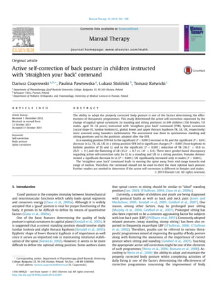
- 4. D. Czaprowski et al. / Manual Therapy 19 (2014) 392e398 395 Fig. 2. A e spontaneous, habitual standing posture, B e standing posture adopted after ‘straighten your back’ command. 3.4. Standing and sitting positions e males In males, in a standing position, a significant (P 0.001) increase of sacral slope was observed after ‘straighten your back’ command. Thoracic kyphosis and its lower and upper parts significantly (P 0.001) decreased after the command. Lumbar lorodosis did not change significantly. In sitting, a significant (P 0.001) change of sacral slope and lumbar lordosis from kyphotic to lordotic position Fig. 3. A e spontaneous, habitual sitting position, B e sitting position adopted after ‘straighten your back’ command.
- 5. 396 D. Czaprowski et al. / Manual Therapy 19 (2014) 392e398 Table 2 Change in spinal curvatures in sagittal plane in standing and sitting - the whole group (n ¼ 249). Parameter Spontaneous standing position Corrected standing position Spontaneous sitting position Corrected sitting position Mean Median Mean Median Mean Median Mean Median Sacral slope (L5/S1-horizontal line) 19.3 6.3 20 21.5 7.1 21** 11.4 9.0 12 7.1 8.2 8** Lumbar lordosis (T12/L1eL5/S1) 33.0 13.0 33 31.3 10.3 31* 17.4 12.1 18 6.6 10.5 6** Thoracic kyphosis (C7/T1eT12/L1) 42.7 9.3 42 33.2 11.6 33** 36.5 10.8 36 23.5 11.7 23** Lower thoracic kyphosis (T6/T7eT12/L1) 9.5 7.7 10 2.6 9.4 3** 15.2 8.7 16 1.0 8.4 1** Upper thoracic kyphosis (C7/T1eT6/T7) 33.2 7.4 33 30.2 10.0 31** 21.1 8.5 21 22.0 11.6 22** was observed. Thoracic kyphosis and its lower part significantly (P 0.001) decreased after ‘straighten your back’ command. Upper thoracic kyphosis significantly (P 0.001) increased (Table 4). 4. Discussion The aim of the study was to determine the change in body posture expressed by the magnitude of sagittal curvatures of the spine in children instructed with ‘straighten your back’ command. Active self-correction was evaluated both in standing and sitting positions. Due to the fact that so far there has been no analysis determining whether this movement is determined by the gender, the study additionally examined it for females and males separately. Apart from the standard assessment of the magnitude of sacral slope, lumbar lordosis and thoracic kyphosis (Saunders, 1998; Andersson and Cocchiarella, 2004), the study also evaluated up-per and lower thoracic kyphosis separately. It stems from the fact that lower thoracic kyphosis is crucial to the rotational stabilisation of the spine and hypokyphosis of this part of thoracic spine is typical for progressive idiopathic scoliosis (Kotwicki, 2002). Studies conducted by O`Sullivan et al. (2006) also assessed the magnitude of lower thoracic kyphosis (T6eT12). 4.1. Standing position In the whole study group, a significant change was observed in the magnitude of all measured parts of the spine in subjects instructed with ‘straighten your back’ command. Thoracic kyphosis as well as its upper and lower parts along with lumbar lordosis decreased whereas sacral slope significantly increased. The obtained results indicate that the ‘straighten your back’ command leads to the extension of the back expressed by the decrease in the magnitude of kyphotic curves and the increase in sacral slope. 4.2. Sitting position It was typical for the whole study group, including both females and males to acquire kyphotic posture (referred to as slump sitting) in a relaxed sitting position (O`Sullivan et al., 2006; Caneiro et al., 2010). The ‘straighten your back’ command brought about a considerable change in position of the spine towards extension what led to the decrease in thoracic kyphosis and its lower part as well as the lordotic alignment of the lumbar spine and sacral slope. However, it seems worth noting that the lumbar spine adopted only a slightly lordotic position (median 6) and lower thoracic kyphosis was flattened (median 1). 4.3. Females and males In our study we have observed a slight discrepancy in active self-correction performed by females and males. Females when instructed with ‘straighten your back’ command demonstrated a significant decrease in lumbar lordosis whereas in males this parameter did not undergo any changes. In turn, upper thoracic kyphosis increased in both females and males. However, this change was statistically significant (P 0.001) in males only. Similar changes were observed in other parts of the spine in both groups. However, it is worth noting that in the present study there was no direct comparison between genders. Therefore, further studies are needed to determine whether the active self-correction is the same or different in females and males. 4.4. Clinical relevance Sitting is one of the risk factors contributing to low back pain. Therefore, re-education of sitting posture may be one of the stra-tegies of preventing and treating it (O`Sullivan et al., 2012). Yet, the optimal sitting position is still the subject of ongoing discussions (Claus et al., 2009a; O`Sullivan et al., 2010). Unquestionably, various sitting and standing postures affect the activity of trunk muscles and spinal load differently (O`Sullivan et al., 2002; O`Sullivan et al., 2006; Claus et al., 2009b). A number of authors recommend acquiring neutral spine position involving slight lumbar lordosis and a relaxed thorax for those LBP subjects who are sensitive to lumbar spine flexion and extension. This position enables subjects to avoid pain resulting from adopting end-range positions and it facilitates the adoption of the most desirable pattern of key trunk muscles activation (Scannell and McGill, 2003; O`Sullivan et al., *p 0.01; **p 0.001; ‘e’ lordotic curve. Table 3 Change in spinal curvatures in sagittal plane in standing and sitting e females (n ¼ 136). Parameter Spontaneous standing position Corrected standing position Spontaneous sitting position Corrected sitting position Mean Median Mean Median Mean Median Mean Median Sacral slope (L5/S1-horizontal line) 21.3 6.0 21.5 22.7 6.7 22.5** 9.4 8.4 10 8.3 8.2 9** Lumbar lordosis (T12/L1eL5/S1) 34.5 8.7 34 31.9 10.1 31** 15.2 12.1 16 7.6 10.6 7.5** Thoracic kyphosis (C7/T1eT12/L1) 42.6 9.9 42 33.1 12.0 32** 36.6 11.4 37 22.8 11.6 21** Lower thoracic kyphosis (T6/T7eT12/L1) 9.3 7.8 10 2.0 10.6 2.5** 15.5 9.2 16 0.5 8.7 1** Upper thoracic kyphosis (C7/T1eT6/T7) 33.4 7.9 33 30.0 11.2 31.5** 20.7 8.7 21 20.7 12.4 21.5 **p 0.001; ‘e’ lorodotic curve.
- 6. D. Czaprowski et al. / Manual Therapy 19 (2014) 392e398 397 Table 4 Change in spinal curvatures in sagittal plane in standing and sitting e males (n ¼ 113). Parameter Spontaneous standing position Corrected standing position Spontaneous sitting position Corrected sitting position Mean Median Mean Median Mean Median Mean Median Sacral slope (L5/S1-horizontal line) 17.1 6.1 17 20.2 7.3 19** 13.8 9.2 15 5.5 8.0 5** Lumbar lordosis (T12/L1eL5/S1) 31.2 8.5 31 30.7 10.6 31 20.1 11.5 20 5.4 10.2 5** Thoracic kyphosis (C7/T1eT12/L1) 42.9 8.7 43 33.5 11.4 36** 36.5 10.1 36 24.3 11.8 24** Lower thoracic kyphosis (T6/T7eT12/L1) 9.8 7.6 10 3.4 7.7 3** 14.9 8.2 16 1.4 8.1 1** Upper thoracic kyphosis (C7/T1eT6/T7) 33.1 7.0 32 30.4 8.4 31** 21.6 8.2 21 23.5 10.4 24** **p 0.001; ‘e’ lorodotic curve. 2006; Claus et al., 2009b). However, according to Claus et al. (2009a) the adoption of such a position might prove difficult and therefore it calls into question whether it might be used in clinical practice. According to Claus et al. (2009a), four types of sitting postures can be distinguished by the direction of curve at thoraco-lumbar and lumbar angles: (1) slump (thoraco-lumbar and lumbar spine in a kyphotic position), (2) flat (thoraco-lumbar and lumbar spine in a vertical position), (3) long lordosis (thoraco-lumbar and lumbar spine in a lordotic position) and (4) short lordosis (thoracic kyphosis and lumbar lordosis). Short lordosis is suggested as ‘ideal’ since it helps achieve proper spinal curves in standing (Claus et al., 2009a). This position divides the direction of spinal curvatures between thoracic and lumbar spine. Caneiro et al. (2010) and O`Sullivan et al. (2006), in turn, proposed three thoraco-lumbar sitting postures: (1) slump sitting (posterior rotation of the pelvis, thoraco-lumbar spine relaxed while looking straight ahead), (2) lumbo-pelvic upright sitting (anterior rotation of the pelvis in order to achieve a neutral lordosis of the lumbar spine and relaxation of the thorax) and (3) thoracic upright sitting (anterior rotation of the pelvis, thoraco-lumbar spine extended with shoulder blades slightly retracted). Taking into consideration the above mentioned descriptions it can be assumed that two types of postures appeared in our study, namely slump sitting adopted before and long lordosis adopted after the ‘straighten your back’ command (Claus et al., 2009a) or thoracic upright sitting (O`Sullivan et al., 2006; Caneiro et al., 2010). The thoracic upright position is connected with an increased ac-tivity of thoracic erector spinae at T4 level and iliocostalis long-issimus pars thoracis. Therefore, it might lead to the higher risk of greater stress to articular and ligamentous structures, greater compression load on cervico-thoracic spine as well as potential discomfort (Lander et al., 1987; O`Sullivan et al., 2006; Claus et al., 2009a, 2009b; Caneiro et al., 2010). Neutral position of the spine, in turn, increases trunk muscles activity without activating large, torque-producing muscles (O`Sullivan et al., 2006; Claus et al., 2009b; Reeve and Dilley, 2009; O`Sullivan et al., 2010). Such a position also modifies the activity of key cervico-thoracic muscles which might be of importance in maintaining the correct sitting posture without the excessive muscle activity (O`Sullivan et al., 2006; Falla et al., 2007; Claus et al., 2009b; Caneiro et al., 2010). What is also interesting is that physiotherapists most frequently (54.9%) indicate that lordosed lumbar spine posture together with relaxed thoracic spine is the best sitting position (O`Sullivan et al., 2012). Our study found that the ‘straighten your back’ command brings about the adoption of a different position. Subjects assumed the posture in which lumbar and thoracic spine was extended. Taking into consideration the results of this study, it seems that the ‘straighten your back’ command should not be used to elicit the most optimal posture. Children who are not provided with any guidance on the appropriate active self-correction are not able to adopt a neutral spine position when instructed with the command. Moreover, the assumed posture is characterised by the reduction of lower thoracic kyphosis which might mean moving further from mid-range and towards end range of motion. It is also confirmed by observations made by Claus et al. (2009a) and O`Sullivan et al. (2010) who claim that the majority of people are not able to ac-quire short lordosis curves without facilitation and feedback and if the correction is made independently (without a therapist’s assis-tance) it is performed by extending the thoracic spine. This is all the more important that even slight changes of spinal curvatures in sagittal plane and the subsequent deviations from the neutral po-sition may lead to the change in muscles activity and consequently to changes in spinal load (O`Sullivan et al., 2006; Claus et al., 2009b; Reeve and Dilley, 2009). 4.5. Limitations The current study evaluated habitual standing and sitting po-sitions and an actively corrected posture adopted after ‘straighten your back’ command. These positions might have been interpreted differently by individual subjects. However, the aim of our study was not to determine the standard magnitude of spinal curvatures in the adopted postures but to evaluate the change in sagittal spinal curves in subjects instructed with the command. Therefore, we believe that it did not influence the obtained results. It is also worth noting that the extent of the differences between some values, despite being statistically significant, were small and in some cases only slightly over the standard error of measurement (SEM 2.8e3.8 standing posture; 1.7e2.5 sitting posture). Spe-cifically those for sacral slope, lumbar lordosis and upper thoracic kyphosis performed in standing and upper thoracic kyphosis measured in a sitting position (whole group) (Table 2). Therefore, these differences may not be clinically meaningful. Hence further research is needed to investigate whether the reported significant differences in those instances are merely the effect of the phe-nomenon of statistics or a true picture of ongoing changes. In the instance of other measurements, namely thoracic kyphosis and its lower part in standing as well as sacral slope, lumbar lordosis, global thoracic kyphosis and lower thoracic kyphosis in sitting, the differences between individual measure-ments were large and they considerably exceeded the magnitude of measurement errors. In our opinion it gives reason to believe that these differences may be clinically significant. Therefore, the fundamental observation we made concerning the reduction of thoracic kyphosis and flattening of its lower part which occur in individuals instructed with ‘straighten your back’ command both in standing and sitting positions is significant and may be applied in clinical practice. The present study was conducted in a group of asymptomatic children aged 10e14 years. Therefore, caution is advised when transferring the obtained results (especially the potential clinical meaningfulness) to other populations, e.g. children with
- 7. 398 D. Czaprowski et al. / Manual Therapy 19 (2014) 392e398 musculoskeletal pain or fatigue. Thus it is essential to undertake further studies to confirm the current study findings in pathological populations as well as those of younger or older children/youth. Our study did not concentrate on evaluating the change in the position of the head or trunk and neck muscles activity in the posture assumed after ‘straighten your back’ command. However, taking into consideration the relationship between head/neck po-sition and thoraco-lumbar posture it seems that a further study should be undertaken to determine the influence of the change in position of one area of musculoskeletal system on the position and functioning of body parts further away. Especially as Caneiro et al. (2010) claim that management isolated in one segment might prove less effective. Further studies might also be supplemented with observations of changes in other than sagittal planes. The majority of studies concerning various sitting and standing postures concentrate on adults (O`Sullivan et al., 2002; O`Sullivan et al., 2006; Womersley and May, 2006; Claus et al., 2009a; Caneiro et al., 2010; O`Sullivan et al., 2010). Therefore, we should be cautions when comparing the results of this study with results presented by other authors without previously comparing the active self-correction between children and adults. In spite of the limitations, we believe that the results of the study may have relevance to the clinical approach. Especially as the current study included a large, homogeneous group of children and it is them who are frequently referred to various preventive and therapeutic programmes (Geldhof et al., 2007). 5. Conclusions The ‘straighten your back’ command brings about the extension of the entire spine. This active self-correction appears in both standing and sitting positions. The reduction of global kyphosis together with the flattening of its lower part, which was the result of the command, calls into question whether the command should be used to improve the body posture in children who were not provided with any guidance on the correct shape of sagittal spinal curves. After ‘straighten your back’ command in a standing position, a lumbar lordosis significantly changed (decreased) only in females, while upper thoracic kyphosis significantly changed (increased) in sitting only in males. Therefore, further studies are needed to determine whether the active self-correction is different in females and males. References Andersson GBJ, Cocchiarella L, American Medical Association. Guides to the eval-uation of permanent impairments. 5th ed. Chicago: American Medical Associ-ation; 2004. p. 373e432. Bland JM, Altman DG. Statistics notes: Cronbach’s alpha. BMJ 1997;314:572. Bulinska K. Program profilaktyczny “Wyprostuj sie˛ ” [The prevention program “Straighten your back”]. Phys Health Educ 2005;8/9:5e9 [article in Polish]. Caneiro JP, O’Sullivan P, Burnett A, Barach A, O’Neil D, Tveit O, et al. The influence of different sitting postures on head/neck posture and muscle activity. Man Ther 2010;15(1):54e60. Claus AP, Hides JA, Moseley GL, Hodges PW. Is “ideal” sitting real?: measurement of spinal curves in four sitting postures. Man Ther 2009a;14(4):404e8. Claus AP, Hides JA, Moseley GL, Hodges PW. Different ways to balance the spine: subtle changes in sagittal spinal curves affect regional muscle activity. Spine 2009b;34(6):E208e14. Czaprowski D, Paw1owska P, Ge˛bicka A, Sitarski D, Kotwicki T. Intra- and interob-server repeatability of the assessment of anteroposterior curvatures of the spine using Saunders digital inclinometer. Ortop Traumatol Rehabil 2012;14(2): 145e53. Falla D, O’Leary S, Fagan A, Jull G. Recruitment of the deep cervical flexor muscles during a postural-correction exercise performed in sitting. Man Ther 2007;12(2):139e43. Geldhof E, Cardon G, De Bourdeaudhuij I, De Clercq D. Back posture education in elementary schoolchildren: a 2-year follow-up study. Eur Spine J 2007;16(6): 841e50. Jones GT, Macfarlane GJ. Epidemiology of low back pain in children and adolescents. Arch Dis Child 2005;90(3):312e6. Kendall FP, Kendall McCreary E, Provance PG, McIntyre Rodgers M, Romani WA. Muscles: testing and function with posture and pain. 5th ed. Baltimore: Lip-pincott Williams Wilkins; 2005. p. 49e117. Kotwicki T. Sagittal and transversal plane deformity in thoracic scoliosis. Stud Health Technol Inform 2002;91:251e6. Lander C, Korbon GA, DeGood DE, Rowlingson JC. The Balans chair and its semi-kneeling position: an ergonomic comparison with the conventional sitting position. Spine 1987;12(3):269e72. Lee L. Ch 7: restoring force closure/motor control of the thorax. In: Lee D, editor. The thorax: an integrated approach. 2nd ed. Minneapolis: OPTP; 2003. p. 103e35. Murphy S, Buckle P, Stubbs D. Classroom posture and self-reported back and neck pain in schoolchildren. Appl Ergon 2004;35(2):113e20. Muscolino JE. The muscle and bone palpation manual with trigger points, referral patterns and stretching. St. Louis: Mosby Elsevier; 2008. p. 106e7. O’Sullivan K, O’Dea P, Dankaerts W, O’Sullivan P, Clifford A, O’Sullivan L. Neutral lumbar spine sitting position in pain-free subjects. Man Ther 2010;15(6):557e 61. O’Sullivan K, O’Sullivan P, O’Sullivan L, Dankaerts W. What do physiotherapists consider to be the best sitting spinal posture? Man Ther 2012;17(5):432e7. O’Sullivan PB. “Clinical instability” of the lumbar spine: its pathological basis, diagnosis and conservative management. In: Boyling JD, Jull GA, editors. Grieve’s modern manual therapy: the vertebral column. 3rd ed. Edinburgh: Churchill Livingstone; 2004. p. 311e31. O’Sullivan PB. Lumbar segmental “instability”: clinical presentation and specific stabilizing exercise management. Man Ther 2000;5(1):2e12. O’Sullivan PB, DankaertsW, Burnett AF, Farrell GT, Jefford E, Naylor CS, et al. Effect of different upright sitting postures on spinal-pelvic curvature and trunk muscle activation in a pain-free population. Spine 2006;31(19):E707e12. O’Sullivan PB, Grahamslaw KM, Kendell M, Lapenskie SC, Moller NE, Richards KV. The effect of different standing and sitting postures on trunk muscle activity in a pain-free population. Spine 2002;27(11):1238e44. Reeve A, Dilley A. Effects of posture on the thickness of transversus abdominis in pain-free subjects. Man Ther 2009;14(6):679e84. Romano M, Negrini A, Parzini S, Negrini S. Scientific exercises approach to scoliosis (SEAS): efficacy, efficiency and innovation. In: Grivas TB, editor. The conserva-tive scoliosis treatment. Amsterdam: IOS Press; 2008. p. 191e207. Saunders HD. Saunders digital inclinometer. User’s guide. Chaska, MN, USA: The Saunders Group Inc.; 1998. p. 5e19. Scannell JP, McGill SM. Lumbar posture: should it and can it be modified? A study of passive tissue stiffness and lumbar position during activities of daily living. Phys Ther 2003;83(10):907e17. Weiss HR, Hollaender M, Klein R. ADL based scoliosis rehabilitation e the key to an improvement of time efficiency? Stud Health Technol Inform 2006;123:594e8. Williams MM, Hawley JA, McKenzie RA, Van Wijmen PM. A comparison of the ef-fects of two sitting postures on back and referred pain. Spine 1991;16(10): 1185e91. Womersley L, May S. Sitting posture of subjects with postural backache. J Manipulative Physiol Ther 2006;29(3):213e8. Zaina F, Negrini S, Atanasio S, Fusco C, Romano M, Negrini A. Specific exercises performed in the period of brace weaning can avoid loss of correction in adolescent idiopathic scoliosis (AIS) patients: winner of SOSORT’s 2008 award for best clinical paper. Scoliosis 2009;4:8.