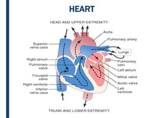
SlideShare a Scribd company logo
1 of 35
HEART
Heart
Preload and Afterload
■ Preload Volume of blood in the ventricle at end diastole
(producing a stretch of ventricular muscle cells)
■ Afterload Resistance the heart must overcome to eject blood
from the ventricle
Cardiac Output and Venous Return
• Cardiac output is the quantity of blood pumped into the
aorta each minute.
• Cardiac output = stroke volume x heart rate
• Venous return is the quantity of blood flowing from the veins
to the right atrium.
• Except for temporary moments, the cardiac output should
equal the venous return
Heart
■ Stroke Volume = the volume of blood pumped by either
the right or left ventricle during single ventricular
contraction.
SV = EDV – ESV
70 = 125 – 55
CO = SV x HR
Regulation of Stroke volume
• Preload: Degree of stretch of heart muscle (Frank-
Starling) – greatest factor influencing stretch is venous
return (see Below)
• Contractility – Strength of contraction
Increased Ca2+ is the result of sympathetic nervous system
Heart Diseases
1. Heart failure
2. Congenital heart diseases
3. Ischaemic heart disease
4. Hypertensive heart disease
5. Cor pulmonale
6. Rheumatic fever and rheumatic heart disease
7. Non-rheumatic endocarditis
8. Valvular diseases and deformities
9. Myocardial disease
10. Pericardial disease
11. Tumor's of the heart
Heart Failure
■ Heart failure is defined as the pathophysiologic state
in which impaired cardiac function and unable to
maintain an adequate circulation blood for the
metabolic needs of the tissues of the body.
■ It is also known as congestive heart failure
■ It may be acute or chronic
■ HF is a complex clinical syndrome that can result
from any structural or functional cardiac disorder
that impairs the ability of the ventricle to fill with or
eject blood.
• Heart failure may result from systolic or diastolic
dysfunction.
• Systolic dysfunction results from inadequate
myocardial contractile function, usually as a
consequence of ischemic heart disease or
hypertension.
• Diastolic dysfunction refers to an inability of the heart
to adequately relax and fill, which may be a
consequence of massive left ventricular hypertrophy,
myocardial fibrosis, amyloid deposition,
Heart Failure
Acute heart failure. Sudden and rapid development of heart
failure occurs in the following conditions:
a) Larger myocardial infarction
b) Valve rupture
c) Massive pulmonary embolism
d) Acute viral myocarditis
e) Acute bacterial toxaemia.
■ In acute heart failure, there is sudden reduction in cardiac
output resulting in systemic hypotension. Instead, a state of
cardiogenic shock and cerebral hypoxia develops.
Heart Failure
Chronic heart failure. More often, heart failure develops slowly
as observed in the following states:
a) Myocardial ischemia from atherosclerotic coronary artery
disease
b) Multivalvular heart disease
c) Systemic arterial hypertension
d) Chronic lung diseases resulting in hypoxia and pulmonary
arterial hypertension
e) Progression of acute into chronic failure.
■ In chronic heart failure, compensatory mechanisms like
tachycardia, cardiac dilatation and cardiac hypertrophy try to
make adjustments so as to maintain adequate cardiac output.
This often results in well-maintained arterial pressure and
there is accumulation of edemas.
Heart Failure
Etiology
Heart failure may be caused by one of the following factors, either singly
or in combination:
1. INTRINSIC PUMP FAILURE.
■ The most common and most important cause of heart failure is
weakening of the ventricular muscle due to disease so that the
heart fails to act as an efficient pump. The various diseases which
may culminate in pump failure by this mechanisms are :
a) Ischemic heart disease
b) Myocarditis
c) Cardiomyopathies
d) Metabolic disorders e.g. beriberi
e) Disorders of the rhythm e.g. atrial fibrillation and flutter.
2. INCREASED WORKLOAD ON THE HEART.
■ Increased mechanical load on the heart results in
increased myocardial demand resulting in myocardial
failure. Increased load on the heart may be in the form of
pressure load or volume load.
i. Increased pressure load may occur in the following
states:
a)Systemic and pulmonary arterial hypertension.
b)Valvular disease e.g. mitral stenosis, aortic stenosis,
pulmonary stenosis.
c) Chronic lung diseases.
Etiology
ii. Increased volume load occurs when a ventricle is required to
eject more than normal volume of the blood resulting in cardiac
failure. This is seen in the following conditions:
a)Valvular insufficiency
b)Severe anemia
c) Thyrotoxicosis
d)Arteriovenous shunts
e) Hypoxia due to lung diseases.
3. IMPAIRED FILLING OF CARDIAC CHAMBERS.
■ Decreased cardiac output and cardiac failure may result from
extra-cardiac causes or defect in filling of the heart:
a)Cardiac tamponade e.g. haemopericardium, hydropericardium
b)Constrictive pericarditis.
Etiology
RISK FACTORS
 Coronary Artery Diseases
 Myocardial Infraction
 Cigarette Smoking
 Hypertension
 Obesity
 Age
 Diabetes
 Heart Valve Disease (e.g. Aortic Stenosis)
 Cardiomyopathy (Heart Muscle Disease)
 Dilated Cardiomyopathy (e.g. Alcohol & Viral Infection)
 Hypertrophic Cardiomyopathy
 Restrictive Cardiomyopathy
Causes of left-sided heart failure:
It is initiated by stress to the left heart. Left-sided heart
failure causes are as follows:
1. Systemic hypertension
2. Mitral or aortic valve disease (stenosis)
3. Ischemic heart disease
4. Myocardial diseases e.g. cardiomyopathies, myocarditis.
5. Restrictive pericarditis.
Types of Congestive Heart Failure
Left-sided failure
• Most common form
• Blood backs up through the left atrium into the pulmonary veins
• Pulmonary congestion and edema
• Eventually leads to biventricular failure
Clinical manifestations:
The clinical manifestations of left-sided heart failure result
from decreased left ventricular output and hence there is
accumulation of fluid upstream in the lungs.
Accordingly, the major pathologic changes are as under:
1. Pulmonary congestion and edema causes dyspnea and
orthopnea
2. Decreased left ventricular output causing hypo-
perfusion and diminished oxygenation of tissues e.g.
a) In kidneys causing ischemic acute tubular necrosis,
b) In brain causing hypoxic encephalopathy,
c) And in skeletal muscles causing muscular weakness
3. Fatigue.
Causes of right-sided heart failure:
Right-sided heart failure occurs more often as a consequence of
left-sided heart failure. The causes of right-sided heart failure are
–
As a consequence of left ventricular failure.
1. Cor pulmonale in which right heart failure occurs due to
intrinsic lung diseases
2. Pulmonary or tricuspid valvular disease.
3. Pulmonary hypertension secondary to pulmonary
thromboembolism.
4. Myocardial disease affecting right heart.
5. Congenital heart disease with left-to-right shunt
Right-sided failure
• Results from diseased right ventricle
• Blood backs up into right atrium and venous circulation
Clinical manifestations:
The clinical manifestations of right-sided heart failure are
upstream of the right heart such as systemic (due to caval
blood) and portal venous congestion, and reduced cardiac
output.
Accordingly, the pathologic changes are as under:
1. Systemic venous congestion in different tissues and organs
e.g.
Subcutaneous edema on dependent parts
Passive congestion of the liver, spleen, and kidney, ascites,
hydrothorax,
Congestion of leg veins and neck veins.
2. Reduced cardiac output resulting in circulatory stagnation
causing anoxia, cyanosis and coldness of extremities
Schematic evolution of congestive heart failure and its
effects.
Schematic pathophysiology of compensatory
mechanisms in cardiac failure
Hypertrophy of the heart is defined as an increase in size and weight of the
myocardium. It generally results from increased pressure load while
increased volume load (e.g. valvular incompetence) results in hypertrophy
with dilatation of the affected chamber due to regurgitation of the blood
through incompetent valve. The atria may also undergo compensatory
changes due to increased workload.
Hypertrophy
Left ventricular hypertrophy.
The common causes are as under:
i) Systemic hypertension
ii) Aortic stenosis and insufficiency
iii) Mitral insufficiency
iv) Occlusive coronary artery disease
v) Congenital anomalies like septal defects and patent
ductus arteriosus
vi) Conditions with increased cardiac output e.g.
thyrotoxicosis, anemia, arteriovenous fistulae.
 Right ventricular hypertrophy
Most of the causes of right ventricular hypertrophy are
due to pulmonary arterial hypertension. These are as
follows:
i) Pulmonary stenosis and insufficiency
ii) Tricuspid insufficiency
iii) Mitral stenosis and/or insufficiency
iv) Chronic lung diseases e.g. chronic emphysema,
bronchiectasis, pneumoconiosis, pulmonary vascular
disease etc.
v) Left ventricular hypertrophy and failure of the left
ventricle.
ISCHAEMIC HEART DISEASE
■ Ischaemic heart disease (IHD) is defined as acute or
chronic form of cardiac disability arising from imbalance
between the myocardial supply and demand for
oxygenated blood.
■ Since narrowing or obstruction of the coronary arterial
system is the most common cause of myocardial anoxia,
the alternate term ‘coronary artery disease (CAD)
• IHD is invariably caused by disease affecting the
coronary arteries, the most prevalent being
atherosclerosis accounting for more than 90% cases,
• While other causes are responsible for less than 10%
cases of IHD.
• Therefore, it is convenient to consider the etiology of
IHD under three broad headings:
i) coronary atherosclerosis;
ii) superadded changes in coronary atherosclerosis; and
iii) non-atherosclerotic causes.
ISCHAEMIC HEART DISEASE
Coronary atherosclerosis: It resulting in ‘fixed’ obstruction is
the major cause of IHD in more than 90% cases. A brief
account of the specific features in pathology of lesions in
atherosclerotic coronary artery disease in particular are
presented.
1) Distribution: Atherosclerotic lesions in coronary arteries
are distributed in one or more of the three major coronary
arterial trunks, the highest incidence being in the anterior
descending branch of the left coronary, followed in
decreasing frequency, by the right coronary artery and still
less in circumflex branch of the left coronary. About one third
of cases have single-vessel disease, most often left anterior
descending arterial involvement; another one-third have two
vessel disease, and the remainder have three major vessel
disease.
2) Location: Almost all adults show atherosclerotic plaques
scattered throughout the coronary arterial system. However,
significant stenotic lesions that may produce chronic myocardial
ischemia show more than 75% (three-fourth) reduction in the
cross-sectional area of a coronary artery or its branch. The
area of severest involvement is about 3 to 4 cm from the
coronary ostia, more often at or near the bifurcation of the
arteries, suggesting the role of haemodynamic forces in
atherogenesis.
3) Fixed atherosclerotic plaques: The atherosclerotic plaques
in the coronaries are more often unusually located bulging into
the lumen from one side. Occasionally, there may be concentric
thickening of the wall of the artery. Atherosclerosis produces
gradual luminal narrowing that may eventually lead to ‘fixed’
coronary obstruction. The general features of atheromas of
coronary arteries are similar to those affecting elsewhere in the
body and may develop similar complications like calcification,
coronary thrombosis, ulceration, haemorrhage, and rupture and
aneurysm formation
Angina Pectoris:
• Angina pectoris is a clinical syndrome of IHD
resulting from transient myocardial ischaemia.
• It is characterized by paroxysmal pain in the
sub sternal or precordial region of the chest
which is aggravated by an increase in the
demand of the heart and relieved by a decrease
in the work of the heart.
• Often, the pain radiates to the left arm, neck,
jaw or right arm.
Classification: There are 3 overlapping clinical
patterns of angina pectoris with some differences
in their pathogenesis:
A. Stable or typical angina
B. Variant angina
C. Unstable angina
Stable or Typical Angina:
• This is the most common pattern. Stable or typical
angina is characterized by attacks of pain following
physical exertion or emotional excitement and is
relieved by rest.
• The pathogenesis of condition lies in chronic stenosing
coronary atherosclerosis that cannot perfuse the
myocardium adequately when the workload on the
heart increases.
• During the attacks, there is depression of ST segment
in the ECG due to poor perfusion of the subendocardial
region of the left ventricle but there is no elevation of
enzymes in the blood as there is no irreversible
myocardial injury.
Variant Angina:
• This pattern of angina is characterized by pain at rest
and has no relationship with physical activity.
• The exact pathogenesis of variant angina is not
known.
• It may occur due to sudden Vasospasm of a coronary
trunk induced by coronary atherosclerosis, or may be
due to release of humoral vasoconstrictors by mast
cells in the coronary adventitia.
• ECG shows ST segment elevation due to Trans mural
ischaemia.
• These patients respond well to vasodilators like
nitroglycerin.
Unstable Angina.
• Also referred to as ‘pre-infarction angina’ or
‘acute coronary insufficiency’, this is the most
serious pattern of angina.
• It is characterized by more frequent onset of
pain of prolonged duration and occurring often
at rest.
• It is thus indicative of an impending acute
myocardial infarction
• Myocardial infarction (MI), also commonly referred to
as “heart attack,” is necrosis of the heart muscle
resulting from ischemia.
• The major underlying cause of IHD is atherosclerosis;
while MIs can occur at virtually any age, the frequency
rises progressively with aging and with increasing risk
factors for atherosclerosis.
• Nevertheless, approximately 10% of MIs occur before 40
years of age, and 45% occur before 65 years of age.
• Blacks and whites are equally affected.
• Men are at greater risk than women, although the gap
progressively narrows with age.
Myocardial Infarction
Acute Myocardial Infarction: Acute myocardial
infarction (MI) is the most important and feared
consequence of coronary artery disease. Many patients
may die within the first few hours of the onset,
while remainder suffer from effects of impaired cardiac
function.
A significant factor that may prevent or diminish the
myocardial damage is the development of collateral
circulation through anastomotic channels over a period
of time.
a) Incidence: In developed countries, acute MI
accounts for 10-25% of all deaths. Due to the
dominant etiologic role of coronary atherosclerosis
in acute MI, the incidence of acute MI correlates well
with the incidence of atherosclerosis in a geographic
area.
Factors associated with acute M. I.
b) Age: Acute MI may virtually occur at all ages, though
the incidence is higher in the elderly. About 5% of
heart attacks occur in young people under the age of
40 years, particularly in those with major risk factors
to develop atherosclerosis like hypertension, diabetes
mellitus, cigarette smoking and dyslipidemia with
familial hypercholesterolemia.
c) Sex: Males throughout their life are at a significantly
higher risk of developing acute MI as compared to
females. Women during reproductive period have
remarkably low incidence of acute MI, probably due to
the protective influence of estrogen. The use of oral
contraceptives is associated with high risk of
developing acute MI. After menopause, this sex
difference gradually declines but the incidence of
disease among women never reaches that among men
of the same age
Factors associated with acute M. I.
Markers: Important myocardial markers in use nowadays are –
(1) Creatine kinase (CK) and CK-MB: CK has three forms—
o CK-MM derived from skeletal muscle;
o CK-BB derived from brain and lungs; and
o CK-MB, mainly from cardiac muscles and insignificant amount
from extra cardiac tissue.
Thus total CK estimation lacks specificity while elevation of CK-
MB iso-enzyme is considerably specific for myocardial damage.
CK-MB has further 2 forms—CK-MB2 is the myocardial form
while CK-MB1 is extra cardiac form. A ratio of CK-MB2: CK-MB1
above 1.5 is highly sensitive for the diagnosis of acute MI after 4-6
hours of onset of myocardial ischaemia. CK-MB disappears from
blood by 48 hours.
(2) Lactic dehydrogenase (LDH): Total LDH estimation also lacks
specificity since this enzyme is present in various tissues besides
myocardium such as in skeletal muscle, kidneys, liver, lungs and red
blood cells. However, like CK, LDH too has two isoforms of which LDH-1
is myocardial-specific.
(3) Cardiac-specific troponins (cTn): Immunoassay of cTn as a serum
cardiac marker has rendered LDH estimation obsolete. Troponins are
contractile muscle proteins present in human cardiac and skeletal
muscle but cardiac troponins are specific for myocardium. There are two
types of cTn:
o Cardiac troponin T (cTnT); and
o Cardiac troponin I (cTnI).
Both cTnT and cTnI are not found in the blood normally, but after
myocardial injury their levels rise very high around the same time when
CK-MB is elevated (i.e. after 4-6 hours).
Both troponin levels remain high for much longer duration; cTnI for 7-
10 days and cTnT for 10-14 days.
(4) Myoglobin: Though myoglobin is the first cardiac marker to become
elevated after myocardial infarction, it lacks cardiac specificity and is
excreted in the urine rapidly. Its levels, thus, return to normal within 24
hours of attack of acute MI.
Why is not the serum myoglobin used routinely
in more cardiac enzyme panels?
• There are two limitations to the use of serum
myoglobin for the diagnosis of acute MI. First, the
rapid release and metabolism of myoglobin can result
in an undulating or “staccato” pattern characterized
by increases and decreases in the plasma myoglobin
concentration that can lead to clinical confusion.
• The second problem is that, like LD, it lacks specificity
for the heart. Serum concentrations are elevated after
injury to a variety of tissues (especially skeletal
muscle) or recent cocaine use and in patients with
impaired renal function due to decreased clearance.
Because of these limitations and lack of apparent
advantage over troponins and CK-MB, serum
myoglobin is not routinely measured in patients with
suspected MI.

More Related Content

Similar to heart disease.pptx

ilovepdf_merged (1).pdf
ilovepdf_merged (1).pdfilovepdf_merged (1).pdf
ilovepdf_merged (1).pdfssuser702574
 
Cardiovascular Pathophysiology- Congestive Cardiac Failure (CCF)/Congestive H...
Cardiovascular Pathophysiology- Congestive Cardiac Failure (CCF)/Congestive H...Cardiovascular Pathophysiology- Congestive Cardiac Failure (CCF)/Congestive H...
Cardiovascular Pathophysiology- Congestive Cardiac Failure (CCF)/Congestive H...VISHALJADHAV100
 
heart failure in children 2015
heart failure in children 2015heart failure in children 2015
heart failure in children 2015Azad Haleem
 
HEART lecture MBChB III 2024.pdfnnnnnnnnnnnnnnnnnnnnnnnnnnnnnnnnnnn
HEART lecture MBChB III 2024.pdfnnnnnnnnnnnnnnnnnnnnnnnnnnnnnnnnnnnHEART lecture MBChB III 2024.pdfnnnnnnnnnnnnnnnnnnnnnnnnnnnnnnnnnnn
HEART lecture MBChB III 2024.pdfnnnnnnnnnnnnnnnnnnnnnnnnnnnnnnnnnnn202112360
 
CVS lssn 2.pptx
CVS lssn 2.pptxCVS lssn 2.pptx
CVS lssn 2.pptxMishiSoza
 
HEART FAILURE.pptx
HEART FAILURE.pptxHEART FAILURE.pptx
HEART FAILURE.pptxKALEDICKSAM1
 
Cardiac Failure, Cardiac Shock, Cardiac Arrest, PPT
Cardiac Failure, Cardiac Shock, Cardiac Arrest,  PPTCardiac Failure, Cardiac Shock, Cardiac Arrest,  PPT
Cardiac Failure, Cardiac Shock, Cardiac Arrest, PPTDR .PALLAVI PATHANIA
 
heartfailure-190730143120.pdf
heartfailure-190730143120.pdfheartfailure-190730143120.pdf
heartfailure-190730143120.pdfJeenaRaj10
 
Pathology of CVS-PR.pptx
Pathology of CVS-PR.pptxPathology of CVS-PR.pptx
Pathology of CVS-PR.pptxImtiyaz60
 
Congestive cardiac failure
Congestive cardiac failureCongestive cardiac failure
Congestive cardiac failureSudeep Kashyap
 
ALTERATIONS IN OXYGENATION CMBC.pptx
ALTERATIONS IN OXYGENATION CMBC.pptxALTERATIONS IN OXYGENATION CMBC.pptx
ALTERATIONS IN OXYGENATION CMBC.pptxAmirAhmedGeza
 
Congestive heart failure 2
Congestive heart failure 2Congestive heart failure 2
Congestive heart failure 2ShivaneeKushwaha
 

Similar to heart disease.pptx (20)

Congestive heart failure
Congestive heart failureCongestive heart failure
Congestive heart failure
 
ilovepdf_merged (1).pdf
ilovepdf_merged (1).pdfilovepdf_merged (1).pdf
ilovepdf_merged (1).pdf
 
Heart failure.ppt
Heart failure.pptHeart failure.ppt
Heart failure.ppt
 
Cardiovascular Pathophysiology- Congestive Cardiac Failure (CCF)/Congestive H...
Cardiovascular Pathophysiology- Congestive Cardiac Failure (CCF)/Congestive H...Cardiovascular Pathophysiology- Congestive Cardiac Failure (CCF)/Congestive H...
Cardiovascular Pathophysiology- Congestive Cardiac Failure (CCF)/Congestive H...
 
heart failure in children 2015
heart failure in children 2015heart failure in children 2015
heart failure in children 2015
 
CCF- B.M.pptx
CCF- B.M.pptxCCF- B.M.pptx
CCF- B.M.pptx
 
Myocardial ischemia
Myocardial ischemiaMyocardial ischemia
Myocardial ischemia
 
CARDIAC FAILURE (2).ppt
CARDIAC FAILURE (2).pptCARDIAC FAILURE (2).ppt
CARDIAC FAILURE (2).ppt
 
heartfailure.pptx
heartfailure.pptxheartfailure.pptx
heartfailure.pptx
 
HEART lecture MBChB III 2024.pdfnnnnnnnnnnnnnnnnnnnnnnnnnnnnnnnnnnn
HEART lecture MBChB III 2024.pdfnnnnnnnnnnnnnnnnnnnnnnnnnnnnnnnnnnnHEART lecture MBChB III 2024.pdfnnnnnnnnnnnnnnnnnnnnnnnnnnnnnnnnnnn
HEART lecture MBChB III 2024.pdfnnnnnnnnnnnnnnnnnnnnnnnnnnnnnnnnnnn
 
CVS lssn 2.pptx
CVS lssn 2.pptxCVS lssn 2.pptx
CVS lssn 2.pptx
 
HEART FAILURE.pptx
HEART FAILURE.pptxHEART FAILURE.pptx
HEART FAILURE.pptx
 
Cardiac Failure, Cardiac Shock, Cardiac Arrest, PPT
Cardiac Failure, Cardiac Shock, Cardiac Arrest,  PPTCardiac Failure, Cardiac Shock, Cardiac Arrest,  PPT
Cardiac Failure, Cardiac Shock, Cardiac Arrest, PPT
 
Heart failure
Heart failureHeart failure
Heart failure
 
heartfailure-190730143120.pdf
heartfailure-190730143120.pdfheartfailure-190730143120.pdf
heartfailure-190730143120.pdf
 
Pathology of CVS-PR.pptx
Pathology of CVS-PR.pptxPathology of CVS-PR.pptx
Pathology of CVS-PR.pptx
 
Congestive cardiac failure
Congestive cardiac failureCongestive cardiac failure
Congestive cardiac failure
 
ALTERATIONS IN OXYGENATION CMBC.pptx
ALTERATIONS IN OXYGENATION CMBC.pptxALTERATIONS IN OXYGENATION CMBC.pptx
ALTERATIONS IN OXYGENATION CMBC.pptx
 
Congestive heart failure 2
Congestive heart failure 2Congestive heart failure 2
Congestive heart failure 2
 
Heart failure...
Heart failure...Heart failure...
Heart failure...
 

Recently uploaded

Cara Menggugurkan Kandungan Dengan Cepat Selesai Dalam 24 Jam Secara Alami Bu...
Cara Menggugurkan Kandungan Dengan Cepat Selesai Dalam 24 Jam Secara Alami Bu...Cara Menggugurkan Kandungan Dengan Cepat Selesai Dalam 24 Jam Secara Alami Bu...
Cara Menggugurkan Kandungan Dengan Cepat Selesai Dalam 24 Jam Secara Alami Bu...Cara Menggugurkan Kandungan 087776558899
 
Test bank for critical care nursing a holistic approach 11th edition morton f...
Test bank for critical care nursing a holistic approach 11th edition morton f...Test bank for critical care nursing a holistic approach 11th edition morton f...
Test bank for critical care nursing a holistic approach 11th edition morton f...robinsonayot
 
TEST BANK For Guyton and Hall Textbook of Medical Physiology, 14th Edition by...
TEST BANK For Guyton and Hall Textbook of Medical Physiology, 14th Edition by...TEST BANK For Guyton and Hall Textbook of Medical Physiology, 14th Edition by...
TEST BANK For Guyton and Hall Textbook of Medical Physiology, 14th Edition by...rightmanforbloodline
 
Top 10 Most Beautiful Russian Pornstars List 2024
Top 10 Most Beautiful Russian Pornstars List 2024Top 10 Most Beautiful Russian Pornstars List 2024
Top 10 Most Beautiful Russian Pornstars List 2024locantocallgirl01
 
7 steps How to prevent Thalassemia : Dr Sharda Jain & Vandana Gupta
7 steps How to prevent Thalassemia : Dr Sharda Jain & Vandana Gupta7 steps How to prevent Thalassemia : Dr Sharda Jain & Vandana Gupta
7 steps How to prevent Thalassemia : Dr Sharda Jain & Vandana GuptaLifecare Centre
 
ABO Blood grouping in-compatibility in pregnancy
ABO Blood grouping in-compatibility in pregnancyABO Blood grouping in-compatibility in pregnancy
ABO Blood grouping in-compatibility in pregnancyMs. Sapna Pal
 
spinal cord disorders and paraplegia .
spinal cord disorders  and  paraplegia .spinal cord disorders  and  paraplegia .
spinal cord disorders and paraplegia .Mohamed Rizk Khodair
 
Physiologic Anatomy of Heart_AntiCopy.pdf
Physiologic Anatomy of Heart_AntiCopy.pdfPhysiologic Anatomy of Heart_AntiCopy.pdf
Physiologic Anatomy of Heart_AntiCopy.pdfMedicoseAcademics
 
VIP ℂall Girls Arekere Bangalore 6378878445 WhatsApp: Me All Time Serviℂe Ava...
VIP ℂall Girls Arekere Bangalore 6378878445 WhatsApp: Me All Time Serviℂe Ava...VIP ℂall Girls Arekere Bangalore 6378878445 WhatsApp: Me All Time Serviℂe Ava...
VIP ℂall Girls Arekere Bangalore 6378878445 WhatsApp: Me All Time Serviℂe Ava...deepakkumar115120
 
Difference Between Skeletal Smooth and Cardiac Muscles
Difference Between Skeletal Smooth and Cardiac MusclesDifference Between Skeletal Smooth and Cardiac Muscles
Difference Between Skeletal Smooth and Cardiac MusclesMedicoseAcademics
 
Part I - Anticipatory Grief: Experiencing grief before the loss has happened
Part I - Anticipatory Grief: Experiencing grief before the loss has happenedPart I - Anticipatory Grief: Experiencing grief before the loss has happened
Part I - Anticipatory Grief: Experiencing grief before the loss has happenedbkling
 
Top 10 Most Beautiful Chinese Pornstars List 2024
Top 10 Most Beautiful Chinese Pornstars List 2024Top 10 Most Beautiful Chinese Pornstars List 2024
Top 10 Most Beautiful Chinese Pornstars List 2024locantocallgirl01
 
Face and Muscles of facial expression.pptx
Face and Muscles of facial expression.pptxFace and Muscles of facial expression.pptx
Face and Muscles of facial expression.pptxDr. Rabia Inam Gandapore
 
VIP ℂall Girls Thane West Mumbai 9930245274 WhatsApp: Me All Time Serviℂe Ava...
VIP ℂall Girls Thane West Mumbai 9930245274 WhatsApp: Me All Time Serviℂe Ava...VIP ℂall Girls Thane West Mumbai 9930245274 WhatsApp: Me All Time Serviℂe Ava...
VIP ℂall Girls Thane West Mumbai 9930245274 WhatsApp: Me All Time Serviℂe Ava...poonam rawat$V15
 
Physicochemical properties (descriptors) in QSAR.pdf
Physicochemical properties (descriptors) in QSAR.pdfPhysicochemical properties (descriptors) in QSAR.pdf
Physicochemical properties (descriptors) in QSAR.pdfRAJ K. MAURYA
 
Obat Aborsi Ampuh Usia 1,2,3,4,5,6,7 Bulan 081901222272 Obat Penggugur Kandu...
Obat Aborsi Ampuh Usia 1,2,3,4,5,6,7 Bulan  081901222272 Obat Penggugur Kandu...Obat Aborsi Ampuh Usia 1,2,3,4,5,6,7 Bulan  081901222272 Obat Penggugur Kandu...
Obat Aborsi Ampuh Usia 1,2,3,4,5,6,7 Bulan 081901222272 Obat Penggugur Kandu...Halo Docter
 
TEST BANK For Porth's Essentials of Pathophysiology, 5th Edition by Tommie L ...
TEST BANK For Porth's Essentials of Pathophysiology, 5th Edition by Tommie L ...TEST BANK For Porth's Essentials of Pathophysiology, 5th Edition by Tommie L ...
TEST BANK For Porth's Essentials of Pathophysiology, 5th Edition by Tommie L ...rightmanforbloodline
 
SEMESTER-V CHILD HEALTH NURSING-UNIT-1-INTRODUCTION.pdf
SEMESTER-V CHILD HEALTH NURSING-UNIT-1-INTRODUCTION.pdfSEMESTER-V CHILD HEALTH NURSING-UNIT-1-INTRODUCTION.pdf
SEMESTER-V CHILD HEALTH NURSING-UNIT-1-INTRODUCTION.pdfSachin Sharma
 
Creeping Stroke - Venous thrombosis presenting with pc-stroke.pptx
Creeping Stroke - Venous thrombosis presenting with pc-stroke.pptxCreeping Stroke - Venous thrombosis presenting with pc-stroke.pptx
Creeping Stroke - Venous thrombosis presenting with pc-stroke.pptxYasser Alzainy
 
Intro to disinformation and public health
Intro to disinformation and public healthIntro to disinformation and public health
Intro to disinformation and public healthTina Purnat
 

Recently uploaded (20)

Cara Menggugurkan Kandungan Dengan Cepat Selesai Dalam 24 Jam Secara Alami Bu...
Cara Menggugurkan Kandungan Dengan Cepat Selesai Dalam 24 Jam Secara Alami Bu...Cara Menggugurkan Kandungan Dengan Cepat Selesai Dalam 24 Jam Secara Alami Bu...
Cara Menggugurkan Kandungan Dengan Cepat Selesai Dalam 24 Jam Secara Alami Bu...
 
Test bank for critical care nursing a holistic approach 11th edition morton f...
Test bank for critical care nursing a holistic approach 11th edition morton f...Test bank for critical care nursing a holistic approach 11th edition morton f...
Test bank for critical care nursing a holistic approach 11th edition morton f...
 
TEST BANK For Guyton and Hall Textbook of Medical Physiology, 14th Edition by...
TEST BANK For Guyton and Hall Textbook of Medical Physiology, 14th Edition by...TEST BANK For Guyton and Hall Textbook of Medical Physiology, 14th Edition by...
TEST BANK For Guyton and Hall Textbook of Medical Physiology, 14th Edition by...
 
Top 10 Most Beautiful Russian Pornstars List 2024
Top 10 Most Beautiful Russian Pornstars List 2024Top 10 Most Beautiful Russian Pornstars List 2024
Top 10 Most Beautiful Russian Pornstars List 2024
 
7 steps How to prevent Thalassemia : Dr Sharda Jain & Vandana Gupta
7 steps How to prevent Thalassemia : Dr Sharda Jain & Vandana Gupta7 steps How to prevent Thalassemia : Dr Sharda Jain & Vandana Gupta
7 steps How to prevent Thalassemia : Dr Sharda Jain & Vandana Gupta
 
ABO Blood grouping in-compatibility in pregnancy
ABO Blood grouping in-compatibility in pregnancyABO Blood grouping in-compatibility in pregnancy
ABO Blood grouping in-compatibility in pregnancy
 
spinal cord disorders and paraplegia .
spinal cord disorders  and  paraplegia .spinal cord disorders  and  paraplegia .
spinal cord disorders and paraplegia .
 
Physiologic Anatomy of Heart_AntiCopy.pdf
Physiologic Anatomy of Heart_AntiCopy.pdfPhysiologic Anatomy of Heart_AntiCopy.pdf
Physiologic Anatomy of Heart_AntiCopy.pdf
 
VIP ℂall Girls Arekere Bangalore 6378878445 WhatsApp: Me All Time Serviℂe Ava...
VIP ℂall Girls Arekere Bangalore 6378878445 WhatsApp: Me All Time Serviℂe Ava...VIP ℂall Girls Arekere Bangalore 6378878445 WhatsApp: Me All Time Serviℂe Ava...
VIP ℂall Girls Arekere Bangalore 6378878445 WhatsApp: Me All Time Serviℂe Ava...
 
Difference Between Skeletal Smooth and Cardiac Muscles
Difference Between Skeletal Smooth and Cardiac MusclesDifference Between Skeletal Smooth and Cardiac Muscles
Difference Between Skeletal Smooth and Cardiac Muscles
 
Part I - Anticipatory Grief: Experiencing grief before the loss has happened
Part I - Anticipatory Grief: Experiencing grief before the loss has happenedPart I - Anticipatory Grief: Experiencing grief before the loss has happened
Part I - Anticipatory Grief: Experiencing grief before the loss has happened
 
Top 10 Most Beautiful Chinese Pornstars List 2024
Top 10 Most Beautiful Chinese Pornstars List 2024Top 10 Most Beautiful Chinese Pornstars List 2024
Top 10 Most Beautiful Chinese Pornstars List 2024
 
Face and Muscles of facial expression.pptx
Face and Muscles of facial expression.pptxFace and Muscles of facial expression.pptx
Face and Muscles of facial expression.pptx
 
VIP ℂall Girls Thane West Mumbai 9930245274 WhatsApp: Me All Time Serviℂe Ava...
VIP ℂall Girls Thane West Mumbai 9930245274 WhatsApp: Me All Time Serviℂe Ava...VIP ℂall Girls Thane West Mumbai 9930245274 WhatsApp: Me All Time Serviℂe Ava...
VIP ℂall Girls Thane West Mumbai 9930245274 WhatsApp: Me All Time Serviℂe Ava...
 
Physicochemical properties (descriptors) in QSAR.pdf
Physicochemical properties (descriptors) in QSAR.pdfPhysicochemical properties (descriptors) in QSAR.pdf
Physicochemical properties (descriptors) in QSAR.pdf
 
Obat Aborsi Ampuh Usia 1,2,3,4,5,6,7 Bulan 081901222272 Obat Penggugur Kandu...
Obat Aborsi Ampuh Usia 1,2,3,4,5,6,7 Bulan  081901222272 Obat Penggugur Kandu...Obat Aborsi Ampuh Usia 1,2,3,4,5,6,7 Bulan  081901222272 Obat Penggugur Kandu...
Obat Aborsi Ampuh Usia 1,2,3,4,5,6,7 Bulan 081901222272 Obat Penggugur Kandu...
 
TEST BANK For Porth's Essentials of Pathophysiology, 5th Edition by Tommie L ...
TEST BANK For Porth's Essentials of Pathophysiology, 5th Edition by Tommie L ...TEST BANK For Porth's Essentials of Pathophysiology, 5th Edition by Tommie L ...
TEST BANK For Porth's Essentials of Pathophysiology, 5th Edition by Tommie L ...
 
SEMESTER-V CHILD HEALTH NURSING-UNIT-1-INTRODUCTION.pdf
SEMESTER-V CHILD HEALTH NURSING-UNIT-1-INTRODUCTION.pdfSEMESTER-V CHILD HEALTH NURSING-UNIT-1-INTRODUCTION.pdf
SEMESTER-V CHILD HEALTH NURSING-UNIT-1-INTRODUCTION.pdf
 
Creeping Stroke - Venous thrombosis presenting with pc-stroke.pptx
Creeping Stroke - Venous thrombosis presenting with pc-stroke.pptxCreeping Stroke - Venous thrombosis presenting with pc-stroke.pptx
Creeping Stroke - Venous thrombosis presenting with pc-stroke.pptx
 
Intro to disinformation and public health
Intro to disinformation and public healthIntro to disinformation and public health
Intro to disinformation and public health
 

heart disease.pptx

  • 2. Heart Preload and Afterload ■ Preload Volume of blood in the ventricle at end diastole (producing a stretch of ventricular muscle cells) ■ Afterload Resistance the heart must overcome to eject blood from the ventricle Cardiac Output and Venous Return • Cardiac output is the quantity of blood pumped into the aorta each minute. • Cardiac output = stroke volume x heart rate • Venous return is the quantity of blood flowing from the veins to the right atrium. • Except for temporary moments, the cardiac output should equal the venous return
  • 3. Heart ■ Stroke Volume = the volume of blood pumped by either the right or left ventricle during single ventricular contraction. SV = EDV – ESV 70 = 125 – 55 CO = SV x HR Regulation of Stroke volume • Preload: Degree of stretch of heart muscle (Frank- Starling) – greatest factor influencing stretch is venous return (see Below) • Contractility – Strength of contraction Increased Ca2+ is the result of sympathetic nervous system
  • 4. Heart Diseases 1. Heart failure 2. Congenital heart diseases 3. Ischaemic heart disease 4. Hypertensive heart disease 5. Cor pulmonale 6. Rheumatic fever and rheumatic heart disease 7. Non-rheumatic endocarditis 8. Valvular diseases and deformities 9. Myocardial disease 10. Pericardial disease 11. Tumor's of the heart
  • 5. Heart Failure ■ Heart failure is defined as the pathophysiologic state in which impaired cardiac function and unable to maintain an adequate circulation blood for the metabolic needs of the tissues of the body. ■ It is also known as congestive heart failure ■ It may be acute or chronic ■ HF is a complex clinical syndrome that can result from any structural or functional cardiac disorder that impairs the ability of the ventricle to fill with or eject blood.
  • 6. • Heart failure may result from systolic or diastolic dysfunction. • Systolic dysfunction results from inadequate myocardial contractile function, usually as a consequence of ischemic heart disease or hypertension. • Diastolic dysfunction refers to an inability of the heart to adequately relax and fill, which may be a consequence of massive left ventricular hypertrophy, myocardial fibrosis, amyloid deposition, Heart Failure
  • 7. Acute heart failure. Sudden and rapid development of heart failure occurs in the following conditions: a) Larger myocardial infarction b) Valve rupture c) Massive pulmonary embolism d) Acute viral myocarditis e) Acute bacterial toxaemia. ■ In acute heart failure, there is sudden reduction in cardiac output resulting in systemic hypotension. Instead, a state of cardiogenic shock and cerebral hypoxia develops. Heart Failure
  • 8. Chronic heart failure. More often, heart failure develops slowly as observed in the following states: a) Myocardial ischemia from atherosclerotic coronary artery disease b) Multivalvular heart disease c) Systemic arterial hypertension d) Chronic lung diseases resulting in hypoxia and pulmonary arterial hypertension e) Progression of acute into chronic failure. ■ In chronic heart failure, compensatory mechanisms like tachycardia, cardiac dilatation and cardiac hypertrophy try to make adjustments so as to maintain adequate cardiac output. This often results in well-maintained arterial pressure and there is accumulation of edemas. Heart Failure
  • 9. Etiology Heart failure may be caused by one of the following factors, either singly or in combination: 1. INTRINSIC PUMP FAILURE. ■ The most common and most important cause of heart failure is weakening of the ventricular muscle due to disease so that the heart fails to act as an efficient pump. The various diseases which may culminate in pump failure by this mechanisms are : a) Ischemic heart disease b) Myocarditis c) Cardiomyopathies d) Metabolic disorders e.g. beriberi e) Disorders of the rhythm e.g. atrial fibrillation and flutter.
  • 10. 2. INCREASED WORKLOAD ON THE HEART. ■ Increased mechanical load on the heart results in increased myocardial demand resulting in myocardial failure. Increased load on the heart may be in the form of pressure load or volume load. i. Increased pressure load may occur in the following states: a)Systemic and pulmonary arterial hypertension. b)Valvular disease e.g. mitral stenosis, aortic stenosis, pulmonary stenosis. c) Chronic lung diseases. Etiology
  • 11. ii. Increased volume load occurs when a ventricle is required to eject more than normal volume of the blood resulting in cardiac failure. This is seen in the following conditions: a)Valvular insufficiency b)Severe anemia c) Thyrotoxicosis d)Arteriovenous shunts e) Hypoxia due to lung diseases. 3. IMPAIRED FILLING OF CARDIAC CHAMBERS. ■ Decreased cardiac output and cardiac failure may result from extra-cardiac causes or defect in filling of the heart: a)Cardiac tamponade e.g. haemopericardium, hydropericardium b)Constrictive pericarditis. Etiology
  • 12. RISK FACTORS  Coronary Artery Diseases  Myocardial Infraction  Cigarette Smoking  Hypertension  Obesity  Age  Diabetes  Heart Valve Disease (e.g. Aortic Stenosis)  Cardiomyopathy (Heart Muscle Disease)  Dilated Cardiomyopathy (e.g. Alcohol & Viral Infection)  Hypertrophic Cardiomyopathy  Restrictive Cardiomyopathy
  • 13. Causes of left-sided heart failure: It is initiated by stress to the left heart. Left-sided heart failure causes are as follows: 1. Systemic hypertension 2. Mitral or aortic valve disease (stenosis) 3. Ischemic heart disease 4. Myocardial diseases e.g. cardiomyopathies, myocarditis. 5. Restrictive pericarditis. Types of Congestive Heart Failure Left-sided failure • Most common form • Blood backs up through the left atrium into the pulmonary veins • Pulmonary congestion and edema • Eventually leads to biventricular failure
  • 14. Clinical manifestations: The clinical manifestations of left-sided heart failure result from decreased left ventricular output and hence there is accumulation of fluid upstream in the lungs. Accordingly, the major pathologic changes are as under: 1. Pulmonary congestion and edema causes dyspnea and orthopnea 2. Decreased left ventricular output causing hypo- perfusion and diminished oxygenation of tissues e.g. a) In kidneys causing ischemic acute tubular necrosis, b) In brain causing hypoxic encephalopathy, c) And in skeletal muscles causing muscular weakness 3. Fatigue.
  • 15. Causes of right-sided heart failure: Right-sided heart failure occurs more often as a consequence of left-sided heart failure. The causes of right-sided heart failure are – As a consequence of left ventricular failure. 1. Cor pulmonale in which right heart failure occurs due to intrinsic lung diseases 2. Pulmonary or tricuspid valvular disease. 3. Pulmonary hypertension secondary to pulmonary thromboembolism. 4. Myocardial disease affecting right heart. 5. Congenital heart disease with left-to-right shunt Right-sided failure • Results from diseased right ventricle • Blood backs up into right atrium and venous circulation
  • 16. Clinical manifestations: The clinical manifestations of right-sided heart failure are upstream of the right heart such as systemic (due to caval blood) and portal venous congestion, and reduced cardiac output. Accordingly, the pathologic changes are as under: 1. Systemic venous congestion in different tissues and organs e.g. Subcutaneous edema on dependent parts Passive congestion of the liver, spleen, and kidney, ascites, hydrothorax, Congestion of leg veins and neck veins. 2. Reduced cardiac output resulting in circulatory stagnation causing anoxia, cyanosis and coldness of extremities
  • 17. Schematic evolution of congestive heart failure and its effects.
  • 18. Schematic pathophysiology of compensatory mechanisms in cardiac failure
  • 19. Hypertrophy of the heart is defined as an increase in size and weight of the myocardium. It generally results from increased pressure load while increased volume load (e.g. valvular incompetence) results in hypertrophy with dilatation of the affected chamber due to regurgitation of the blood through incompetent valve. The atria may also undergo compensatory changes due to increased workload. Hypertrophy
  • 20. Left ventricular hypertrophy. The common causes are as under: i) Systemic hypertension ii) Aortic stenosis and insufficiency iii) Mitral insufficiency iv) Occlusive coronary artery disease v) Congenital anomalies like septal defects and patent ductus arteriosus vi) Conditions with increased cardiac output e.g. thyrotoxicosis, anemia, arteriovenous fistulae.
  • 21.  Right ventricular hypertrophy Most of the causes of right ventricular hypertrophy are due to pulmonary arterial hypertension. These are as follows: i) Pulmonary stenosis and insufficiency ii) Tricuspid insufficiency iii) Mitral stenosis and/or insufficiency iv) Chronic lung diseases e.g. chronic emphysema, bronchiectasis, pneumoconiosis, pulmonary vascular disease etc. v) Left ventricular hypertrophy and failure of the left ventricle.
  • 22. ISCHAEMIC HEART DISEASE ■ Ischaemic heart disease (IHD) is defined as acute or chronic form of cardiac disability arising from imbalance between the myocardial supply and demand for oxygenated blood. ■ Since narrowing or obstruction of the coronary arterial system is the most common cause of myocardial anoxia, the alternate term ‘coronary artery disease (CAD)
  • 23. • IHD is invariably caused by disease affecting the coronary arteries, the most prevalent being atherosclerosis accounting for more than 90% cases, • While other causes are responsible for less than 10% cases of IHD. • Therefore, it is convenient to consider the etiology of IHD under three broad headings: i) coronary atherosclerosis; ii) superadded changes in coronary atherosclerosis; and iii) non-atherosclerotic causes. ISCHAEMIC HEART DISEASE
  • 24. Coronary atherosclerosis: It resulting in ‘fixed’ obstruction is the major cause of IHD in more than 90% cases. A brief account of the specific features in pathology of lesions in atherosclerotic coronary artery disease in particular are presented. 1) Distribution: Atherosclerotic lesions in coronary arteries are distributed in one or more of the three major coronary arterial trunks, the highest incidence being in the anterior descending branch of the left coronary, followed in decreasing frequency, by the right coronary artery and still less in circumflex branch of the left coronary. About one third of cases have single-vessel disease, most often left anterior descending arterial involvement; another one-third have two vessel disease, and the remainder have three major vessel disease.
  • 25. 2) Location: Almost all adults show atherosclerotic plaques scattered throughout the coronary arterial system. However, significant stenotic lesions that may produce chronic myocardial ischemia show more than 75% (three-fourth) reduction in the cross-sectional area of a coronary artery or its branch. The area of severest involvement is about 3 to 4 cm from the coronary ostia, more often at or near the bifurcation of the arteries, suggesting the role of haemodynamic forces in atherogenesis. 3) Fixed atherosclerotic plaques: The atherosclerotic plaques in the coronaries are more often unusually located bulging into the lumen from one side. Occasionally, there may be concentric thickening of the wall of the artery. Atherosclerosis produces gradual luminal narrowing that may eventually lead to ‘fixed’ coronary obstruction. The general features of atheromas of coronary arteries are similar to those affecting elsewhere in the body and may develop similar complications like calcification, coronary thrombosis, ulceration, haemorrhage, and rupture and aneurysm formation
  • 26. Angina Pectoris: • Angina pectoris is a clinical syndrome of IHD resulting from transient myocardial ischaemia. • It is characterized by paroxysmal pain in the sub sternal or precordial region of the chest which is aggravated by an increase in the demand of the heart and relieved by a decrease in the work of the heart. • Often, the pain radiates to the left arm, neck, jaw or right arm. Classification: There are 3 overlapping clinical patterns of angina pectoris with some differences in their pathogenesis: A. Stable or typical angina B. Variant angina C. Unstable angina
  • 27. Stable or Typical Angina: • This is the most common pattern. Stable or typical angina is characterized by attacks of pain following physical exertion or emotional excitement and is relieved by rest. • The pathogenesis of condition lies in chronic stenosing coronary atherosclerosis that cannot perfuse the myocardium adequately when the workload on the heart increases. • During the attacks, there is depression of ST segment in the ECG due to poor perfusion of the subendocardial region of the left ventricle but there is no elevation of enzymes in the blood as there is no irreversible myocardial injury.
  • 28. Variant Angina: • This pattern of angina is characterized by pain at rest and has no relationship with physical activity. • The exact pathogenesis of variant angina is not known. • It may occur due to sudden Vasospasm of a coronary trunk induced by coronary atherosclerosis, or may be due to release of humoral vasoconstrictors by mast cells in the coronary adventitia. • ECG shows ST segment elevation due to Trans mural ischaemia. • These patients respond well to vasodilators like nitroglycerin.
  • 29. Unstable Angina. • Also referred to as ‘pre-infarction angina’ or ‘acute coronary insufficiency’, this is the most serious pattern of angina. • It is characterized by more frequent onset of pain of prolonged duration and occurring often at rest. • It is thus indicative of an impending acute myocardial infarction
  • 30. • Myocardial infarction (MI), also commonly referred to as “heart attack,” is necrosis of the heart muscle resulting from ischemia. • The major underlying cause of IHD is atherosclerosis; while MIs can occur at virtually any age, the frequency rises progressively with aging and with increasing risk factors for atherosclerosis. • Nevertheless, approximately 10% of MIs occur before 40 years of age, and 45% occur before 65 years of age. • Blacks and whites are equally affected. • Men are at greater risk than women, although the gap progressively narrows with age. Myocardial Infarction
  • 31. Acute Myocardial Infarction: Acute myocardial infarction (MI) is the most important and feared consequence of coronary artery disease. Many patients may die within the first few hours of the onset, while remainder suffer from effects of impaired cardiac function. A significant factor that may prevent or diminish the myocardial damage is the development of collateral circulation through anastomotic channels over a period of time. a) Incidence: In developed countries, acute MI accounts for 10-25% of all deaths. Due to the dominant etiologic role of coronary atherosclerosis in acute MI, the incidence of acute MI correlates well with the incidence of atherosclerosis in a geographic area. Factors associated with acute M. I.
  • 32. b) Age: Acute MI may virtually occur at all ages, though the incidence is higher in the elderly. About 5% of heart attacks occur in young people under the age of 40 years, particularly in those with major risk factors to develop atherosclerosis like hypertension, diabetes mellitus, cigarette smoking and dyslipidemia with familial hypercholesterolemia. c) Sex: Males throughout their life are at a significantly higher risk of developing acute MI as compared to females. Women during reproductive period have remarkably low incidence of acute MI, probably due to the protective influence of estrogen. The use of oral contraceptives is associated with high risk of developing acute MI. After menopause, this sex difference gradually declines but the incidence of disease among women never reaches that among men of the same age Factors associated with acute M. I.
  • 33. Markers: Important myocardial markers in use nowadays are – (1) Creatine kinase (CK) and CK-MB: CK has three forms— o CK-MM derived from skeletal muscle; o CK-BB derived from brain and lungs; and o CK-MB, mainly from cardiac muscles and insignificant amount from extra cardiac tissue. Thus total CK estimation lacks specificity while elevation of CK- MB iso-enzyme is considerably specific for myocardial damage. CK-MB has further 2 forms—CK-MB2 is the myocardial form while CK-MB1 is extra cardiac form. A ratio of CK-MB2: CK-MB1 above 1.5 is highly sensitive for the diagnosis of acute MI after 4-6 hours of onset of myocardial ischaemia. CK-MB disappears from blood by 48 hours.
  • 34. (2) Lactic dehydrogenase (LDH): Total LDH estimation also lacks specificity since this enzyme is present in various tissues besides myocardium such as in skeletal muscle, kidneys, liver, lungs and red blood cells. However, like CK, LDH too has two isoforms of which LDH-1 is myocardial-specific. (3) Cardiac-specific troponins (cTn): Immunoassay of cTn as a serum cardiac marker has rendered LDH estimation obsolete. Troponins are contractile muscle proteins present in human cardiac and skeletal muscle but cardiac troponins are specific for myocardium. There are two types of cTn: o Cardiac troponin T (cTnT); and o Cardiac troponin I (cTnI). Both cTnT and cTnI are not found in the blood normally, but after myocardial injury their levels rise very high around the same time when CK-MB is elevated (i.e. after 4-6 hours). Both troponin levels remain high for much longer duration; cTnI for 7- 10 days and cTnT for 10-14 days. (4) Myoglobin: Though myoglobin is the first cardiac marker to become elevated after myocardial infarction, it lacks cardiac specificity and is excreted in the urine rapidly. Its levels, thus, return to normal within 24 hours of attack of acute MI.
  • 35. Why is not the serum myoglobin used routinely in more cardiac enzyme panels? • There are two limitations to the use of serum myoglobin for the diagnosis of acute MI. First, the rapid release and metabolism of myoglobin can result in an undulating or “staccato” pattern characterized by increases and decreases in the plasma myoglobin concentration that can lead to clinical confusion. • The second problem is that, like LD, it lacks specificity for the heart. Serum concentrations are elevated after injury to a variety of tissues (especially skeletal muscle) or recent cocaine use and in patients with impaired renal function due to decreased clearance. Because of these limitations and lack of apparent advantage over troponins and CK-MB, serum myoglobin is not routinely measured in patients with suspected MI.