2. presence of large numbers and extensive areas of addi-
tional deposits (named basal linear deposits and drusen)
beneath the RPE and within the inner collagenous layer of
Bruch’s membrane (BrM) is a defining histopathological
landmark associated with dry AMD,6
the most common
type of the disease that affects the greatest number of
people. Until now, the underlying pathogenic mecha-
nisms of sub-RPE deposits formation and accumulation
as well as their relative contribution to the pathogenesis
of AMD are still obscure. However, a growing body of
evidence suggests that cumulative oxidative injury is im-
plicated in the pathogenesis of the disease7–9
and plays
a critical role in deposits formation.10
Cigarette smoking
is a major source of oxidative stress and has unequivo-
cally been established as the single greatest environ-
mental risk factor in the onset and development of
AMD.11–14
It has been suggested that cigarette smoking
might contribute to the pathogenesis of AMD by causing
oxidative damage to the RPE.15
Tar within cigarette smoke
contains a large number of pro-oxidant compounds among
which hydroquinone (HQ) is the most abundant. HQ is an
oxidant of special relevance due to its presence not only in
cigarette smoke but also in processed foods, plastic con-
tainers, and atmospheric pollutants as well as its wide-
spread occurrence in nature.16,17
Detectable levels of HQ
have been measured in the plasma of many nonsmoker
individuals living a Western life style.16
We embrace the pathogenic paradigm based on the
response-to-injury hypothesis, which proposes that sub-
RPE deposits originate from RPE-derived cell membrane
blebs18,19
induced by chronic nonlethal injury to the RPE
in response to oxidative damage. Clinical studies per-
formed on eyes from patients with AMD have revealed
that membranous debris from the basolateral RPE sur-
face traversed the RPE membrane and deposited in the
inner and outer collagenous layers of BrM.20
We previ-
ously found that exposure to cigarette smoke and HQ
resulted in RPE membrane blebbing and sub-RPE de-
posits in mice.21,22
We also demonstrated that nonlethal
HQ-induced oxidative injury in cultured RPE cells results
in reorganization of actin cytoskeleton and blebs forma-
tion22–25
relevant to the accumulation of deposits. Bleb-
bing of the plasma membrane is an early morphological
sign of cell injury, which occurs immediately after expo-
sure to a wide variety of toxic agents. Oxidative stress
induces profound rearrangements of the actin cytoskel-
eton26 –28
leading to membrane blebbing through the
activation of p38 mitogen activated protein kinase
(MAPK)/Heat shock protein 27 (Hsp27) pathway.26,29
Hsp27 (Hsp25 in mice) is a phosphorylation-regulated
filamentous actin (F-actin) cap binding protein that plays
a key role in modulating actin microfilaments dynamics.30
We previously reported that ARPE-19 cells constitutively
express high levels of Hsp27, which are regulated by
oxidant-mediated injury.31
We also found that Hsp27 is
expressed in extruded blebs confirming that Hsp27 plays
a role in actin filaments dynamics. In addition, we dem-
onstrated that prolonged exposure of RPE cells to HQ
results in actin rearrangements into aggregates and
membrane blebbing, with a concomitant activation of the
p38 MAPK pathway showed by immunohistochemistry.25
However, whether HQ-induced oxidative injury can acti-
vate other MAPK signaling pathways, including extracel-
lular signal-regulated kinase 1/2 (ERK1/2) cascade, has
not been investigated. In addition, this study did not
address the question of any posttranslational modifica-
tions such as dimerization and phosphorylation of Hsp27
known to regulate F-actin polymerization. Taken together,
these data suggest that Hsp27 may be an important
mediator of RPE response to HQ-induced oxidative dam-
age and may contribute to injury-induced actin rear-
rangement and blebbing, which may be crucial in the
formation of sub-RPE deposits. The aim of this study was
to evaluate the expression of phosphorylated Hsp27 in
RPE from patients with AMD and further characterize the
contribution of phosphorylated Hsp27 in mediating F-
actin cytoskeleton reorganization during nonlethal HQ-
induced oxidative stress as well as the involvement of
p38 and ERK MAPK pathways. A greater understanding
of the sequence of early cellular events associated with
HQ-induced F-actin dysregulation in RPE cells is of crit-
ical importance to further identify its role in the regulation
of RPE blebbing and define its contribution to the accu-
mulation of sub-RPE deposits relevant to the pathophys-
iology of AMD.
Materials and Methods
Compounds and Drugs
Hydroquinone and dispase were purchased from Sigma-
Aldrich (St. Louis, MO). Mouse monoclonal anti-endothe-
lial cell (CD146) antibody was purchased from Chemi-
con International (Temecula, CA). Mouse monoclonal
Hsp25/27 and phospho-Hsp25/27 (Ser82) antibodies
were purchased from Stressgen (Ann Arbor, MI). Rabbit
p38, mouse phospho-p38 (Thr180/182), mouse monoclo-
nal -actin, and rabbit monoclonal glyceraldehyde-3-
phosphate dehydrogenase (GAPDH) antibodies were
purchased from Cell Signaling (Danvers, MA). Rabbit
ERK1/2 antibody and PCR Master Mix were purchased
from Promega (Madison, WI). Mouse phospho-ERK1/2
antibody, horseradish-peroxidase-linked donkey anti-
rabbit or anti-mouse antibody, the chemiluminescent
solution for Western blots, mouse monoclonal anti-cytok-
eratin 18 antibody, and Hsp27 small-interfering RNA
(siRNA) were purchased from Santa Cruz Biotechnology,
Inc. (Santa Cruz, CA). Detergent compatible (DC) protein
quantification kit, agarose, and iQ SybrGreen Supermix
were purchased from BioRad (Hercules, CA). Rhodamine-
phalloidin and Alexa Fluor 488 goat anti-mouse IgG anti-
bodies were purchased from Molecular Probes (Eugene,
OR). Protein lysis buffer M-PER was purchased from Pierce
(Rockford, IL). SB203580 was purchased from Upstate
(Lake Placid, NY). Okadaic acid, PD98059, ethidium bro-
mide, bovine serum albumin, SDS, Tris, TRIreagent, and all
PCR primers were purchased from Sigma-Aldrich (St.
Louis, MO). High-Capacity cDNA Archive Kit was pur-
chased from Applied Biosystems (Foster City, CA). PBS,
Dulbecco’s modified Eagle’s medium-Ham’s F12, fetal bo-
vine serum (FBS), L-glutamine, penicillin/streptomycin, Tris-
Hsp27 and Oxidative Stress in RPE 1199
AJP September 2010, Vol. 177, No. 3
3. buffered saline, Na2HCO3, and 4, 6-diamidino-2-phenylin-
dole-2-HCL were purchased from Invitrogen-GIBCO
(Carlsbad, CA).
Human Tissue Samples
Human RPE lysates were prepared from isolated RPE
sheets kindly provided by the Lions Eye Bank (Miami, FL)
and Dr. George Inana (University of Miami, Miami, FL;
n ϭ 4 patients given a diagnosis of dry AMD and n ϭ 4
control donors with no known history of eye disease). In
addition, paraffin-embedded eye cross-sections from do-
nors (n ϭ 3 patients given a diagnosis of AMD and n ϭ 3
controls with no known history of eye disease) were ob-
tained from the National Disease Research Interchange
(Philadelphia, PA). Table 1 provides available information
about the eye donors. The present study adhered to the
tenets of the Declaration of Helsinki.
Mice
Eighteen-month-old female C57BL/6 mice (Jackson Lab-
oratories, Bar Harbor, MN) were used in this study. All
experiments were conducted according to the Associa-
tion for Research Vision and Ophthalmology Statement
for the Use of Animals in Ophthalmic and Vision Research
and were approved by the University of Miami Care and
Use Committee. Mice were divided into two groups.
Group 1 consisted of control mice receiving regular
drinking tap water (n ϭ 6) and group 2 consisted of mice
receiving HQ orally in drinking water (0.8% HQ) for 7
months (n ϭ 6). Mice were housed in plastic cages with
free access to food and water, and were kept on a
12-hour light–dark cycle. At the end of the experimental
period, mice were euthanized and eyes were immedi-
ately removed for recovery of the RPE sheets.
RPE Isolation from Mice and Human Donors
RPE sheets were isolated as previously described.32
Briefly, the eyes were enucleated, rinsed with 10% gen-
tamicin for sterilization and twice with PBS 1X, and
placed in a dish containing PBS 1X. Using a dissecting
microscope, eyes were opened by a circumferential in-
cision at the ora serrata. The anterior segment was re-
moved, and the vitreous-retina was separated from the
RPE and choroid eyecup with a round-tipped disposable
blade (K20-1504; Katena Products, Inc., Denville, NJ)
and Tennant forceps (K5-5230; Katena Products, Inc.).
The remaining eyecup was incubated with dispase in
PBS 1X at 37°C for 30 minutes. Then, using a Barraquer
spatula (K3-2310; Katena Products, Inc.), the RPE mono-
layer was dissected from BrM and choroid. The RPE
sheets were transferred into individual tubes (Eppendorf,
Fremont, CA) and homogenized on ice with a pestle.
Subsequently, total RNA and protein were extracted and
stored at Ϫ80°C. Aliquots from the isolated RPE sheets
were stained with an anti-cytokeratin 18 antibody as a
positive control for epithelial cells and with an anti-endo-
thelial (CD146) monoclonal antibody to ensure that there
was no contamination with endothelial cells.
Cell Culture and Treatments
ARPE-19 cells, a human retinal pigment epithelial cell
line33
obtained from American Type Culture Collection
(Manassas, VA), and ARPE-19 cells stably expressing
green fluorescent protein (GFP) targeted to the inner
leaflet of the plasma membrane (GFP-cЈ-rRas-ARPE-19)
as described previously31
were maintained and grown to
confluence in Dulbecco’s modified Eagle’s medium-
Ham’s F12 (1:1 v/v) medium supplemented with 10%
FBS, 1 mmol/L L-glutamine, 100 g/ml penicillin/strepto-
mycin, and 0.348% Na2CO3 in a 5% CO2 humidified air
Table 1. Human Donor Eyes Information
Samples
Control donors Dry AMD donors
Age, year Sex Smoker Age, year Sex Smoker
Ocular slices
1 82 F No
2 80 M No
3 79 F No
4 87 F Yes
5 76 F Yes
6 81 M Quit at 69 (45 pack/year)
Isolated RPE sheets
1 64 M No
2 85 F No
3 69 M ?
4 74 M ?
5 70 M Yes
6 68 M Yes
7 81 M ?
8 75 M Yes
F, female; M, male.
? denotes information on smoking status was unavailable.
1200 Pons et al
AJP September 2010, Vol. 177, No. 3
4. incubator at 37°C. For experiments, cells were split and
plated at subconfluent density in six-well plates or two-
well chamber slides and grown to confluence. At the time
of confluence, the medium was replaced for 48 hours
with phenol red-free 10% FBS medium and then with
phenol red-free 1% FBS medium for another 24 hours. At
this time, cells were treated with 100 mol/L HQ for
various periods of time in phenol red-free 0.1% FBS
medium. In some experiments, cells were pretreated for 1
hour with either 20 mol/L of SB203580, a pyridinyl imi-
dazole highly selective inhibitor of the kinase activity of
p38␣ and p38 MAPK isoforms to block Hsp27 phos-
phorylation or 60 nmol/L of okadaic acid, a specific in-
hibitor of protein phosphatases (PP) 1 and 2A used to
prevent Hsp27 dephosphorylation, or 40 mol/L of the
ERK inhibitor PD98059. After treatment, cells were
washed with PBS and harvested for protein (with lysis
buffer M-PER) or RNA (TRI reagent) assessment. All ex-
periments (triplicate wells for each condition) were per-
formed at least in triplicate.
RPE Membrane Blebbing
Confluent GFP-ARPE-19 cells were split and plated onto
six-well plates at a density of 200,000 cells per well. At
the time of confluence, cells were prepared for the ex-
periments as described previously, and then incubated
for 1 hour with or without 20 mol/L of SB203580 or 60
nmol/L of okadaic acid, and then exposed to 100 mol/L
of HQ for 6 hours in assay medium supplemented with
0.1% FBS. After exposure to HQ, cells were examined
under a fluorescence microscope (Axiophot; Carl Zeiss
Medith, Inc., Oberkochen, Germany) equipped with the
appropriate filters. Images were digitally acquired and
the figures composed using image-processing software
(Photoshop CS2, San Jose, CA).
Western Blot Analysis
Lysates from ARPE-19 cells, mice, and human RPE
sheets were harvested, and protein concentration was
determined by a DC protein assay (Bio-Rad, Hercules,
CA). Thirty micrograms of protein were resolved on a 4%
to 20% SDS polyacrylamide gel (Invitrogen) and trans-
ferred in 25 mmol/L Tris, 192 mmol/L glycine, 0.1% SDS,
and 20% methanol, pH ϳ8.4 to a nitrocellulose mem-
brane (Hybond-ECL; GE Health Care; Piscataway, NJ).
The membranes were then blocked in Tris-buffered
saline–Tween 20 buffer containing 5% nonfat dry milk for
1 hour at room temperature. The blots were incubated
overnight at 4°C with one of the following primary anti-
bodies: Hsp25/27, phospho-Hsp25/27, p38, phospho-
p38, ERK1/2, phospho-ERK1/2, and GAPDH or -actin as
loading controls. Since the total and phosphorylated
forms of Hsp27, p38, and ERK have similar molecular
weight, separate membranes were used for loading con-
trols in some cases. Membranes were washed with Tris-
buffered saline–Tween 20 and incubated with horserad-
ish-peroxidase-linked donkey anti-rabbit or anti-mouse
antibody for 1 hour at room temperature. After final
washes, immunofluorescence bands were detected by
exposing the blots to a chemiluminescent solution and
exposure to autoradiograph film (Amersham Hyperfilm
ECL, GE Health Care Limited, Buckinghamshire, UK).
The bands were scanned and quantified by densitometry
(ImageJ version 1.34s software available at http://rsb.info.
nih.gov/ij/index.html, last accessed May 31, 2010; devel-
oped by Wayne Rasband, NIH, Bethesda, MD).
Immunohistochemistry for Phosphorylated
Hsp27
Paraffin-embedded cross-sections were deparaffinized
and rehydrated from xylene through a graded ethanol
series to distilled water. Sections were washed with PBS
and then blocked in 1% Triton X-100 containing 5% bo-
vine serum albumin for 1 hour. After washes in PBS,
sections were incubated overnight at 4°C with mouse
monoclonal anti-phospho-Hsp25/27 (dilution 1:200) anti-
body. Negative controls were included, omitting the pri-
mary antibody to p-Hsp25/27. Subsequently, washes in
PBS were followed by incubation with Alexa Fluor 488
goat anti-mouse IgG (dilution 1:500) for 2 hours at room
temperature. Cell nuclei were stained with 4, 6-diamidino-
2-phenylindole-2-HCL (dilution 1:5000). After several
washes in PBS, the slides were mounted in PBS:glycerol
1:1 and examined at ϫ40 magnification by using a dual-
channel laser scanning confocal microscope (Leica, Ex-
ton, PA) equipped with the appropriate filters. Images
were digitally acquired and the figures were composed
by using image-processing software (Photoshop CS2).
Immunostaining was performed in three different eye
sections per donor.
Immunofluorescence Staining for F-Actin
Cytoskeleton and Quantification of Actin
Aggregates
ARPE-19 cells grown and treated on chamber slides
were fixed in 4% formaldehyde in PBS for 10 minutes at
room temperature, washed with PBS, permeabilized in
0.1% Triton X-100 in PBS for 20 minutes at room temper-
ature, and washed with PBS. F-actin was visualized with
rhodamine-phalloidin. After several washes in PBS, the
slides were mounted in PBS:glycerol 1:1 and examined at
ϫ40 magnification by using a dual-channel laser scan-
ning confocal microscope (Leica) equipped with the ap-
propriate filters. Images were digitally acquired and the
figures were composed by using image-processing soft-
ware (Photoshop CS2). Captured images were imported
into NIH Image J version 1.34s software to digitally quan-
tify the actin aggregates by using the particle analysis
commands of ImageJ. For each experiment, five images
were obtained from three independent experiments.
Once images were uploaded onto ImageJ, they were all
changed to eight-bit binary images. Particles (aggre-
gates) were extracted from grayscale image by thresh-
olding the image into background and foreground states.
Threshold range was adjusted manually for all images
Hsp27 and Oxidative Stress in RPE 1201
AJP September 2010, Vol. 177, No. 3
5. and pixel in the image whose values lies in this range was
converted to black. Pixels with values outside this range
were converted to white. Segmentation settings for image
analysis were set so that F-actin aggregates were de-
fined as those having pixel intensity values between 5
and 500, allowing the exclusion of nonaggregate parti-
cles. Results were expressed as percentage of either HQ
alone or scrambled siRNA.
RNA Extraction and Real-Time PCR
Total RNA was extracted by using TRI reagent (Sigma-
Aldrich). RNA concentrations were measured spec-
trophotometrically (Nanodrop 2000, ThermoScientific,
Waltham, MA), and the quality of each RNA sample
was confirmed by calculating the ratio of optical den-
sity at 260:280 nm. Reverse transcription was per-
formed with random primers on 1.25 g of total RNA in
a final reaction volume of 50 l by using the High-
Capacity cDNA Archive Kit. Quantitative real-time PCR
was performed on the BioRad iCycler iQ system by using
iQ SybrGreen Supermix as specified by the manufactur-
ers’ instructions, and the fluorescence threshold values
were calculated by using iCycle iQ system software.
Primer sequences for human Hsp27 were published pre-
viously:34
forward 5Ј-AGGATGGCGTGGTGGAGAT-3Ј
and reverse 5Ј-GATGTAGCCATGCTCGTCCTG-3Ј, yield-
ing a 64-bp product. Primer sequences for mouse Hsp25
were published previously:35
forward 5Ј-GAGAAC-
CGAACGACCGTCC-3Ј and reverse 5Ј-CCCAATCCTTT-
GACCTAACGC-3Ј, yielding a 100-bp product. GAPDH
was used as the endogenous control gene. GAPDH
primer sequences are available in the public RTPrim-
erDB database (http://medgen.UGent.be/rtprimerdb/, last
accessed May 31, 2010).36
The expected size of the prod-
uct is 87 bp for human GAPDH and 55 bp for mouse
GAPDH. The amplification program consisted of 1 cycle at
95°C for 15 minutes, followed by 40 cycles at 95°C for 15
seconds and 65°C for 1 minute. Regular PCR was per-
formed with the primer sets, and products were analyzed on
an ethidium bromide-stained 2% agarose gel to confirm the
product size and specificity (data not shown). Melting
curves were also acquired and analyzed to ensure speci-
ficity of the reaction. Each sample was run in duplicate and
normalized to the GAPDH transcript content. The fold
change in mRNA levels was calculated by using the 2Ϫ⌬⌬CT
method, with corrections for the housekeeping gene
GAPDH.37
Separate control experiments using serial dilu-
tions of cDNA demonstrated that the efficiencies of Hsp25,
Hsp27, and GAPDH amplification were similar, hence vali-
dating the use of comparative Ct method to determine
relative expression of Hsp27 (data not shown).
Transfection with Small Interfering RNA Against
Hsp27
ARPE-19 cells grown in six-well plates or two-well cham-
ber slides were allowed to reach ϳ90% confluence be-
fore transfection. Then, cells were transiently transfected
with 60 pmol of either Hsp27 siRNA or scrambled control
siRNA according to the manufacturer’s protocol. Twenty-
four hours after transfection, cells were prepared for the
experiment by replacing the medium with phenol red-free
1% FBS medium for 24 hours. Subsequently, cells were
treated with 100 mol/L of HQ for either 5 minutes or 6
hours in phenol red-free medium supplemented with
0.1% FBS. The Hsp27 siRNA and control siRNA are a
pool of four siRNAs targeting Hsp27 or a pool of four
nontargeting siRNAs, respectively. The efficiency of
siRNA in reducing endogenous Hsp27 protein was con-
firmed by performing Western blot analysis.
Statistical Analysis
Data are expressed as percentage of control. Results are
the mean Ϯ SE of three to six experiments performed in
triplicate. Student’s t-test and one-way analysis of vari-
ance with the Dunnett multiple comparison posthoc test
were performed. A Kruskal-Wallis test and post hoc Wil-
coxon-Mann-Whitney U-test were used for Western blot
densitometric analysis of the time-course experiments.
All analyses were performed with GraphPad Prism ver-
sion 4.00 (GraphPad Software, San Diego, CA). P Ͻ 0.05
was considered statistically significant.
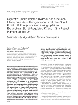
Results
Phosphorylated Hsp27 Is Up-Regulated in RPE
from Patients with AMD
Expression of phosphorylated Hsp27 has not been stud-
ied in human RPE. With the use of Western blot analysis,
we investigated the expression of phosphorylated Hsp27
in RPE from patients who were given a diagnosis of dry
AMD and control donors. As shown in Figure 1A, we
showed for the first time that phosphorylated Hsp27 ex-
pression is robustly up-regulated in RPE from patients
with AMD compared with controls. As well, clear expres-
sion of phosphorylated Hsp27 was evidenced in human
RPE monolayer by immunostaining on paraffin-embed-
ded eye cross-sections from control eyes (Figure 1, D
and F). We confirmed that phosphorylated Hsp27 ex-
pression was increased in the RPE from patients with
AMD (Figure 1, E and G). All of the sections were posi-
tively stained for phosphorylated Hsp27. Negative con-
trols did not show any staining for phosphorylated Hsp27
(Figure 1, B and C). These findings demonstrate for the
first time that RPE monolayer constitutively expresses
phosphorylated Hsp27 and that increased Hsp27 phos-
phorylation may play a critical role in the pathogenesis of
dry AMD.
Exposure of RPE Cells to HQ Induces Hsp27
Dimerization and Up-Regulates Hsp27 mRNA
Expression
Hsp27 in unstressed cells exists as large oligomers,
whereas on phosphorylation, Hsp27 reorganizes into
small dimeric units. Oxidative injury has been previously
1202 Pons et al
AJP September 2010, Vol. 177, No. 3
6. shown to regulate Hsp27 expression in RPE cells.31
In the
present study, we treated the cells with 100 mol/L of HQ
for 6 hours and investigated whether HQ induces Hsp27
dimerization and modulates Hsp27 expression in cul-
tured ARPE-19 cells by Western blot and real-time PCR,
respectively. The Hsp27 antibody detected a band with a
molecular weight 54 kDa recognizing Hsp27 dimers (Fig-
ure 2A). A basal level of dimerized Hsp27 was expressed
in control ARPE-19 cells. Moreover, we found that expo-
sure of cells to 100 mol/L of HQ for 6 hours resulted in
ϳ8.9-fold (P Ͻ 0.001) increase in Hsp27 dimer formation
(Figure 2A). Treatment with 100 mol/L of HQ for 6 hours
also increased Hsp27 mRNA levels by ϳ38.7% com-
pared with control cells in response to HQ (P Ͻ 0.0001;
Figure 2B). These results show that HQ-induced nonle-
thal oxidative injury up-regulates Hsp27 expression in
RPE cells.
HQ Induces p38 MAPK-Mediated Hsp27
Phosphorylation
Next, we sought to elucidate the signaling pathway by
which HQ exerts these effects on Hsp27 in ARPE-19
cells. Hsp27 undergoes posttranslational modifications,
phosphorylation being the best characterized. The activ-
ity of Hsp27 likely depends on the phosphorylation state
of serine residues at positions 15, 78, and 82. To inves-
tigate whether HQ can regulate this type of modification,
we performed a time-course analysis of Hsp27 phosphor-
ylation by Western blot on cell lysates from ARPE-19 cells
exposed to 100 mol/L of HQ for various periods of time
by using a specific antibody that recognizes phosphory-
Figure 1. Increased phosphorylated Hsp27 (p-Hsp27) expression in human
RPE from patient donors with dry AMD. A: Representative Western blot for
p-Hsp27, total Hsp27, and GAPDH on RPE lysates from three donors with dry
AMD and three controls with no known history of eye disease. B–G: Repre-
sentative immunofluorescent double staining of p-Hsp27 (green) and nuclei
(bleu) in retina sections from human donor eyes with no known eye disease
(D), and human donor eyes with dry AMD (E). Negative controls were
generated by omission of the primary antibody (B and C). Higher magnifi-
cation showing RPE and BrM in control (F) and patients with AMD (G).
Sections were analyzed by using confocal microscopy (original magnifica-
tion, ϫ40 and ϫ400). INL, inner nuclear layer; ONL, outer nuclear layer; PIS,
photoreceptor inner segments; POS, photoreceptor outer segments; Ch,
choroid.
Figure 2. Effect of HQ on Hsp27 in ARPE-19 cells. Confluent serum-starved
ARPE-19 cells were treated with or without 100 mol/L HQ for six hours. A:
Hsp27 monomer and dimer expression in ARPE-19 cells before and after
treatment with HQ for six hours. Hsp27 expression was assessed by Western
blot analysis. Top: Western blot from a representative experiment showing
change in Hsp27 dimer protein expression. Number on the left represents
protein molecular weight in kilodaltons. Bottom: Average results of three
independent experiments run in duplicate. Total Hsp27 protein expression
was normalized to -actin. B: Up-regulation of Hsp27 mRNA expression in
ARPE-19 cells exposed to HQ for six hours. Total RNA was extracted and
Hsp27 mRNA expression was analyzed by real-time PCR. GAPDH was used
as the housekeeping gene. Data represent average results of five indepen-
dent experiments run in duplicate. Results are expressed as mean Ϯ SE.
***P Ͻ 0.001 versus control (ϪHQ).
Hsp27 and Oxidative Stress in RPE 1203
AJP September 2010, Vol. 177, No. 3
7. lated Hsp27 at Serine 82. HQ time-dependently induced
a rapid and robust phosphorylation of Hsp27 in ARPE-19
cells that began within 1 minute (147 Ϯ 18.2%, P Ͻ 0.05)
with a maximum response observed at 5 minutes (321 Ϯ
42.5%, P Ͻ 0.001; Figure 3A).
It is widely recognized that Hsp27 phosphorylation is
mediated by the p38 MAPK cascade typically activated
on cellular stress. Therefore, we next examined the role of
p38 MAPK signaling pathway in HQ-induced Hsp27
phosphorylation in ARPE-19 cells. A time-course study of
p38 MAPK activation was first determined by Western
blot analysis for phosphorylated p38 MAPK by using a
specific antibody that recognizes the dually phosphory-
lated p38 at threonine 180/tyrosine 182. Exposure to HQ
resulted in a rapid increase in p38 MAPK phosphoryla-
tion, concomitant to Hsp27 that was maximal at 5 minutes
(334.6 Ϯ 44.7%, P Ͻ 0.001) and started to decline pro-
gressively thereafter (Figure 3B). These data suggest that
p38 MAPK mediates HQ-induced Hsp27 phosphorylation
in RPE cells exposed to oxidative stress.
Inhibition of Hsp27 Phosphorylation Decreases
HQ-Induced Disruption of the F-Actin
Cytoskeleton
To confirm that Hsp27 phosphorylation by HQ was de-
pendent on p38 MAPK signaling pathway, ARPE-19 cells
were pretreated for 1 hour with 20 mol/L of SB203580, a
pyridinyl imidazole highly selective inhibitor of the kinase
activity of p38␣ and p38 MAPK isoforms, and then
exposed to 100 mol/L of HQ for 5 minutes (based on the
kinetics of Hsp27 phosphorylation reported above).
Hsp27 phosphorylation was determined by Western blot
analysis. Exposure to HQ for 5 minutes resulted in ϳ48%
(147.6 Ϯ 6.5%, P Ͻ 0.001) increase in Hsp27 phosphor-
ylation compared with control cells (Figure 4A). Interest-
ingly, HQ-induced Hsp27 phosphorylation was ϳ2.9-fold
decreased in response to SB203580 compared with cells
treated with HQ alone (P Ͻ 0.001; Figure 4A).
It has been well documented that phosphorylation of
Hsp27 modulates the polymerization of actin and is pro-
posed to play a key role in actin cytoskeleton remodeling
during cellular stress. Therefore, to test the biological role
of Hsp27 and p38 MAPK in actin cytoskeleton rearrange-
ment in ARPE-19 cells exposed to oxidative injury, cells
pretreated with SB203580 before being exposed to HQ
for 6 hours were stained for F-actin by using rhodamin-
phalloidin and then examined by confocal fluorescence
microscopy. As shown in Figure 4B, control cells dem-
onstrated normal actin filaments. Also, nonlethal oxidative
injury with HQ preserved most of the cytoskeleton but
resulted in formation of focal aggregates compared with
the control cells (Figure 4B). Inhibiting p38 MAPK activity
and Hsp27 phosphorylation with the specific inhibitor
SB203580 almost totally blocked the reorganization of
F-actin microfilaments and formation of aggregates in
response to HQ as shown by a ϳ75% decrease com-
pared with cells treated with HQ alone (P Ͻ 0.0001;
Figure 4, B and C). Taken together, these results dem-
onstrate that phosphorylated Hsp27 is a key mediator in
HQ-induced actin aggregates formation in RPE cells.
Inhibition of Hsp27 Dephosphorylation
Increases HQ-Induced Reorganization of
F-Actin Filaments
PP2A is responsible for Hsp27 dephosphorylation in
vivo.38
To further determine whether phosphorylation of
Hsp27 is necessary for its effects on HQ-induced F-actin
reorganization, ARPE-19 cells were pretreated for 1 hour
with 60 nmol/L of okadaic acid, a potent and specific
inhibitor of PP1 and 2A, and then exposed to 100 mol/L
of HQ for 5 minutes. Hsp27 phosphorylation was exam-
Figure 3. HQ activates Hsp27 and p38 MAPK phosphorylation. A: Time-
dependent Hsp27 phosphorylation assessed by Western blot on cell lysates
from ARPE-19 cells treated with 100 mol/L HQ for various periods of time.
Top: Western blot from a representative experiment. Number on the left
represents protein molecular weight in kilodaltons. Bottom: Average results
of three independent experiments run in duplicate. Phospho-Hsp27 (p-
Hsp27) protein expression was normalized to total Hsp27. GAPDH was used
as loading control for total Hsp27. B: Time-dependent p38 MAPK phosphor-
ylation assessed by Western blot on cell lysates from ARPE-19 exposed to 100
mol/L HQ for various periods of time. Top: Western blot from a represen-
tative experiment. Number on the left represents protein molecular weight in
kilodaltons. Bottom: Average results of three independent experiments run
in duplicate on ARPE-19 cells. Phospho-p38 (p-p38) MAPK protein expres-
sion was normalized to GAPDH. Data are expressed as percentage of control
and are means Ϯ SE of three independent experiments run in duplicate. *P Ͻ
0.05, **P Ͻ 0.01, and ***P Ͻ 0.001 versus control.
1204 Pons et al
AJP September 2010, Vol. 177, No. 3
8. ined by Western blot analysis. As shown in Figure 5A,
inhibition of PP2A activity with okadaic acid resulted in
increased phosphorylation of Hsp27 in ARPE-19 cells
exposed to HQ for 5 minutes by ϳ29% (128.68 Ϯ 16.4%,
Figure 5. Inhibition of Hsp27 dephosphorylation increases HQ-induced
Hsp27 phosphorylation and reorganization of F-actin filaments in ARPE-19
cells. A: Increased HQ-induced Hsp27 phosphorylation by okadaic acid.
Confluent serum-starved ARPE-19 cells were pretreated for one hour with 60
nmol/L of okadaic acid, and then exposed to 100 mol/L HQ for five
minutes. Total Hsp27, p-Hsp27, and GAPDH protein expression was evalu-
ated by Western blot analysis. Top: Western blot from a representative
experiment. Numbers on the left represent protein molecular mass in kilo-
daltons. p-Hsp27 protein expression was normalized to total Hsp27 protein.
Bottom: Average densitometry results of five independent experiments run
in duplicate. Data are expressed as percentage of control and are means Ϯ
SE. **P Ͻ 0.01 and ***P Ͻ 0.001 versus control; *P Ͻ 0.05 versus okadaic acid
alone; ****P Ͻ 0.01 versus HQ alone. B: Increased formation of F-actin
aggregates showed by staining for F-actin in ARPE-19 cells pretreated for on
hour with or without 60 nmol/L of okadaic acid and then exposed to 100
mol/L HQ for six hours. Cells were stained with rhodamine-phalloidin and
examined by confocal microscopy by using magnification ϫ40. White ar-
rowheads show formation of focal aggregates. C: Quantification of F-acting
aggregates from three independent experiments run in duplicate. Data are
expressed as a percentage of HQ-treated cells and are means Ϯ SE. ***P Ͻ
0.001 versus HQ-treated cells.
Figure 4. Inhibition of p38 MAPK blocks HQ-induced Hsp27 phosphorylation
and focal aggregates formation in ARPE-19 cells. A: Inhibition of HQ-induced
Hsp27 phosphorylation by the selective inhibitor of the kinase activity of p38␣
and p38 MAPK isoforms, SB203580 (SB). ARPE-19 cells were pretreated for one
hour with 20 mol/L of SB, and then exposed to 100 mol/L HQ for five
minutes. Total Hsp27, p-Hsp27, and GAPDH protein expression was assessed by
Western blot analysis. Top: Representative Western blot for p-Hsp27. Numbers
on the left represent protein molecular mass in kilodaltons. p-Hsp27 protein
expression was normalized to total Hsp27 protein. GAPDH was used as loading
control for total Hsp27. Bottom: Average densitometry results of three indepen-
dent experiments run in duplicate. Data are expressed as percentage of control
and are means Ϯ SE. **P Ͻ 0.01 versus control; ***P Ͻ 0.001 versus HQ alone.
B: Decreased formation of F-actin aggregates showed by staining for F-actin in
ARPE-19 cells pretreated for one hour with or without 20 mol/L of SB, before
they were exposed to 100 mol/L HQ for six hours. Cells were stained with
rhodamine-phalloidin and examined by confocal microscopy by using magni-
fication ϫ40. White arrowheads show formation of focal aggregates. C: Quan-
tification of F-actin aggregates from three independent experiments run in
duplicate. Data are expressed as a percentage of HQ-treated cells and are
means Ϯ SE. ***P Ͻ 0.001 versus HQ-treated cells.
Hsp27 and Oxidative Stress in RPE 1205
AJP September 2010, Vol. 177, No. 3
9. P Ͻ 0.05) compared with cells treated with HQ alone.
Also, as shown in Figure 5, B and C, HQ-induced F-actin
aggregates formation was robustly increased by ϳ75%
in the presence of okadaic acid (P Ͻ 0.0001). These
results confirm that phosphorylated Hsp27 is a key me-
diator in HQ-induced actin aggregates formation in RPE
cells.
Inhibition of Hsp27 Phosphorylation Decreases
HQ-Induced Blebbing Whereas Inhibition of
Hsp27 Dephosphorylation Increases Blebs
Formation in Response to HQ
Our group previously showed that ARPE-19 cells exhibit
a distinct set of physiological responses, including mem-
brane blebbing, when subjected to nonlethal oxidative
injury.24
GFP-ARPE-19 cells were pretreated for 1 hour
with or without 20 mol/L of SB203580 or 60 nmol/L of
okadaic acid, and then exposed to 100 mol/L of HQ for
6 hours. Figure 6, A and B, shows the extensive blebbing
that occurs in GFP-ARPE-19 cells after exposure to non-
lethal concentrations of HQ for 6 hours. Inhibition of
p38 MAPK activity and Hsp27 phosphorylation with
SB203580 partially blocked bleb formation (Figure 6C).
On the other hand, inhibition of PP2 activity with okadaic
acid resulted in a large production of cell membrane
blebs (Figure 6D). Figure 6, E and F, shows the extensive
F-actin aggregates that occur in GFP-ARPE-19 cells after
exposure to nonlethal concentrations of HQ for 6 hours.
HQ-induced F-actin aggregates formation was partially
blocked by SB203580 (Figure 6G) and increased in the
presence of okadaic acid (Figure 6H) suggesting a rela-
tionship between bleb formation regulation and actin cy-
toskeleton modifications mediated by phosphorylated
Hsp27. These data suggest that regulation of actin cy-
toskeleton dynamics by phosphorylated Hsp27 plays a
key role in the regulation of blebbing.
Hsp27 siRNA Decreases HQ-Induced Hsp27
Phosphorylation and F-Actin Microfilaments
Disruption
To further evaluate the functional significance of Hsp27 in
HQ-induced F-actin rearrangement by another strategy,
ARPE-19 cells were transfected with siRNA against
Hsp27 to decrease endogenous Hsp27 protein expres-
sion. Cells transfected with scrambled siRNA were used
as controls. Forty-eight hours later, cells were exposed to
100 mol/L of HQ for either 5 minutes or 6 hours. The
constitutive expression of Hsp27 as evaluated by West-
ern blot was reduced by ϳ60% in ARPE 19 cells trans-
fected with Hsp27 siRNA compared with cells trans-
fected with scrambled siRNA (41.0 Ϯ 5.1% versus
100.0 Ϯ 5.7%, P Ͻ 0.01; Figure 7A). Next, we tested the
effect of Hsp27 silencing on HQ-induced Hsp27 phos-
phorylation. ARPE-19 cells transfected with Hsp27 siRNA
or scrambled siRNA were treated with 100 mol/L of HQ
for 5 minutes, and HQ-induced Hsp27 phosphorylation
was examined by Western blot analysis. Treatment with
HQ resulted in ϳ45% increase in Hsp27 phosphorylation
in cells transfected with scrambled siRNA compared with
unstimulated cells (P Ͻ 0.05). Interestingly, HQ-induced
Hsp27 phosphorylation was completely abolished in cells
transfected with Hsp27 siRNA (Figure 7B). Finally, rho-
damin-phalloidin staining on siRNA transfected cells
demonstrated that cells transfected with scrambled
siRNA displayed actin aggregates on HQ exposure for 6
hours. Also as shown in Figure 7, C and D, HQ-induced
F-actin aggregates formation was robustly decreased by
ϳ90% when the Hsp27 protein levels were depleted by
Figure 6. Induction of cellular changes in GFP-ARPE-19 cells exposed to HQ. Membrane blebbing (A–D) and formation of F-actin aggregates (E–H). Confluent
GFP-ARPE-19 cells were pretreated with or without 20 mol/L of SB203580 (SB) or 60 nmol/L of okadaic acid, and then exposed to 100 mol/L HQ for six hours.
Cells were directly observed under a fluorescence microscope (top) or stained with rhodamin-phalloidin and then examined under a fluorescent microscope
(bottom). A: Control GFP-ARPE-19 cells in which GFP was localized to the membrane. Membrane-derived blebs (thin arrows) after exposure to 100 mol/L
HQ for six hours (B), 20 mol/L of SB for one hour followed by 100 mol/L HQ for six hours (C), and 60 nmol/L of okadaic acid for one hour followed by 100
mol/L HQ for six hours (D). E: Control GFP-ARPE-19 cells showing normal F-actin filaments. F-actin aggregates (thick arrows) after exposure to 100 mol/L
HQ for six hours (F), 20 mol/L of SB for one hour followed by 100 mol/L HQ for six hours (G), and 60 nmol/L of okadaic acid for one hour followed by 100
mol/L HQ for six hours (H). Images are representative of three independent experiments. Original magnification, ϫ400.
1206 Pons et al
AJP September 2010, Vol. 177, No. 3
10. transfection with Hsp27 siRNA. These data demonstrate
that phosphorylated Hsp27 plays an essential role in the
regulation of actin cytoskeleton dynamics in response to
HQ-induced oxidative injury in RPE cells.
HQ Activates ERK1/2 MAPK Signaling Pathway
H2O2-induced oxidative stress has been shown to cause
ERK1/2 phosphorylation and reorganization of actin cy-
toskeleton in ARPE-19 cells.39
Although an important
contribution of p38 MAPK in HQ-induced Hsp27 phos-
phorylation and F-actin rearrangement has been evi-
denced in the present study, we sought to also measure
ERK1/2 activation to determine whether HQ-induced
phosphorylation of Hsp27 was restricted to the p38
MAPK pathway. Therefore, a time-course of ERK1/2
phosphorylation was analyzed by Western blot in cell
lysates from ARPE-19 cells exposed to 100 mol/L of HQ
for various periods of time as described in Material and
Methods. Exposure of ARPE 19 cells to HQ induced a
rapid and robust ERK1/2 phosphorylation starting at 1
minute (215.32 Ϯ 29.2%, P Ͻ 0.01), reaching a peak
between 5 (340.05 Ϯ 32.4%, P Ͻ 0.001) and 20 minutes
(305.6 Ϯ 38.6, P Ͻ 0.01), and declining quickly afterward
(Figure 8). These results show for the first time that HQ-
induced oxidative stress leads to a dual activation of p38
and ERK pathways in RPE cells.
A Cross Talk between p38 and ERK1/2 MAPK
Signaling Pathways Mediates HQ Effects
A substantial body of evidence suggests a cross talk
between p38 and ERK1/2 MAPK pathways in a variety of
cells.40 – 42
Enhancement of ERK1/2 phosphorylation by
treatment with pharmacological inhibitor of p38 MAPK
kinase SB203580 has been reported in PC12 cells in
response to epidermal growth factor.43
Moreover,
Figure 7. Effect of Hsp27 gene silencing on HQ-in-
duced Hsp27 phosphorylation and F-actin aggregates
formation. ARPE-19 cells were transfected with siRNA
against Hsp27 or scrambled siRNA. Forty-eight hours
later, cells were exposed to 100 mol/L HQ for either
five minutes or six hours. A: Reduction in endogenous
Hsp27 protein expression by siRNA against Hsp27. The
efficiency of siRNA against Hsp27 in reducing endoge-
nous Hsp27 protein was confirmed by Western blot
analysis. Top: Western blot from a representative exper-
iment. Numbers on the left represent protein molecular
mass in kilodaltons. Hsp27 protein expression was nor-
malized to GAPDH. Bottom: Average densitometry re-
sults of four independent experiments run in duplicate.
Data are expressed as percentage of scrambled siRNA
and are means Ϯ SE. *P Ͻ 0.05 versus scrambled siRNA.
B: Effect of Hsp27 siRNA on HQ-induced Hsp27 phos-
phorylation. Hsp27 phosphorylation was assessed by
Western blot analysis and p-Hsp27 protein expression
normalized to GAPDH. Top: Western blot from a repre-
sentative experiment. Number on the left represents pro-
tein molecular weight in kilodaltons. Bottom: Average
densitometry results of four independent experiments
run in duplicate. Data are expressed as percentage of
control (ϪHQ) and are means Ϯ SE. *P Ͻ 0.05 versus
control (ϪHQ). C: Inhibition of focal aggregates forma-
tion by Hsp27 gene silencing in ARPE-19 cells treated
with HQ. Cells were stained with rhodamine-phalloidin
and examined by confocal microscopy by using magni-
fication ϫ40. Arrows show formation of focal aggre-
gates. D: Quantification of F-acting aggregates from
three independent experiments run in duplicate. Data
are expressed as a percentage of HQ-treated cells trans-
fected with scrambled siRNA and are means Ϯ SE. ***P Ͻ
0.001 versus scrambled siRNA.
Hsp27 and Oxidative Stress in RPE 1207
AJP September 2010, Vol. 177, No. 3
11. SB203580 has been shown to activate ERK1/2 transduc-
tion pathway in primary cultures of human hepatocytes44
as well as in monocytic cells.45
These observations
prompted us to examine the possibility of similar interac-
tions between p38 and ERK1/2 MAPK in ARPE 19 cells
treated with HQ by determining whether changes in the
activity of either the ERK1/2 or the p38 pathways had a
feedback effect on the other parallel pathway. First, we
sought to establish whether ERK1/2 activation could be
modulated by p38 MAPK. For this purpose, ERK1/2 phos-
phorylation status was analyzed by Western blot in
ARPE-19 cells pretreated with 20 mol/L of SB203580,
then exposed to 100 mol/L of HQ for 5 minutes. As
shown in Figure 9A, HQ-induced phosphorylation of
ERK1/2 was dramatically enhanced in the presence of
p38 kinase inhibitor SB203580 (323.4 Ϯ 36.4%, P Ͻ
0.001) compared with HQ alone (191.6 Ϯ 33.6%, P Ͻ
0.01). These data strongly suggest a negative regulation
of ERK signaling by p38 MAPK in human RPE cells ex-
posed to HQ-induced oxidative injury.
To further investigate the potential interactions be-
tween these two MAPK pathways, we next evaluated the
ability of the ERK pathway to modulate p38. ARPE-19
cells were pretreated with 40 mol/L of ERK inhibitor
PD98059 then with HQ for 5 minutes, and p38 phosphor-
ylation was analyzed by Western blot. We observed that
pretreatment with PD98059 partially blocked HQ-induced
p38 MAPK phosphorylation (Figure 9B). These results
suggest a positive regulation of p38 MAPK signaling by
ERK in human RPE cells treated with HQ.
Inhibition of ERK Signaling Pathways Blocked
HQ-Induced Hsp27 Phosphorylation and
F-Actin Reorganization
It is believed that p38 MAPK is the major physiological
activator of Hsp27. However, a recent study by Hong et
al46
revealed that both p38 and ERK MAPK are upstream
regulators of Hsp27 phosphorylation in Glial cell-derived
neurotrophic factor (GDNF)-induced neurite outgrowth of
dopaminergic neurons. Based on these observations and
the cross talk of p38 and ERK MAPK pathways reported
above, we next examined whether ERK1/2 could be an
upstream regulator of Hsp27 phosphorylation in re-
Figure 8. HQ activates ERK1/2 phosphorylation. Time-dependent ERK1/2
phosphorylation assessed by Western blot on cell lysates from ARPE-19 cells
treated with 100 mol/L HQ for various periods of time. Top: Western blot
from a representative experiment. Number on the left represents protein
molecular weight in kilodaltons. Bottom: Average densitometry results of
three independent experiments run in duplicate. Phospho-ERK (p-ERK)
protein expression was normalized to total ERK. GAPDH was used as loading
control for total ERK. Data are expressed as percentage of control and are
means Ϯ SE. *P Ͻ 0.05, **P Ͻ 0.01, and ***P Ͼ 0.001 versus control.
Figure 9. Cross talk between p38 and ERK1/2 MAPK signaling pathways in
ARPE-19 cells treated with HQ. A: Inhibition of p38 MAPK pathway poten-
tiates HQ-induced ERK1/2 phosphorylation in ARPE-19 cells. Confluent se-
rum-starved ARPE-19 cells were pretreated for one hour with 20 mol/L of
p38 MAPK inhibitor SB203580 (SB), and then exposed to 100 mol/L HQ for
five minutes. Phospho-ERK (p-ERK) was examined by Western blot and
normalized to total ERK. GAPDH was used as loading control for total ERK.
B: Inhibition of ERK MAPK pathway with PD98059 (PD) leads to a decrease
in HQ-induced p38 phosphorylation in ARPE-19 cells. ARPE-19 cells were
pretreated for one hour with 40 mol/L of ERK inhibitor PD, and then
exposed to 100 mol/L HQ for five minutes. Phospho-p38 (p-p38) was
examined by Western blot and normalized to total p38. GAPDH was used as
loading control for total p38. Top: Western blot from a representative ex-
periment. Number on the left represents protein molecular weight in kilo-
daltons. Bottom: Average densitometry results of three independent exper-
iments run in duplicate. Data are expressed as percentage of control and are
means Ϯ SE. *P Ͻ 0.05, **P Ͻ 0.01, and ***P Ͻ 0.001 versus control; ****P Ͻ
0.01 and *P Ͻ 0.05 versus HQ alone.
1208 Pons et al
AJP September 2010, Vol. 177, No. 3
12. sponse to HQ-induced oxidative injury in ARPE-19 cells.
To address this question, induction of Hsp27 phosphor-
ylation was analyzed by Western blot in ARPE-19 cells
pretreated with 40 mol/L of the specific ERK inhibitor
PD98059, then exposed to 100 mol/L of HQ for 5 min-
utes. As shown in Figure 10A, inhibition of ERK signaling
pathway with PD98059 totally abolished HQ-induced
Hsp27 phosphorylation. These data suggest that Hsp27
might be a downstream common activated protein of p38
and ERK1/2 transduction pathways.
Since we observed an induction of ERK1/2 phosphor-
ylation in response to HQ that was increased by
SB203580, we were interested in studying the potential
role of ERK in HQ-induced actin cytoskeletal alterations in
ARPE-19 cells. Therefore, to determine the role of the
ERK pathway in mediating the formation of actin aggre-
gates in response to oxidative stress, we examined F-
actin immunostaining in cells pretreated with 40 mol/L of
ERK inhibitor PD98059 for 1 hour before being exposed
to 100 mol/L of HQ for 6 hours. PD98059 almost com-
pletely abolished F-actin rearrangement induced by HQ
(Figure 10, B and C). These data show that F-actin cy-
toskeleton rearrangement induced by HQ is also modu-
lated by the ERK pathway.
HQ Induces Hsp25, p38, and ERK MAPK
Phosphorylation but Decreased Hsp25
Expression in Mice
We used the experimental model for sub-RPE deposits of
mice chronically exposed to oral HQ described else-
where.22,23
As reported earlier, HQ-treated mice had in-
creased blood levels of HQ (10.4 Ϯ 0.7 ng/ml) relative to
control mice that showed nondetectable levels.22
Here,
we demonstrated for the first time that Hsp25 phosphor-
ylation was increased in RPE from mice exposed to HQ
as shown by Western blot (ϳ1.5-fold, P Ͻ 0.0001; Figure
11B). Furthermore, in agreement with our in vitro results,
we confirmed that treatment with HQ induced a concom-
itant robust phosphorylation of p38 (ϳ4.0-fold increase,
P Ͻ 0.0001; Figure 12A) and ERK (1.5-fold increase, P Ͻ
0.001; Figure 12C) MAPK with no change in total p38
(Figure 12B) and total ERK (Figure 12D). However, we
found that HQ administered chronically to mice for 7
months resulted in a dramatic 69% decrease in Hsp25
mRNA (Figure 11A) and 63% decline in Hsp25 protein
expression compared with control mice (P Ͻ 0.0001;
Figure 11C). Together, these data demonstrate that in
conjunction with sub-RPE deposits formation reported
previously,21
exposure to HQ induces Hsp25 phosphor-
ylation with concomitant activation of both p38 and ERK
MAPK pathways in mouse RPE, therefore suggesting a
role of phosphorylated Hsp27/25 in mediating deposits
formation.
Discussion
The present study shows for the first time that phosphor-
ylated Hsp27 expression is increased in RPE from pa-
tients with AMD. We also demonstrate that HQ-induced
Hsp27 phosphorylation and F-actin reorganization re-
quired for RPE-derived bleb formation are mediated by a
cross talk between p38 and ERK MAPK in ARPE-19 cells.
Moreover, we show that mice exposed to HQ in drinking
water for 7 months exhibit increased phosphorylated
Hsp25, p38, and ERK, and decreased Hsp25 mRNA and
protein expression in the RPE.
We previously reported that ARPE-19 cells constitu-
tively express high levels of Hsp27.31
Although Hsp27
expression has been reported in human cornea,47,48
in
Figure 10. Inhibition of ERK MAPK pathway blocks HQ-induced Hsp27 phosphorylation and focal aggregates formation. A: Abrogation of HQ-induced Hsp27
phosphorylation by PD98059. Confluent serum-starved ARPE-19 cells were pretreated for one hour with 40 mol/L of ERK inhibitor PD98059 (PD), and then
exposed to 100 mol/L HQ for five minutes. Total Hsp27, p-Hsp27, and GAPDH protein expression was evaluated by Western blot analysis. Top: Western blot
from a representative experiment. Numbers on the left represent protein molecular mass in kilodaltons. p-Hsp27 protein expression was normalized to total Hsp27
protein. Bottom: Average densitometry results of three independent experiments run in duplicate. Data are expressed as percentage of control and are means Ϯ
SE. *P Ͻ 0.01 versus HQ alone; **P Ͻ 0.01 versus control. B: Decreased formation of F-actin aggregates showed by staining for F-actin in ARPE-19 cells pretreated
for one hour with or without 40 mol/L of PD and then exposed to 100 mol/L HQ for six hours. Cells were stained with rhodamine-phalloidin and examined
by confocal microscopy by using magnification ϫ40. Arrows show formation of focal aggregates. C: Quantification of F-acting aggregates from three independent
experiments run in duplicate. Data are expressed as percentage of HQ-treated cells and are means Ϯ SE. ***P Ͻ 0.001 versus HQ-treated cells.
Hsp27 and Oxidative Stress in RPE 1209
AJP September 2010, Vol. 177, No. 3
13. vivo expression of phosphorylated Hsp27 has not been
explored in human RPE. Our results show for the first time
that RPE from human eye donors constitutively express
phosphorylated Hsp27, and that phosphorylated Hsp27
expression is increased in patients with dry AMD. Hsp27
phosphorylation has been abundantly described in sev-
eral human diseases.49
Yet, there is a complete lack of
information regarding the possible association between
phosphorylated Hsp27 and AMD. Increased Hsp27 pro-
tein content along with evidence of cellular oxidative
stress was reported in human eyes with AMD, but Hsp27
phosphorylation was not investigated.50
Our findings pro-
vide novel evidence that phosphorylated Hsp27 may play
a major role in the pathogenesis of AMD.
In response to stress, phosphorylated Hsp27 under-
goes conformational changes and reorganizes into
dimeric units.26,51–54
Phosphorylated Hsp27 regulates
actin filaments dynamics by repressing the ability of
Hsp27 to block actin polymerization.55
We previously
showed that exposure of ARPE-19 cells to oxidative injury
with myeloperoxidase and hydrogen peroxide resulted in
a marked increase in Hsp27 mRNA and protein expres-
sion.31
The results presented here extend these previous
observations by showing that exposure of ARPE-19 cells
to a 6-hour HQ nonlethal injury induced the transcrip-
tional activation of Hsp27, accumulation of Hsp27
dimers, and a rapid phosphorylation of Hsp27. Our find-
ings are in agreement with previous studies revealing
Hsp27 phosphorylation in different cellular systems in
response to oxidative stress.26,29,51,52,56
Several reports
have shown that PP2A is involved in Hsp27 dephosphor-
ylation.38,49
Using okadaic acid as an inhibitor of Hsp27
dephosphorylation by PP1 and 2A, we observed an in-
crease in Hsp27 phosphorylation in HQ-stimulated
ARPE-19 cells as well as F-actin reorganization and blebs
formation. Moreover, we demonstrate that Hsp27 phos-
phorylation and F-actin aggregates formation are almost
completely abolished in cells transfected with siRNA
against Hsp27 following treatment with HQ. These data
establish a direct correlation between levels of phosphor-
ylated Hsp27 and actin cytoskeleton reorganization in
response to HQ-induced oxidative injury in ARPE-19
cells. Collectively, these findings give support to a key
role of phosphorylated Hsp27 in the regulation of F-actin
filaments dynamics and blebs formation following HQ-
induced oxidative stress in RPE cells. Furthermore, in an
effort to correlate our in vitro observations to a more
physiological environment in vivo, we used the experi-
mental model for sub-RPE deposits.21,22
We previously
reported that mice chronically exposed to HQ developed
sub-RPE deposits and BrM thickening consistent with
changes in early stages of human AMD.57
Here, we
found that RPE from these mice showed increased levels
of phosphorylated Hsp25. However, in contrast to
ARPE-19 cells, our results indicate that Hsp25 mRNA and
protein levels were dramatically decreased in mice
chronically exposed to HQ. One possible explanation
might be that human RPE cells were transiently exposed
to HQ for 24 hours, whereas mice were chronically
treated for 7 months, which might result in a differential
transcriptional regulation of Hsp25/27. Another possibility
is that mice might not be able to tolerate chronic activa-
tion of the Hsp25 signaling pathway resulting from sus-
tained exposure to HQ. Sustained treatment with HQ
might provide a negative feed-back loop that down-reg-
ulates Hsp25 expression to accommodate the chronic
phosphorylation of Hsp25. Together, these data confirm
that phosphorylated Hsp25/27 plays a major role in HQ-
induced oxidative injury in the RPE, the key target cell in
the pathogenesis of AMD.
Figure 11. Regulation of Hsp25 and phosphorylated Hsp27 (p-Hsp25) in
dissected RPE sheets from mice treated with HQ (0.8%) in drinking water for
seven months. Protein and total RNA were extracted from RPE sheets (n ϭ 6
eyes/group). A: Hsp25 mRNA expression analyzed by real-time PCR. GAPDH
was used as the housekeeping gene. Data represent average results from six
eyes normalized to the housekeeping gene. Results are expressed as mean Ϯ
SE. ***P Ͻ 0.001 versus control. B: Ratio p-Hsp25/Hsp25 protein expression
evaluated by Western blot. Top: Western blot from a representative exper-
iment. Numbers on the left represent protein molecular mass in kilodaltons.
p-Hsp25 protein expression was normalized to total Hsp25 protein. Bottom:
Data are the average results from six eyes. Data are expressed as percentage
of control and shown are mean results Ϯ SE. ***P Ͻ 0.001 versus control. C:
Ratio Hsp25/GAPDH protein expression evaluated by Western blot. Top:
Western blot from a representative experiment. Numbers on the left repre-
sent protein molecular mass in kilodaltons. Hsp25 protein expression was
normalized to GAPDH. Bottom: Data are the average results from six eyes.
Data are expressed as percentage of control and shown are mean results Ϯ
SE. ***P Ͻ 0.001 versus control.
1210 Pons et al
AJP September 2010, Vol. 177, No. 3
14. Next, we demonstrated that HQ induced a rapid and
robust activation of p38 MAPK signaling pathway, which
parallels Hsp27 phosphorylation in ARPE-19 cells. Chronic
exposure to HQ also resulted in increased p38 phosphor-
ylation in mice. These results together with the observation
that SB203580, a specific pharmacological inhibitor of p38
kinase activity,58
efficiently blocked Hsp27 phosphorylation
as well as actin cytoskeleton remodeling and blebs forma-
tion in response to HQ in RPE cells strongly suggest that
p38 activation by HQ modulates F-actin aggregates forma-
tion and membrane blebbing through Hsp27 phosphoryla-
tion in RPE cells. Our findings are in agreement with prior
studies revealing p38 MAPK signaling pathway as an up-
stream mediator in oxidative stress-induced actin reorgani-
zation and Hsp27 phosphorylation.26,29,52
The ERK cascade participates in numerous intracellular
signaling pathways in response to environmental stimuli,
such as oxidative stress. H2O2-induced oxidative stress
causes ERK phosphorylation and reorganization of actin
cytoskeleton in ARPE-19 cells.39
In the present study, we
found that treatment with HQ led to a robust activation of
ERK signaling pathway in ARPE-19 cells as well as in mice.
These results together with the observation that PD98059, a
specific pharmacological inhibitor of Mitogen-activated
ERK Kinase (MEK), completely abolished Hsp27 phosphor-
ylation as well as actin cytoskeleton remodeling in response
to HQ strongly suggest that ERK is also a key upstream
activator of HQ-induced Hsp27 phosphorylation in RPE
cells. Our results not only showed that kinetics of p38 and
ERK phosphorylation correlated well with that of Hsp27, but
also that Hsp27 phosphorylation and F-actin aggregates
formation were decreased after inhibition of either p38 or
ERK signaling cascades. These observations suggest that
p38 as well as ERK MAPK pathways are required for the
optimal activation of Hsp27 leading to F-actin rearrange-
ment and bleb formation in RPE cells in response to HQ.
Van Gorp et al59
have reported that H2O2-induced oxidative
stress results in p38-mediated membrane blebbing in en-
dothelial cells. However, activation of both p38 and ERK
pathways has been described in endothelial cells in re-
sponse to H2O2-induced oxidative injury.29
But our data are
not in agreement with the former study, which revealed that
inhibition of the ERK pathway led to an increase in bleb
formation after exposure to H2O2. Cell-type specific differ-
ences in oxidative stress response may account for such a
discrepancy.
Given the concomitant activation of p38 and ERK in
response to HQ, we were interested in determining whether
interactions might occur between these two signaling path-
ways. A substantial body of evidence suggests a cross talk
between p38 and ERK MAPK cascades in a variety of
cells.40–42,60
Treatment with p38 inhibitor SB203580 has
been shown to enhance ERK phosphorylation in a number
of cell lines.43–45
Here, we provide evidence that p38 MAPK
negatively regulates HQ-induced ERK activation in
ARPE-19 cells. HQ-induced ERK phosphorylation was en-
hanced in response to SB203580. We also showed that p38
MAPK inhibition had the ability to activate the ERK pathway
in cells treated with SB203580 alone, suggesting that p38
exerts a tonic inhibition on the ERK pathway under basal
conditions. The signaling events responsible for the cross
talk between p38 and ERK signaling cascades are un-
known. The regulation of MAPK pathways involves a com-
plex interplay between kinases and phosphatases. There-
fore, the dynamic balance and the potential interactions
between p38 and ERK are important in determining how
RPE cells respond to HQ-induced oxidative stress. In this
regard, PP2A appears to be the major kinase phosphatase
Figure 12. Regulation of phosphorylated p38 (p-
p38), and phosphorylated ERK (p-ERK) in dis-
sected RPE sheets from mice treated with HQ
(0.8%) in drinking water for seven months. Protein
was extracted from RPE sheets (n ϭ 6 eyes/
group). A: p-p38 protein expression determined
by Western blot. Top: Western blot from a repre-
sentative experiment. Numbers on the left repre-
sent protein molecular mass in kilodaltons. p-p38
protein expression was normalized to total p38
protein. Bottom: Data are the average results from
six eyes. Data are expressed as percentage of con-
trol and shown are mean results Ϯ SE. ***P Ͻ 0.001
versus control. B: Ratio p38/GAPDH protein ex-
pression evaluated by Western blot. Top: Western
blot from a representative experiment. Numbers
on the left represent protein molecular mass in
kilodaltons. Bottom: Data are the average results
from six eyes. Data are expressed as percentage of
control and shown are mean results Ϯ SE. C: pERK
protein expression determined by Western blot.
Top: Western blot from a representative experi-
ment. Numbers on the left represent protein mo-
lecular mass in kilodaltons. p-ERK protein expres-
sion was normalized to total ERK protein. Bottom:
Data are the average results from six eyes. Data are
expressed as percentage of control and shown are
mean results Ϯ SE. ***P Ͻ 0.001 versus control. D:
Ratio ERK/GAPDH protein expression evaluated
by Western blot. Top: Western blot from a repre-
sentative experiment. Numbers on the left repre-
sent protein molecular mass in kilodaltons. Bot-
tom: Data are the average results from six eyes.
Data are expressed as percentage of control and
shown are mean results Ϯ SE.
Hsp27 and Oxidative Stress in RPE 1211
AJP September 2010, Vol. 177, No. 3
15. in eukaryotic cells that may play an important role by acting
on ERK signaling cascade at multiple levels.61
Interestingly,
it has been demonstrated that activation of p38 MAPK can
block ERK pathway through activation of protein phos-
phates 1 and 2A.62
Moreover, PP2A mediates a cross talk
between p38 MAPK and ERK in cardiac myocytes, as
shown by increased ERK phosphorylation in H2O2-stimu-
lated ventricular myocytes in response to okadaic acid.60
Based on these observations, it can be imagined that p38
MAPK might regulate HQ-induced ERK activation through
PP2A in ARPE-19 cells. Future studies will test this hypoth-
esis. Another possibility to be considered is a direct phys-
ical interaction between phosphorylated p38 and ERK.63
One-way cross talk between p38 and ERK has been
reported in various cells.60,64,65
To further investigate the
relationship between p38 and ERK pathways in response to
HQ in ARPE-19 cells, we examined the effects of MEK
inhibitor PD98059 on HQ-induced phosphorylation of p38.
We found that PD98059 decreased HQ-induced p38 MAPK
activation indicating that ERK1/2 pathway positively modu-
lates p38 signaling and that ERK is required for HQ-induced
activation of p38 MAPK signaling pathway in human RPE
cells. These data present evidence of a previously unre-
ported interaction between ERK and p38 during HQ-in-
duced oxidative stress in RPE cells.
In summary, our observations demonstrate that phos-
phorylated Hsp27 might be a key mediator in human AMD
and cigarette smoke-related HQ-induced oxidative injury in
RPE. Our study also provides evidence that HQ-induced
oxidative stress leads to a dual activation of p38 and ERK
pathways, which contribute to HQ-induced Hsp27 phos-
phorylation, actin rearrangement, and blebs formation in
ARPE-19 cells. Furthermore, this study presents ERK as a
novel upstream positive regulator of Hsp27 and actin ag-
gregates formation in response to HQ-induced oxidative
injury in RPE cells. The cellular mechanisms underlying the
regulation of these signaling events have yet to be deter-
mined. Given that there is no effective treatment for dry
AMD, this study highlights Hsp27 as a potential, disease-
related protein as well as biochemical pathways for poten-
tial therapeutic strategies.
Acknowledgments
We thank Dr. George Inana, Dr. Sander Dubovy, and
co-workers at the Lions Eye Bank, Miami, for donating the
human tissues. We also would like to acknowledge Dr.
William Feuer for his technical assistance with statistical
analysis, and Dr. Gabriel Gaidosh for his expertise and
technical assistance with confocal microscopy.
References
1. Augood CA, Vingerling JR, de Jong PT, Chakravarthy U, Seland J,
Soubrane G, Tomazzoli L, Topouzis F, Bentham G, Rahu M, Vioque J,
Young IS, Fletcher AE: Prevalence of age-related maculopathy in
older Europeans: the European Eye Study (EUREYE). Arch Ophthal-
mol 2006, 124:529–535
2. Evans JR: Risk factors for age-related macular degeneration. Prog
Retin Eye Res 2001, 20:227–253
3. Javitt JC, Zhou Z, Maguire MG, Fine SL, Willke RJ: Incidence of
exudative age-related macular degeneration among elderly Ameri-
cans. Ophthalmology 2003, 110:1534–1539
4. Klein R, Peto T, Bird A, Vannewkirk MR: The epidemiology of age-
related macular degeneration. Am J Ophthalmol 2004, 137:486–495
5. Katta S, Kaur I, Chakrabarti S: The molecular genetic basis of age-
related macular degeneration: an overview. J Genet 2009, 88:425–449
6. Green WR: Histopathology of age-related macular degeneration. Mol
Vis 1999, 5:27–36
7. Shen JK, Dong A, Hackett SF, Bell WR, Green WR, Campochiaro PA:
Oxidative damage in age-related macular degeneration. Histol His-
topathol 2007, 22:1301–1308
8. Beatty S, Koh H, Phil M, Henson D, Boulton M: The role of oxidative
stress in the pathogenesis of age-related macular degeneration. Surv
Ophthalmol 2000, 45:115–134
9. Clemons TE, Milton RC, Klein R, Seddon JM, Ferris FL, 3rd: Risk
factors for the incidence of advanced age-related macular degener-
ation in the Age-Related Eye Disease Study (AREDS) AREDS report
no. 19. Ophthalmology 2005, 112:533–539
10. Crabb JW, Miyagi M, Gu X, Shadrach K, West KA, Sakaguchi H,
Kamei M, Hasan A, Yan L, Rayborn ME, Salomon RG, Hollyfield JG:
Drusen proteome analysis: an approach to the etiology of age-
related macular degeneration. Proc Natl Acad Sci USA 2002,
99:14682–14687
11. Christen WG, Glynn RJ, Manson JE, Ajani UA, Buring JE: A prospec-
tive study of cigarette smoking and risk of age-related macular de-
generation in men. JAMA 1996, 276:1147–1151
12. Seddon JM, Willett WC, Speizer FE, Hankinson SE: A prospective
study of cigarette smoking and age-related macular degeneration in
women. JAMA 1996, 276:1141–1146
13. Klein R, Klein BE, Moss SE: Relation of smoking to the incidence of
age-related maculopathy: the Beaver Dam Eye Study. Am J Epide-
miol 1998, 147:103–110
14. Solberg Y, Rosner M, Belkin M: The association between cigarette
smoking and ocular diseases. Surv Ophthalmol 1998, 42:535–547
15. Bertram KM, Baglole CJ, Phipps RP, Libby RT: Molecular regulation
of cigarette smoke induced-oxidative stress in human retinal pigment
epithelial cells: implications for age-related macular degeneration.
Am J Physiol Cell Physiol 2009, 297:C1200–C1210
16. Deisinger PJ, Hill TS, English JC: Human exposure to naturally oc-
curring hydroquinone. J Toxicol Environ Health 1996, 47:31–46
17. DeCaprio AP: The toxicology of hydroquinone: relevance to occupa-
tional and environmental exposure. Crit Rev Toxicol 1999, 29:283–330
18. Burns RP, Feeney-Burns L: Clinico-morphologic correlations of
drusen of Bruch’s membrane. Trans Am Ophthalmol Soc 1980,
78:206–225
19. Ishibashi T, Patterson R, Ohnishi Y, Inomata H, Ryan SJ: Formation of
drusen in the human eye. Am J Ophthalmol 1986, 101:342–353
20. Sarks S, Cherepanoff S, Killingsworth M, Sarks J: Relationship of
Basal laminar deposit and membranous debris to the clinical presen-
tation of early age-related macular degeneration. Invest Ophthalmol
Vis Sci 2007, 48:968–977
21. Espinosa-Heidmann DG, Suner IJ, Catanuto P, Hernandez EP, Marin-
Castano ME, Cousins SW: Cigarette smoke-related oxidants and the
development of sub-RPE deposits in an experimental animal model of
dry AMD. Invest Ophthalmol Vis Sci 2006, 47:729–737
22. Marin-Castano ME, Striker GE, Alcazar O, Catanuto P, Espinosa-
Heidmann DG, Cousins SW: Repetitive nonlethal oxidant injury to
retinal pigment epithelium decreased extracellular matrix turnover in
vitro and induced sub-RPE deposits in vivo. Invest Ophthalmol Vis Sci
2006, 47:4098–4112
23. Alcazar O, Cousins SW, Marin-Castano ME: MMP-14 and TIMP-2
overexpression protects against hydroquinone-induced oxidant in-
jury in RPE: implications for extracellular matrix turnover. Invest Oph-
thalmol Vis Sci 2007, 48:5662–5670
24. Marin-Castano ME, Csaky KG, Cousins SW: Nonlethal oxidant injury
to human retinal pigment epithelium cells causes cell membrane
blebbing but decreased MMP-2 activity. Invest Ophthalmol Vis Sci
2005, 46:3331–3340
25. Strunnikova N, Zhang C, Teichberg D, Cousins SW, Baffi J, Becker
KG, Csaky KG: Survival of retinal pigment epithelium after exposure
to prolonged oxidative injury: a detailed gene expression and cellular
analysis. Invest Ophthalmol Vis Sci 2004, 45:3767–3777
26. Huot J, Houle F, Marceau F, Landry J: Oxidative stress-induced actin
1212 Pons et al
AJP September 2010, Vol. 177, No. 3
16. reorganization mediated by the p38 mitogen-activated protein ki-
nase/heat shock protein 27 pathway in vascular endothelial cells. Circ
Res 1997, 80:383–392
27. Dalle-Donne I, Rossi R, Milzani A, Di Simplicio P, Colombo R: The
actin cytoskeleton response to oxidants: from small heat shock pro-
tein phosphorylation to changes in the redox state of actin itself. Free
Radic Biol Med 2001, 31:1624–1632
28. Milzani A, DalleDonne I, Colombo R: Prolonged oxidative stress on
actin. Arch Biochem Biophys 1997, 339:267–274
29. Huot J, Houle F, Rousseau S, Deschesnes RG, Shah GM, Landry J:
SAPK2/p38-dependent F-actin reorganization regulates early mem-
brane blebbing during stress-induced apoptosis. J Cell Biol 1998,
143:1361–1373
30. Benndorf R, Hayess K, Ryazantsev S, Wieske M, Behlke J, Lutsch G:
Phosphorylation and supramolecular organization of murine small
heat shock protein HSP25 abolish its actin polymerization-inhibiting
activity. J Biol Chem 1994, 269:20780–20784
31. Strunnikova N, Baffi J, Gonzalez A, Silk W, Cousins SW, Csaky KG:
Regulated heat shock protein 27 expression in human retinal pigment
epithelium. Invest Ophthalmol Vis Sci 2001, 42:2130–2138
32. Praddaude F, Cousins SW, Pecher C, Marin-Castano ME: Angioten-
sin II-induced hypertension regulates AT1 receptor subtypes and
extracellular matrix turnover in mouse retinal pigment epithelium. Exp
Eye Res 2009, 89:109–118
33. Dunn KC, Aotaki-Keen AE, Putkey FR, Hjelmeland LM: ARPE-19, a
human retinal pigment epithelial cell line with differentiated proper-
ties. Exp Eye Res 1996, 62:155–169
34. Kassem H, Sangar V, Cowan R, Clarke N, Margison GP: A potential
role of heat shock proteins and nicotinamide N-methyl transferase in
predicting response to radiation in bladder cancer. Int J Cancer
2002, 101:454–460
35. Cunningham LL, Brandon CS: Heat shock inhibits both aminoglyco-
side- and cisplatin-induced sensory hair cell death. J Assoc Res
Otolaryngol 2006, 7:299–307
36. Vandesompele J, De Preter K, Pattyn F, Poppe B, Van Roy N, De
Paepe A, Speleman F: Accurate normalization of real-time quantita-
tive RT-PCR data by geometric averaging of multiple internal control
genes. Genome Biol 2002, 3:RESEARCH0034
37. Livak KJ, Schmittgen TD: Analysis of relative gene expression data
using real-time quantitative PCR and the 2(-Delta Delta C(T)) method.
Methods 2001, 25:402–408
38. Cairns J, Qin S, Philp R, Tan YH, Guy GR: Dephosphorylation of the
small heat shock protein Hsp27 in vivo by protein phosphatase 2A.
J Biol Chem 1994, 269:9176–9183
39. Garg TK, Chang JY: Oxidative stress causes ERK phosphorylation
and cell death in cultured retinal pigment epithelium: prevention of
cell death by AG126 and 15-deoxy-delta 12, 14-PGJ2. BMC Ophthal-
mol 2003, 3:5–20
40. Wang Z, Yang H, Tachado SD, Capo-Aponte JE, Bildin VN, Koziel H,
Reinach PS: Phosphatase-mediated crosstalk control of ERK and p38
MAPK signaling in corneal epithelial cells. Invest Ophthalmol Vis Sci
2006, 47:5267–5275
41. Chen G, Hitomi M, Han J, Stacey DW: The p38 pathway provides
negative feedback for Ras proliferative signaling. J Biol Chem 2000,
275:38973–38980
42. Kogkopoulou O, Tzakos E, Mavrothalassitis G, Baldari CT, Paliogianni
F, Young HA, Thyphronitis G: Conditional up-regulation of IL-2 pro-
duction by p38 MAPK inactivation is mediated by increased Erk1/2
activity. J Leukoc Biol 2006, 79:1052–1060
43. New L, Li Y, Ge B, Zhong H, Mansbridge J, Liu K, Han J: SB203580
promotes EGF-stimulated early morphological differentiation in PC12
cell through activating ERK pathway. J Cell Biochem 2001, 83:585–596
44. Henklova P, Vrzal R, Papouskova B, Bednar P, Jancova P, Anzen-
bacherova E, Ulrichova J, Maurel P, Pavek P, Dvorak Z: SB203580, a
pharmacological inhibitor of p38 MAP kinase transduction pathway
activates ERK and JNK MAP kinases in primary cultures of human
hepatocytes. Eur J Pharmacol 2008, 593:16–23
45. Numazawa S, Watabe M, Nishimura S, Kurosawa M, Izuno M,
Yoshida T: Regulation of ERK-mediated signal transduction by p38
MAP kinase in human monocytic THP-1 cells. J Biochem 2003,
133:599–605
46. Hong Z, Zhang QY, Liu J, Wang ZQ, Zhang Y, Xiao Q, Lu J, Zhou HY,
Chen S: Phosphoproteome study reveals Hsp27 as a novel signaling
molecule involved in GDNF-induced neurite outgrowth. J Proteome
Res 2009, 8:2768–2787
47. Shi B, Han B, Schwab IR, Isseroff RR: UVB irradiation-induced
changes in the 27-kd heat shock protein (HSP27) in human corneal
epithelial cells. Cornea 2006, 25:948–955
48. Gain P, Thuret G, Chiquet C, Dumollard JM, Mosnier JF, Campos L:
In situ immunohistochemical study of Bcl-2 and heat shock proteins in
human corneal endothelial cells during corneal storage. Br J Oph-
thalmol 2001, 85:996–1000
49. Kostenko S, Moens U: Heat shock protein 27 phosphorylation: ki-
nases, phosphatases, functions and pathology. Cell Mol Life Sci
2009, 66:3289–3307
50. Decanini A, Nordgaard CL, Feng X, Ferrington DA, Olsen TW: Changes
in select redox proteins of the retinal pigment epithelium in age-related
macular degeneration. Am J Ophthalmol 2007, 143:607–615
51. Lavoie JN, Lambert H, Hickey E, Weber LA, Landry J: Modulation of
cellular thermoresistance and actin filament stability accompanies
phosphorylation-induced changes in the oligomeric structure of heat
shock protein 27. Mol Cell Biol 1995, 15:505–516
52. Guay J, Lambert H, Gingras-Breton G, Lavoie JN, Huot J, Landry J:
Regulation of actin filament dynamics by p38 map kinase-mediated
phosphorylation of heat shock protein 27. J Cell Sci 1997, 110 (Pt
3):357–368
53. Clerk A, Michael A, Sugden PH: Stimulation of multiple mitogen-
activated protein kinase sub-families by oxidative stress and phos-
phorylation of the small heat shock protein, HSP25/27, in neonatal
ventricular myocytes. Biochem J 1998, 333 (Pt 3):581–589
54. Gusev NB, Bogatcheva NV, Marston SB: Structure and properties of
small heat shock proteins (sHsp) and their interaction with cytoskel-
eton proteins. Biochemistry (Mosc) 2002, 67:511–519
55. Schneider GB, Hamano H, Cooper LF: In vivo evaluation of hsp27 as an
inhibitor of actin polymerization: hsp27 limits actin stress fiber and focal
adhesion formation after heat shock. J Cell Physiol 1998, 177:575–584
56. Huot J, Lambert H, Lavoie JN, Guimond A, Houle F, Landry J: Char-
acterization of 45-kDa/54-kDa HSP27 kinase, a stress-sensitive ki-
nase which may activate the phosphorylation-dependent protective
function of mammalian 27-kDa heat-shock protein HSP27. Eur J Bio-
chem 1995, 227:416–427
57. Curcio CA, Millican CL: Basal linear deposit and large drusen are
specific for early age-related maculopathy. Arch Ophthalmol 1999,
117:329–339
58. Cuenda A, Rouse J, Doza YN, Meier R, Cohen P, Gallagher TF, Young
PR, Lee JC: SB 203580 is a specific inhibitor of a MAP kinase
homologue which is stimulated by cellular stresses and interleukin-1.
FEBS Lett 1995, 364:229–233
59. van Gorp RM, Heeneman S, Broers JL, Bronnenberg NM, van Dam-
Mieras MC, Heemskerk JW: Glutathione oxidation in calcium- and
p38 MAPK-dependent membrane blebbing of endothelial cells. Bio-
chim Biophys Acta 2002, 1591:129–138
60. Liu Q, Hofmann PA: Protein phosphatase 2A-mediated cross-talk
between p38 MAPK and ERK in apoptosis of cardiac myocytes. Am J
Physiol Heart Circ Physiol 2004, 286:H2204–H2212
61. Millward TA, Zolnierowicz S, Hemmings BA: Regulation of protein
kinase cascades by protein phosphatase 2A. Trends Biochem Sci
1999, 24:186–191
62. Westermarck J, Li SP, Kallunki T, Han J, Kahari VM: p38 mitogen-
activated protein kinase-dependent activation of protein phospha-
tases 1 and 2A inhibits MEK1 and MEK2 activity and collagenase
1 (MMP-1) gene expression. Mol Cell Biol 2001, 21:2373–2383
63. Zhang H, Shi X, Hampong M, Blanis L, Pelech S: Stress-induced
inhibition of ERK1 and ERK2 by direct interaction with p38 MAP
kinase. J Biol Chem 2001, 276:6905–6908
64. Aikawa R, Komuro I, Yamazaki T, Zou Y, Kudoh S, Tanaka M, Shiojima
I, Hiroi Y, Yazaki Y: Oxidative stress activates extracellular signal-
regulated kinases through Src and Ras in cultured cardiac myocytes
of neonatal rats. J Clin Invest 1997, 100:1813–1821
65. Singh RP, Dhawan P, Golden C, Kapoor GS, Mehta KD: One-way
cross-talk between p38(MAPK) and p42/44(MAPK). Inhibition of
p38(MAPK) induces low density lipoprotein receptor expression
through activation of the p42/44(MAPK) cascade. J Biol Chem 1999,
274:19593–19600
Hsp27 and Oxidative Stress in RPE 1213
AJP September 2010, Vol. 177, No. 3