
Recommended
Recommended
More Related Content
Similar to Sample atlas of ultrasound application in large vessel vasculitis giant cell arteritis and takayasu arteritis
Similar to Sample atlas of ultrasound application in large vessel vasculitis giant cell arteritis and takayasu arteritis (14)
Sample atlas of ultrasound application in large vessel vasculitis giant cell arteritis and takayasu arteritis
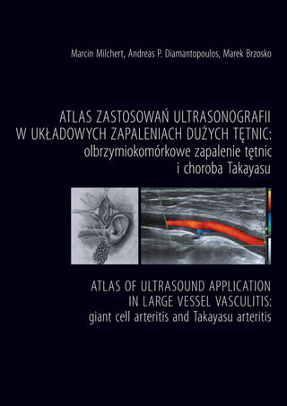
- 1. ATLAS ZASTOSOWAŃ ULTRASONOGRAFII W UKŁADOWYCH ZAPALENIACH DUŻYCH TĘTNIC: olbrzymiokomórkowe zapalenie tętnic i choroba Takayasu ATLAS OF ULTRASOUND APPLICATION IN LARGE VESSEL VASCULITIS: giant cell arteritis and Takayasu arteritis Marcin Milchert, Andreas P. Diamantopoulos, Marek Brzosko
- 2. ATLAS ZASTOSOWAŃ ULTRASONOGRAFII W UKŁADOWYCH ZAPALENIACH DUŻYCH TĘTNIC: olbrzymiokomórkowe zapalenie tętnic i choroba Takayasu ATLAS OF ULTRASOUND APPLICATION IN LARGE VESSEL VASCULITIS: giant cell arteritis and Takayasu arteritis Marcin Milchert, Andreas P. Diamantopoulos, Marek Brzosko Wydawnictwo Pomorskiego Uniwersytetu Medycznego w S z c z e c i n i e 2 0 1 6
- 3. Redaktor naczelny: prof. dr hab. n. med. Ireneusz Kojder Redakcja: Wojciech Markowski Korekta: Emilia Syldatk Waldemar Jachimczak Bernard Piotuch) oraz Andreas Diamantopoulos)
- 6. 7 - - - - - - - - - - giant cell arteritis - - - - - - - (supericial temporal artery - - - - -
- 7. 8 - - Takayasu arteritis - - - - - positron emission tomogra- phy - - - - - - - - - - - - - - - - -
- 8. 9 - pulse repetition frequency color gain ½–⅔ - color gain - ½–⅔ -
- 9. 10 (1a)
- 10. 11 - (1b)
- 11. 12 - - -
- 13. 15 - - Case 1 -
- 14. 16 halo
- 15. 17
- 16. 18
- 17. 19 - - - -
- 19. 125 Case 25
- 20. 126
- 21. 127
- 22. 128
- 23. 129 - -
- 24. 130 -
- 25. 131 Case 26 -
- 26. 132 - -
- 27. ISBN 978-83-64906-04-6ISBN 978-83-64906-04-6 Wydawnictwo Pomorskiego Uniwersytetu Medycznego w S z c z e c i n i e 2 0 1 6 Olbrzymiokomórkowe zapalenie tętnic jest najczęstszym pierwotnym zapaleniem naczyń. W jego diagnostyce kluczową rolę odgrywa potwierdzenie zmian zapalnych w tętnicach, do czego szczególnie dobrze nadaje się badanie ultrasonograficzne. Badanie USG me- todą Dopplera w olbrzymiokomórkowym zapaleniu tętnic ma znaczenie nie tylko dia- gnostyczne, ale i rokownicze, ponieważ szybkie wprowadzenie leczenia redukuje liczbę powikłań niedokrwiennych, natomiast zajęcie wielu tętnic – możliwe do potwierdzenia w USG – jest związane z gorszą odpowiedzią na leczenie. Mamy nadzieję, że książka ta ułatwi lekarzom wykonującym USG metodą Dopplera tętnic różnicowanie zmian miażdżycowych z zapaleniem tętnic. Reumatolodzy wykonujący badania USG układu mięśniowo-szkieletowego znajdą w tej książce praktyczne informacje, jak wykorzystać używane przez nich aparaty USG do diagnostyki zapaleń dużych tętnic i jak wprowadzić ścieżkę szybkiej diagnostyki olbrzymiokomórkowego zapalenia tętnic. Atlas ma formę opartą na przypadkach klinicznych. Dzięki nim chcielibyśmy przekonać lekarzy o znaczeniu stosowania szybko dostępnego badania USG metodą Dopplera w podejmowaniu decyzji leczniczych w reumatologii, czyli o zaletach wykonywaniu tego badania samemu w codziennej praktyce. Autorzy * * * Giant cell arteritis is the most common form of systemic vasculitis. Confirmation of ar- teritis is essential to diagnose large vessel vasculitis, and ultrasound is particularly well suited for that purpose. Doppler ultrasound examination in giant cell arteritis is both diagnostically and prognostically important because the rapid introduction of treatment reduces significantly the number of ischemic complications. In addition, extended disease presented with inflammation of many arteries – that can be assessed by ultrasound – is associated with a prolonged treatment with corticosteroids and higher relapse rates. We hope, that this book will be helpful for specialists performing vascular Doppler ultrasound and particularly Rheumatologists who will find here useful information on how to use their equipment to diagnose inflammation in large arteries and how to introduce a fast-track giant cell arteritis diagnostic clinic. This atlas is based on clinical cases and we hope that will convince physicians about the importance of vascular ultrasound in decision making in rheumatology, and the practical benefits of performing this test on their own. The authors