Chemical Injury & Foreign Body Ocular Injury
•Download as PPTX, PDF•
0 likes•107 views

Chemical Injury & Foreign Body Ocular Injury , G S MEDICAL COLLEGE, DR M SAQUIB

Recommended
More Related Content
What's hot
What's hot (20)
Similar to Chemical Injury & Foreign Body Ocular Injury
Similar to Chemical Injury & Foreign Body Ocular Injury (20)
More from MEDICS india
More from MEDICS india (18)
Recently uploaded
Recently uploaded (20)
Chemical Injury & Foreign Body Ocular Injury
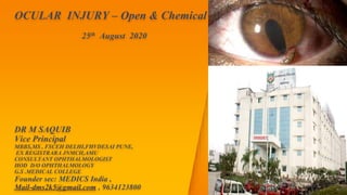
- 1. OCULAR INJURY – Open & Chemical 25th August 2020 DR M SAQUIB Vice Principal MBBS,MS , FSCEH DELHI,FHVDESAI PUNE, EX REGISTRARA JNMCH,AMU CONSULTANT OPHTHALMOLOGIST HOD D/O OPHTHALMOLOGY G.S .MEDICAL COLLEGE Founder sec: MEDICS India , Mail-dms2k5@gmail.com , 9634123800
- 2. TRAUMA EYE LID TRAUMA Laceration Periocular Haematoma ORBITAL FRACTURE Blow out orbital fractures GLOBE TRAUMA Blunt Trauma Penetrating Trauma Foreign Body Shaken baby Syndrome CHEMICAL INJURIES2
- 3. Penetrating Trauma ▸ Young , ▸ Male ▸ Assault ,Accident ,Sports injury ▸ Kinetic Injury of object determine considerable injury 3
- 4. Mechanism of Damage ▸ Mechanical effect ▸ Infection – Abscess, Endophthalmitis, Panophthalmitis ▸ Iridocyclitis – Sympathetic ophthalmitis 4
- 5. ▸ Wound of Conjunctiva ▸ Wound of Cornea ▸ Wound Sclera ▸ Wound of the lens 5
- 6. Traumatic Tractional Retinal Detachment ▸ Penetrating Injury ▸ Vitreous Prolapse & Haemorhage ▸ Vitreoretinal proliferation ▸ Tractional band ▸ Tractional RD 6
- 7. Penetrating wounds with the Retention of Foreign Bodies ▸ Damage by ▸ Mechanical Effect ▸ Introduction of Infection ▸ Specific action (Chemical or otherwise ) 7
- 8. Non -Organic material ▸ Gold ,Plastic ,stone ,Glass ▸ Lead – Gun powder ▸ Aluminium – ▸ Zinc ▸ Iron ▸ Copper 8
- 9. 9
- 10. ▸ Siderosis is a complication due to a magnetic intraocular or intraorbital foreign body. ▸ Generally metals with a low redox potential, such as Fe++and Cu++, have the greatest potential for metallosis. ▸ Siderosis may develop within weeks, but the course is variable depending on the iron content in the foreign body and its location. ▸ Virtually all ocular structures are involved in the siderotic process—Glaucoma, Cataract, iris color changes, mydriasis, retinal function destruction, and optic nerve atrophy. 10
- 11. Iron Foreign body ▸ Iron intraocular foreign bodies can result in siderosis bulbi, a condition characterized by deposition of iron molecules in the trabecular meshwork, lens epithelium, iris, and retina . ▸ Iron make chemical combination with Protein of the cells and produce degenerative changes . 11
- 12. 12
- 13. Lens ▸ In siderosis or hemosiderosis the lens epithelium takes on a yellow-brown or rusty appearance from minute dots of intracellular iron, identifiable by Perls' or other iron stains. ▸ Focal rusty-brown nodules of subcapsular cataract may develop. When the foreign body is in the lens there may be progression to a mature cataract, with diffusion of ionizable iron throughout the lens fibers. 13
- 14. 14
- 15. Retina ▸ The iron concentrates mainly in RPE and inner limiting membrane neurosensory retina. Necrosis of the photoreceptor cells occurs over large areas of the retina. ▸ Functional damage to the retina occurs at a very early stage, before extensive siderosis is apparent, and before stainable iron is detected in retinal tissues. ▸ Retinal degeneration with attenuated blood vessels ▸ ERG- Increased amplitude of the a-wave with normal b-wave . Advanced cases b wave diminishes15
- 16. 16
- 17. ▸ Heterochromia iridis- Staining of Iris greenish –Reddish to brown . ▸ Mydriasis- Deposition of Iron in the sphincter of the Iris leads to Mydriasis . ▸ Secondary Glaucoma ▸ Prussian Blue reaction 17
- 18. Chalcosis ▸ Alloy of copper causes specific changes ▸ Electrolytic dissociation & deposition in membrane ▸ Pure copper cause violent reaction ▸ Suppurative Reaction ▸ Fibrosis …. Globe shrinkage18
- 19. Chalcosis : Copper /Brass Kayser-fleischer Ring- Copper deposition in descmet’ membrane of peripheral cornea ,Golden brown ring 19
- 20. Sunflower Cataract Deposition of copper under posterior capsule of lens. ▸ Golden green in Colour , Arranged like petals of flower ▸ Retina : Deposition of Golden plaques at the posterior pole which reflect the light with a metallic sheen20
- 21. Organic Material ▸ Leaves, Fonds, Thorns , Woods, Vegetables , Eyelashes , Caterpillar hair ▸ Granulation Tissue ▸ Fungal Infection ▸ Caterpillar – Iridocyclitis – ophthalmia nodosa 21
- 22. 22
- 23. Diagnosis ▸ Detail History ▸ Mode of Injury ▸ Hammer chisel injury look for Entry wound ▸ Slit Lamp Examination ▸ Anterior segment ▸ Angle – Gonioscopy ▸ Radiography – X ray – Caldwell & Lateral view ▸ Metal ring suturing at limbus X ray for localisation ▸ CT – 2mm > slices ▸ USG – B scan ▸ MRI ▸ 23
- 24. 24
- 25. Treatment – ▸ Surgical Removal ▸ Unless- Inert ,Little Damage , Removal Is Dangerous For Sight . ▸ Metallic / Magnet Foreign Body – Surgical , Magnet Can Be Used ▸ Followup 25
- 26. Sympathetic Ophthalmitis ▸ Rare bilateral granulomatous inflammation that follows accidental or surgical insult to the uvea of one eye. ▸ Onset -insidious or acute, with recurrent periods of exacerbation. ▸ Clinical presentation –Loss of accomodation.DOV ,9 days-9 year mutton-fat keratic precipitates, choroidal infiltrations, and Dalen-Fuchs nodules. ▸ Histopathology reveals diffuse or nodular granulomatous inflammation of the uvea. ▸ Prevention and treatment: enucleation of the injured eye and immunosuppressive therapy, aimed at controlling inflammation. ▸ The etiology and pathophysiology of the disease is still unclear autoimmune in nature.26
- 27. 27
- 28. Chemical Injuries ▸ Alkalies –Caustic , Lime ( Fresh mortar , white wash ) , Ammonia , Sodium Hydroxide ▸ Acidic –Hydrochloric , sulphuric acid Mode of Injury : ▸ Domestic accident ▸ Agriculture accident – Fertilisers, Insecticide ▸ Chemical laboratory accidents ▸ Deliberate chemical attacks ▸ Chemical warfare Injury ▸ Self Inflicted chemical Injury28
- 29. 29 Alkali Burn Acid Burm Lime ,Caustic Potash , Caustic Soda , Liquid ammonia Sulphuric, Hydrochloric , Nitic acid Saponification of fatty acid of the cell membrane Necrosis – Gelatinisation - Instant coagulation of all protein , make barrier for further penetration Penetraion of Alkali to deeper tissue and for longer period . Superficial tissue ,line of demarcation Stages Acute Ischemic Necrosis- Inflammation,Edema , Discharge of conjuctiva,Cornea, Iris Reparation –Regeneration,Vascularisation Complication –Symblephron ,Recurrent corneal ulcer , Complicated cataract ,Glaucoma Conjuctiva -Necrosis ,Sloughing , Symblephron Corneal edema Sloughing , opacification , Staphyloma
- 30. 30
- 31. 31
- 32. 32
- 33. 33
- 34. Treatment ▸ Prevent Further Damage ▸ Irrigation – 2 Litre – Clean water or saline ▸ Contaminant Removal , Necrosed Conjunctiva removal . ▸ Support Healing and Prevent Inflammation & Infection34
- 35. ▸ Antibiotic Eye Drop ▸ Steroid Eye drop ▸ Cycloplegic – Atropine eye drop ▸ Lubricant Eye drop ▸ Autologous serum ▸ Ascorbic acid ▸ Tab Doxycycline 100 mg bd ▸ PREVENTION OF Symblepahron –Glass shell , Sweeping glass rod ▸ Treat Complication – AMG with or without stem cell transplantation , Glaucoma , Keratoplasty , Keratoprosthesis 35
- 36. 36