
Recommended
More Related Content
What's hot
What's hot (20)
Similar to lecture 9.pptx
Similar to lecture 9.pptx (20)
Recently uploaded
Recently uploaded (20)
lecture 9.pptx
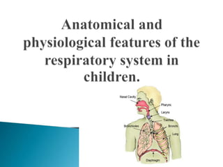
- 2. The respiratory system consists of the airways and the respiratory tract. The laying and formation of the respiratory system begins from the 3rd week of gestation from the protrusion of the anterior intestinal wall and by the 16th week the main structure of the bronchial derev is formed. The fetus performs respiratory movements from the end of the first trimester of intrauterine development. They are performed with the glottis closed, which prevents the penetration of amniotic fluid into the lungs.
- 3. Surfactant (a surfactant - a tension factor that provides the area of the respiratory surface and prevents the collapse of the alveoli) matures at 35-36 weeks of gestation, so the lack of surfactant in premature infants is the main cause of the respiratory disorders syndrome. In violation of embryogenesis 3-7 week of fetal development (at the time of formation of the trachea, the main bronchi, the lobes and main lobes of the lung, the pulmonary arteries), as a result of damage can develop: atresia of the larynx, tracheal stenosis, tracheoesophageal atresia, fistula, pulmonary agenesis, congenital pulmonary cysts (including bronchial).
- 4. Features Value The softness of the ribs and the pliability of the chest Easier passage through the birth canal, but prone to paradoxical breathing The "expiratory" structure of the chest (the ribs are located almost at right angles to the spine) Limited opportunities to increase respiratory volume Poor development of the respiratory muscles Limited ability to increase the respiratory volume, overcome resistance during obstruction, ease of occurrence of weakness and exhaustion of the respiratory muscles High standing of the diaphragm The diaphragmatic type of breathing, a decrease in the respiratory volume with bloating of the intestine Narrow (up to 1 mm) and short nasal passages Difficulty in sucking with rhinitis, deterioration of anti-infective protection, warming, mechanical air purification when breathing through the mouth
- 5. Incomplete development of the paranasal sinuses The rarity of the occurrence of sinusitis at an early age The cartilage of the larynx is tender and pliable mucosa is rich in blood vessels and lymphatic vessels, elastic tissue poorly developed, fiber subglottic apparatus has high porosity. 1 mm of edema of the mucosa of the subclavian space leads to a decrease in its lumen by 50-75% (in adults by 20%) High frequency of occurrence and severe course of laryngitis, often accompanied by the development of laryngeal stenosis The trachea is relatively short, has a funnel- shaped shape, the tracheal mucosa is tender, richly vascularized; 1/3 of the trachea is represented by the membranous part(in adults-1/5), which leads to a decrease in its lumen by one third during the respiratory cycle and when coughing . More frequent than in adults, as an isolated lesion of the trachea, and its involvement in the inflammatory process in laryngitis and bronchitis(laryngotracheitis and tracheobronchitis) The bronchi are narrow, the bronchioles of the newborn are 0.1 mm in diameter (in adults-0.5 mm), edema of the bronchial wall by 1 mm increases the resistance in the airways of the newborn by 16 times (in adults-2-3 times); less elastic tissue, underdevelopment and softness of cartilage; absence (up to 8 years) "auxiliary" air flow between adjacent bronchi; relatively thick, loose, well-vascularized bronchial mucosa, large concentration of mucous glands The tendency to acute and recurrent bronchial obstruction; the predominance of components of hypersecretion and edema in the bronchial obstruction; a greater tendency to reduce the lumen of the respiratory tract with external compression((for example, the lumen of the mid-lobe bronchus with enlarged lymph nodes); a greater tendency to atelectasis, the occurrence of air traps
- 6. The right bronchus is wider and seems to be a continuation of the trachea A greater frequency of foreign body penetration into the right bronchus Lower lung extensibility (1/3 the size of an adult) Relatively large amount of work spent on breathing, high energy costs, especially in tachypnea and shortness of breath The respiratory equivalent in an infant is 2 times greater than in an adult; three times the minute volume of ventilation (0.4 l / kg in a one-month-old child and 0.125 l/kg in a 14-year-old child), which provides greater oxygen absorption (13.2 ml/kg per minute in a newborn compared to 4.3 ml/kg per minute in an adult) The possibility of rapid development of respiratory failure in diseases of the respiratory system, because for the assimilation of 1 liter of oxygen, the child has to do 2 times more work Greater resistance to hypoxia in newborns and infants; reduced intensity of oxidative processes Good tolerance to short-term moderate hypoxia compared to adults; poor tolerance to chronic hypoxia "Primitive" nature of acinus, poor collateral ventilation, greater looseness of interalveolar and interlobular connective tissue, rich lung vascularization Lower diffusion capacity with a lower coefficient of oxygen utilization from the air (in newborns, 1 ml of oxygen is absorbed from 42 ml of air, and in adults-from 16 ml); greater tendency to edema and generalization of infection in the lungs, a decrease in respiratory volume in any tachypnea
- 7. The pleura is thin, tender; the elastic network of the pleura is formed by the age of 7; loose and pliable fiber surrounding the mediastinum Ease of displacement of the mediastinal organs with the accumulation of fluid in the pleural cavity Slightly lower viscosity of bronchial mucus compared to adults Easier evacuation of sputum from the airways Greater mediastinal mobility The possibility of inflection of large major vessels and compression of the lung during mediastinal shift A relatively weak cardiac sphincter of the stomach in well-marked pyloric. Tendency to gastroesophageal reflux, regurgitation, regurgitation followed by aspiration Low overall and local immune response at an early age High frequency, tendency to relapse and more severe course of infectious diseases of the respiratory tract, bronchiolitis; easier occurrence of sensitization to exogenous non-infectious allergens
- 8. Respiratory movements in the fetus occur at the 13th week of intrauterine development, which occur when the glottis is closed. During labor, the fetus significantly reduces the partial pressure of oxygen (pO2), increases the pCO2, and decreases the pH. In this regard, there is an impulse from the aortic and carotid artery receptors to the respiratory center and its excitation. Along with this, there are also signals from the irritation of the skin receptors. After birth and contraction of the diaphragm, a negative intra- thoracic pressure is created, which facilitates the entry of air into the airways and lungs, which are filled with a viscous liquid. With normal expansion of the lung, with the participation of a surfactant, the pulmonary fluid is quickly absorbed by the lymphatic vessels and blood capillaries and gas exchange begins. The regulation of respiration in the future is carried out mainly by the respiratory center.
- 9. When collecting anamnesis, you should pay attention to the most frequent complaints: - difficulty breathing through the nose, the nature of the discharge from it;- - cough its strength, frequency, duration, soreness, presence and nature of sputum, etc.; - change of voice-hoarseness, aphonia; - shortness of breath (shortness of breath), to clarify its nature. Further specify:- - whether there was an increased body temperature; - were there any contacts with infectious patients in the family, in children's institutions; - were there any diseases of the respiratory system that preceded the present, - has he received treatment so far and what was the degree of recovery; - did the child have measles, whooping cough; - the presence of respiratory diseases in the family and in the next of kin;- - allergic history (the presence of allergies in the child, in other family members, in relatives). At the end of the history collection, the data is analyzed and a short conclusion is made.
- 10. During the examination, attention is drawn to: 1. Consciousness, position in bed, reaction to others. 2. The state of physical development. 3. Color of the skin of the face and mucous membranes. 4. Whether the breath is free through the nose, the nature of the discharge. 5. The nature of cough, sputum. 6. The child's voice. 7. The shape and symmetry of the chest. 8. Type of breathing.
- 11. 9. Rhythm, frequency, and depth of breathing. It should be remembered about the average parameters of the respiratory rate in children of different ages: Respiratory rate (per minute) in young children Age sleep Waking up 0 –1 мес. 30 (29 –47) 50 (40 – 60) 1 – 6 мес. 35 (20 – 60) 65 (50 – 75) 6 –12 мес. 27 (22 – 32) 60 (55 – 75) 1 –4-года 20 (16 – 25) 33 (23 – 42) 4 –10 лет 18 (13 – 23) 23 (15 – 36) 10 –14 лет 16 (13 – 19) 21 (15 – 28)
- 12. 10. The presence of shortness of breath; what is the ratio between inhaling and exhaling; 11. Bulging or sinking of the intercostal spaces (especially one-sided). The symmetry of the participation of both halves of the chest in breathing. 12. Inflating the wings of the nose. 13. The ratio between the respiratory rate and pulse. 14. The condition of the pharynx, posterior pharyngeal wall and tonsils.
- 13. When palpating, pay attention to: - elasticity and resistance of the chest; - soreness; - voice tremor; - the symmetry of the participation of both halves of the chest in breathing; - the thickness of the skin-subcutaneous fold on symmetrically locatedareas of the chest. When percussion, pay attention to: - the age of the children, the severity of the condition, which will determine the position of the patient with percussion; - symmetry of both halves of the chest; - features of percussion in young children (less impact force – "quietest" percussion, direct percussion). In comparative percussion, the nature of the percussion sound and the presence of a pathological focus are determined
- 14. In topographic percussion is determined: - height Vistana apices above the clavicles, the margins of Kreniga (in children under 5 years the tops of the lungs do not act outside of the clavicles, normally the upper bounds at this age is not defined); - the lower borders of the lungs; the projection of the lobes of the lungs on the chest; - mobility of the lower edge of the lungs.
- 15. The lower border of the lungs in children Lines From Right To Left Middle clavicle VI rib The lower border of the left lung along the midclavicular line differs in that it forms a notch for the heart and departs from the sternum at the height of the IV rib and steeply descends downwards Middle axillary VII rib IX rib Scapular IX-X rib X rib Paravertebral At the level of the –»»» spinous process of the XI thoracic vertebra
- 16. During auscultation, attention should be paid to: - asymmetry of the auscultative zones; - the nature of the main respiratory noise, for which you need to determine the duration of inspiration and exhalation, their strength and ratio; - places of the most frequent localization of pathological foci – axillary areas, paravertebral spaces, supra-and sub- scapular areas;- - the presence of pathological changes in the main respiratory noise (weakening, strengthening, hard, bronchial breathing, etc.); - the presence of pathological noises (wheezing, pleural friction noise); - changes in bronchophony;- auscultatory signs of improvement in bronchial lymph nodes (symptom D" Espina). After completing the clinical examination of the patient, you need to make a short conclusion about the changes found and the severity of the condition according to the degree of respiratory failure and signs of toxicosis.
- 17. Next, it is necessary to evaluate the results of additional research methods: 1. Acid-base state of blood gases, paying attention to the decrease in PO2 and increase in PCO2. 2. Sputum examination (bacteriological and microscopic). 3. Examination of the pleural punctate (bacteriological, microscopic, protein content). 4. X-ray data. 5. Instrumental studies (bronchoscopy, etc.). 6. Functional studies – spirography, pneumotachometry, etc. results of functional tests. 7. Scarification tests with the most common allergens. 8. Examination of the biopsy of the bronchial mucosa and lung tissue.
- 18. Cough is one of the most characteristic signs of respiratory damage. Cough is especially typical in whooping cough: it occurs with paroxysms (paroxysmal) and with reprises (long, high breath), and is also accompanied by redness of the face and often vomiting. Cough paroxysms are usually observed at night. Cough with laryngeal lesions is usually dry, rough, and barking. It is so characteristic that it makes it possible to suspect a lesion of the larynx (laryngitis or croup) at a distance. Cough with tracheitis is rough (like in a barrel).With bronchitis, the cough can be both dry (at the beginning of the disease) and wet, with sputum separation - during the development of the disease and at the end of it. With bronchial asthma, first the cough is dry, irritable, and then – wet, when the viscous, viscous sputum begins to separate. With pneumonia, also in the first days of the disease, the cough is more often dry, and in the following days it becomes wet. When the pleura is involved in the process, the cough is usually painful (croup pneumonia, pleurisy).
- 19. Bitonal cough - spasmodic cough, having a rough the main tone and musical II high tone, arises from irritation cough zone of bifurcation of the trachea by enlarged lymph nodes (often with bronhoadenita tuberculosis) or observed in lim-agranulocytose, lymphosarcoma, leukemia, tumors of the mediastinum (thymoma, sarcoma, etc.). Painful dry cough occurs in pharyngitis and nasopharyngitis. If adult patients usually expectorate sputum, then young children usually swallow it. Infants do not know how to cough up phlegm at all. Therefore, to solve the question: dry or wet cough, it is necessary to observe a small child, whether he swallows sputum. Copious discharge of sputum (purulent) with a full mouth in young children is observed when an abscess, or a suppurated cyst of the lungs, is emptied into the bronchi. Older children have a lot of sputum in chronic pneumonia, when there are already bronchiectases.
- 20. Inspection. On external examination, cyanosis should be noted, which can be permanent, local or general. The greater the respiratory failure and the lower the oxygen saturation of the body, the stronger and more widespread the cyanosis. In young children (up to 2-3 months of age), foamy discharge can be seen in the corners of the mouth, under the tongue in bronchiolitis and pneumonia. This is due to the penetration of inflammatory exudate from the respiratory tract into the oral cavity. During the inspection of the nose may be noted discharge (serous, mucous, Muco-purulent, sukrovichnye, bloody), and difficulty breathing through the nose. Depending on the nature of the discharge, rhinitis is distinguished: serous, mucous, mucopurulent and hemorrhagic. Rhinitis is most often one of the symptoms of an acute respiratory viral infection (adenovirus, parainfluenza, or influenza), but it is also observed in measles. Sukrovichnye nasal discharge is characteristic of diphtheria of the nose or foreign body. For congenital syphilis, the so-called snoring breath is characteristic.
- 21. During the examination, pay attention to the child's voice, which often changes when the larynx and vocal cords are affected. Laryngitis is clinically manifested by a rough barking cough and a change in voice. Unlike adults, laryngitis in children is often accompanied by difficulty breathing-croup. Croup can be true or false. True croup is observed in diphtheria of the larynx, when there is a croup inflammation of the vocal cords with the formation of a hard-to-separate film. False croup- sublingual laryngitis, most often occurs in acute respiratory viral infections (most often with parainfluenza) and is caused by swelling of the mucous membrane below the vocal cords.
- 22. False croup usually occurs suddenly and usually in the evening and at night. Before that, it is as if a healthy child suddenly wakes up and begins to suffocate. True croup usually develops gradually (within 1-3 days). Unlike the false croup, with the true croup, the voice disappears gradually (aphonia).
- 23. A rough, low voice is one of the signs of myxedema. Nasal tone of the voice occurs in chronic rhinitis, adenoids, pharyngeal abscess, etc. The appearance of nasal tone in diphtheria of the pharynx and encephalopathies indicates paresis of the palatine curtain. In children of preschool and school age with chronic adenoiditis, the face acquires a characteristic appearance. It is pale, puffy, with a slightly open mouth, raised upper lip and upturned nose, often there is an incorrect bite.
- 24. The appearance of a frequently coughing child is typical, especially in whooping cough and chronic non-specific lung lesions. Such children have a pale, pasty face and eyelids (due to a violation of the outflow of lymph- lymphostasis), cyanotic lip mucosa, swollen skin veins, and may experience hemorrhages in the conjunctiva and subcutaneous tissue.
- 25. When examining the oral cavity, it is necessary to pay attention to the condition of the pharynx and tonsils. In children of the first year of life, the tonsils, as a rule, do not extend beyond the front arches. In preschool children, there is hyperplasia of the lymphoid tissue, and the tonsils usually extend beyond the front arches when examined. They are dense and do not differ in color from the mucous membrane of the pharynx. Children often have inflammatory processes of the tonsils - angina, which are divided into catarrhal, follicular, lacunar, as well as specific infec-tions. Angina in scarlet fever differs from the banal angina by sharply limited hyperemia, and in moderate to severe forms-by necrosis of the mucous membrane ("necrotic angina").
- 26. Changes in the shape of the chest in children occur with rickets, pneumothorax, pneumomediastinum, bronchial asthma (barrel-shaped). In exudative pleurisy, there is a swelling of the chest on the side of the lesion, and in chronic pneumonia - a depression. A significant retraction of the intercostal spaces and the jugular fossa in the inhalation phase is typical for stenotic breathing in croup. Restriction of the chest excursion is observed in acute pulmonary distension, bronchial asthma, pulmonary fibrosis, subdiaphragmal abscess, intercostal neuralgia. There may be a change in the rhythm of breathing.
- 27. Rapid breathing (tachypnea) in healthy children occurs with excitement, physical exercise, etc. diseases of the respiratory system, diseases of the cardiovascular system, blood diseases (anemia), febrile diseases, pain, and distress syndrome in newborns. Respiratory depression (bratipnea) is rarely observed in children, indicates exhaustion of the respiratory center and usually occurs in comatose states (uremia), poisoning (for example, with sleeping pills), increased intracranial pressure, and in newborns - in the terminal stages of the distress syndrome. Pathological types of respiration: Kussmaul, Biot, Cheyne-Stokes, reflect the severe degrees of his disorder.
- 28. When examining the child, you should pay attention to the participation in the breathing of auxiliary muscles (rectus abdominis, sternocleidomastoid, thoracic, etc.), which indicates difficulty breathing-shortness of breath. At the same time, young children also experience swelling and tension of the wings of the nose. Dyspnea occurs with hypoxemia, hypercapnia, an excess of various under- oxidized products that accumulate in the blood and brain matter during acidosis
- 29. Inspiratory dyspnea – difficulty in inhaling, observed with obstruction or narrowing of the upper respiratory tract (croup, foreign body, cysts and tumors, congenital narrowing of the larynx, trachea, pharyngeal abscess, due to a drop in intra-thoracic pressure much lower than atmospheric). Clinically, it is manifested by the retraction of the intercostal spaces, the jugular fossa, the supraclavicular regions and the epigastric region. Expiratory dyspnea is characterized by difficulty in inhaling, intra-thoracic pressure, on the contrary, exceeds atmospheric pressure. The chest is raised up and almost does not participate in the act of breathing, the rectus abdominis muscles are tense, the intercostal spaces are flattened. It is observed in bronchial asthma, bronchitis, with compression of large bronchi (tuberculous bronchoadenitis).
- 30. * Mixed shortness of breath (expiratory-inspiratory), manifested by chest swelling and retraction of compliant places. It is observed in pneumonia, pulmonary edema, exudate compression, heart disease, flatulence, ascites, in patients with rickets. * Shortness of breath Chic-expiratory puffing, depends on the compression of the bronchi by tuberculous infiltrate and lymph nodes of the lung root in the lower part of the trachea and bronchi, which freely pass air only when inhaled. *The complete absence of respiratory movements in a newly born child is the main symptom of imaginary death - asphyxia of the newborn. * Congenital stridor-a disease of early age, characterized by inspiratory noise when breathing. The noise is whistling, ringing, reminiscent of the cooing of pigeons, sometimes the purring of a cat, the clucking of a chicken. The noise in stridor is constant, the intensity of the noise decreases during sleep, when the child is transferred from a cold room to a warm one. Stridor begins after birth, decreases in the second half of the year, and is self- cured by 2-3 years.
- 31. Palpation in the diagnosis of respiratory diseases can be used to assess chest deformities, search for pain points and areas (myalgia, intercostal neuralgia, rib fractures). When determining the elasticity and resistance of the chest, in sick children, there is a great resistance to exudate in the pleural cavity, with tumors and strong compaction of the lung tissue. When palpating (feeling) the chest, you can detect soreness. It is necessary to distinguish between superficial soreness (damage to muscles, nerves, bones) and deep - pleural.
- 32. The first one is found: 1) with inflammatory processes in soft tissues; 2) when the intercostal muscles are affected; 3) when the ribs and sternum are affected; 4) for diseases of the intercostal nerves. Pleural (deep) pain usually increases when inhaling and exhaling, often given to the epigastric and subcostal areas, weaken if you squeeze the chest, decrease when bending the body in the affected direction. The palpation method determines the thickness of the skin fold on the symmetrical areas of the chest. Thickening of the skin fold is observed in exudative pleurisy, less pronounced in tuberculous bronchoadenitis on the affected side.
- 33. Voice tremor. Its strengthening is associated with the compaction of the lung tissue, the presence of cavities in the lungs (the distance from the glottis is shortened). Weakening - when the bronchus is blocked (atelectasis of the lung), when the lung is pushed away from the chest wall (exudate, pnevmotorax, pleural tumor).Restriction of the chest excursion is observed in acute pulmonary distension, bronchial asthma, subdiaphragmal abscess, intercostal neuralgia.
- 34. Features of percussion in young children: less impact force, mainly direct percussion is performed. In comparative percussion, the nature of the percussion sound and the presence of a pathological focus are determined. With topographic percussion, determine: the height of standing on the tops of the clavicles, the margins of Kreniga; - the lower borders of the lungs, the projection of the lobes of the lungs on the chest; - mobility of the lower edge of the lungs.
- 35. When the respiratory system is affected, there is a change in the percussion sound of different intensity. The shortening of the percussion sound is noted: 1. When the lung becomes less airy when: - inflammation of the lungs (infiltration and edema of the alveoli and interalveolar septa; - hemorrhages in the lung tissue (lung infarction); - significant pulmonary edema; - scarring of the lungs; - decline of the lung tissue - atelectasis, compression of the lung tissue with pleural fluid, a greatly expanded heart, a tumor in the chest cavity. 2. In case of formation of other airless tissue in the lung cavity: - in case of tumors- with the formation of a cavity in the lungs and the accumulation of fluid in it (sputum, pus, echinococcal cyst), provided that this cavity is more or less filled with fluid
- 36. 3. When filling the pleural space: - exudate (exudative pleurisy) or transudate; - fibrinous overlays on the pleural leaves.In exudative pleurisy, if the fluid does not fill the entire pleural space, it is possible to determine the Elissa-Damoiseau-Sokolov line - the upper border of dullness with the highest point in the posterior axillary line. From here it goes in and down. The line corresponds to the maximum level of fluid standing and is formed due to the displacement of the lungs to their root outlet. On the affected side it is possible to define a shortened tympanitis, which is in the form of a right triangle over exudate (triangle Garland). It corresponds to the location of the compressed lung. Its boundaries are: the hypotenuse-the Elissa-Damoiseau-Sokolov line, the catheters - the spine and the line perpendicular to the upper point of the Elissa-Damoiseau-Sokolov line on the spine. Behind, on the healthy side, due to the movement of the mediastinal organs, a section of blunting of the percussion sound is formed, which has the shape of a right triangle, this is the so-called Grokko-Rauchfus triangle. One of its catheters is the line of the spine, the second is the lower edge of a healthy lung, the hypotenuse is the continuation of the Damoiseau line to the healthy side.
- 37. A tympanic tinge of percussive sound appears: 1. In the formation of pathological, air-containing cavities:- in the lung tissue by destruction during inflammation (cavities in pulmonary tuberculosis, abscess), tumors( decay), cysts; - for diaphragmatic hernia and pneumatization of cysts; - in the pleura, in the form of an accumulation of gas or air in the pleural cavity: pneumothorax (spontaneous pneumothorax, artificial). 2. With some relaxation of the lung tissue due to a decrease in its elastic properties (emphysema), with compression of the lungs above the location of the fluid (exudative pleurisy and other forms of atelectasis). 3. At a certain degree of filling of the alveoli with air with the simultaneous presence of fluid in them-pulmonary edema at the beginning of inflammation, with the dilution of inflammatory exudate in the alveoli.
- 38. Box sound-a loud percussive sound with a tympanic tinge, appears when the elasticity of the lung tissue is weakened, and its airiness is increased (emphysema of the lungs), and is observed in pneumothorax. The noise of a "cracked pot" is a kind of intermittent, rattling sound, similar to the sound when tapping on a cracked pot. It becomes clearer when the patient opens his mouth. It is often obtained by percussion on the chest during screaming in children. In pathology, it occurs in cavities that communicate with the bronchi by a narrow slit.
- 39. A decrease in the height of the standing of the tops can be observed when they are wrinkled on the soil of tuberculosis. Thus there is a reduction in the width field of Kreniga.The lower borders of the lungs are lowered either due to an increase in the volume of the lungs (emphysema, acute pulmonary bloating), or due to a low standing of the diaphragm-with a sharp lowering of the abdominal organs and a decrease in intra- abdominal pressure, as well as paralysis of the diaphragmatic nerve.
- 40. The lower borders of the lungs are raised when: reduction of the lungs, due to their shrinking (more often on one side in chronic inflammatory processes); when pushed back by pleural fluid or lung gas; lifting of the diaphragm due to increased intra-abdominal pressure or pressure of the diaphragm upward by an organ or fluid (flatulence, ascites, enlargement of the liver or spleen, abdominal tumor). The decrease in the mobility of the pulmonary edges is caused by: loss of lung tissue elasticity (emphysema in bronchial asthma); wrinkling of the lung tissue; an inflammatory condition or edema of the lung tissue; the presence of adhesions between the pleural leaves. The complete cessation of mobility occurs when: filling the pleural cavity with fluid (pleurisy, hydrothorax) or gas (pneumothorax); complete overgrowth of the pleural cavity; paralysis of the diaphragm
- 41. Additional respiratory noises. The mechanism of their formation The weakening of vesicular respiration is noted when: • general weakening of the respiratory act with a decrease in the intake of air into the alveoli (severe narrowing of the larynx, trachea, sharp general weakness, diseases of the respiratory muscles, etc.); • closing of air access to a certain part of the lobe or lobe as a result of blockage or compression of the bronchus (foreign body, tumor, etc., atelectasis); pushing away a part of the lung with something: when fluid accumulates in the pleura (exudative pleurisy), air (pneumothorax), the lung moves deeper, the alveoli spread out when breathing; loss of lung tissue elasticity, stretching, i.e. rigidity (low mobility) of the alveolar walls (emphysema); in the initial or final stage of the inflammatory process in the lungs, when there is only a violation of the elastic function of the pulmonary alveoli without infiltration and compaction;
- 42. Increased breathing occurs when: narrowing of the small or minute bronchi (strengthening is due to exhalation) with their inflammation or spasm (asthma attack, bronchiolitis); febrile diseases, with compensatory strengthening on the healthy side in the case of pathological processes on the other. Hard breathing is rough vesicular breathing with an elongated exhalation. There are two options for hard breathing: inhale and exhale are heard equally well and are the same in duration; the inhale and exhale are equally rough, but the exhalation is somewhat longer.
- 43. Hard breathing usually indicates the defeat of small bronchi, occurs in bronchitis and bronchopneumonia. In these diseases, inflammatory exudate reduces the lumen of the bronchi, which creates conditions for the occurrence of this type of breathing. Bronchial respiration is heard only in cases of compaction of the lung tissue (tuberculosis, heart attacks, pneumonia) or the formation of cavities communicating with the bronchi. With bronchial breathing, the inhale and exhale are heard equally well (the exhale is heard even better, the timbre is changed - a rough “x”is heard). Bronchial respiration by the degree of strength can be weakened (when the lung is compressed with exudate)"it sounds like it's coming from far away." If the foci of compaction are located deep in the lung tissue and are normally closed by the lung tissue, a rougher and longer exhalation is heard, approaching the bronchial one (breathing with a bronchial tinge). Bronchial respiration can be of the amphoric type (in smooth-walled cavities- caverns, bronchiectasis, etc.).
- 44. Wheezes are additional noises and are formed when moving or oscillating in the air-bearing cavities of secretions, blood, mucus, edematous fluid, etc. Dry wheezes: whistling - treble, high and bass, low. The first are more common with narrowing of the bronchi, especially small ones, the second are formed from fluctuations in thick sputum, especially in large bronchi, giving resonance. Dry wheezes are called them because the liquid does not play a big role in their formation. In the mechanism of formation of dry wheezes, the bronchial wall itself and the air stream play a role. They are characterized by impermanence and variability. They are found in laryngitis, pharyngitis, bronchitis, emphysema, asthma. Laryngeal and tracheal wheezes are characterized by the fact that they are single-caliber and are heard as if under the ear. Bronchial tubes are divided into large and medium-sized, depending on the place of formation, and their audibility is different, which is associated with the place of their formation.
- 45. Wet wheezes are formed from the passage of air through the liquid. Depending on the caliber of the bronchus, where they are formed, they are small-bubble. It is important to divide them into ringing and non-ringing ones. Ringing sounds are heard when the lung tissue lying next to the bronchus is compacted, which is observed in bronchiolitis, bronchitis, pulmonary edema, atelectasis.
- 46. Pleural friction noise occurs when the visceral and parietal pleural leaves are rubbed and is heard: when the pleura is inflamed, when it is covered with fibrin or infiltration foci are formed on it, which leads to irregularities, roughness of the pleural surface, combined with pain; with the formation of pleural adhesions as a result of inflammation; when the pleura is affected by a tumor; with severe dehydration of the body.
- 47. From small crepitating wheezes, the noise of pleural friction differs in the following signs: 1) the noise of pleural friction is heard in both phases of breathing, and crepitation only at the height of inspiration, when the air reaches the small bronchioles and alveoli; 2) wheezing often disappears after coughing, while the noise of pleural friction remains; 3) wheezing during respiratory movements, with the mouth and nose closed, due to insufficient air movement in the bronchi-do not occur. and the noise of pleural friction continues to be heard; 4). pleural noises when the phonendoscope is pressed on the chest are amplified, while crepitation remains unchanged; 5) pleural noises are heard more superficially than small- bubble wheezes formed in the depth of the lung.
- 48. Increased bronchophonia is noted with infiltrative compaction of the lung (pnev-monia, tuberculosis), atelectasis. Above the caverns and bronchiectatic cavities, if the adductor bronchus is not blocked, the bronchophony is also loud and has a metallic character. When the lung tissue is compacted, increased bronchophony is due to better voice conduction, and in cavities - resonance. For the same reason, bronchophonia may be increased in a patient with an open pneumothorax. With an increase in the bronchial lymph nodes, a symptom of D'espin appears - listening to whispered speech and bronchial breathing below the I thoracic vertebra along the vertebra. In infants, this symptom does not apply. They use the De la Campa symptom - a loud laryngotracheal exhalation is heard above the V and VI thoracic vertebrae. You can identify a symptom of Smith: if you tilt the child's head so that the face is horizontal, then a venous noise is heard near the upper part of the chest. If you slowly lower the child's head down, the noise increases. The intensity of venous noise (in the absence of anemia) depends on the size of the enlarged paratracheal lymph nodes.
- 49. It is necessary to evaluate the results of additional research methods: 1. Acid-base state of blood gases, paying attention to the decrease in PO2 and increase in PCO2. 2. Sputum examination (bacteriological and microscopic). 3. Examination of the pleural punctate (bacteriological, microscopic, protein content). 4. Radiological data 5. Instrumental studies (bronchoscopy, etc.). 6. Functional studies: spirography, pneumotachometry, etc. results of functional tests. 7. Scarification tests with the most common allergens. 8. Examination of the biopsy of the bronchial mucosa and lung tissue. After the summary of the results of additional research methods, a final conclusion is made about the level and severity of the respiratory system damage, for which it is necessary to bring the data of the anamnesis and clinical examination into line and agree
- 50. Obstruction of the airways above the main bronchi. Such obstruction, arising, for example, in the larynx or trachea due to true (with diphtheria) or false croup, Quincke's edema in the larynx or a foreign body at this level, leads primarily to a violation of inspiration and shortness of breath takes on an inspiratory character. For shortness of breath.
- 51. Bronchial obstruction syndrome always indicates the presence of bronchitis, which can be independent or enter into the manifestations of bronchopneumonia, bronchial asthma, foreign body aspiration. Most often, the cause of obstruction is the combined effect of edema and inflammatory infiltration of the bronchial wall, the abundance of secreted mucus with its thickening and stagnation in the lumen of the bronchi, and, finally, spasm of the smooth muscle of the bronchial wall. From the obstruction of the main bronchi and below, first of all, the mechanisms of exhalation suffer, since it occurs under the influence of the lung's own elastic traction, which is still very weak in children. Therefore, one of the most persistent symptoms of bronchial obstruction is the expiratory nature of shortness of breath. In addition, you can hear distant whistling wheezes. With a complete blockage of the bronchi in a certain segment or lobe, the wheezing completely disappears, the respiratory noises weaken and the phenomenon of "silent lung" occurs.
- 52. Respiratory failure (DN) is a condition in which either the normal gas composition of the blood is not maintained, or the latter is achieved due to the abnormal operation of the external respiratory apparatus, which leads to a decrease in the functional capabilities of the body. There are 4 degrees of DN. Causes of respiratory failure: 1. Reduced pO2 in the inhaled air - anoxemic hypoxemia. 2. The defeat of respiratory muscles, impaired air passage through the respiratory tract, obstruction, impaired diffusion of oxygen through the alveolar-capillary membrane, impaired capillary blood flow as a result of distension of the alveoli (emphysema of bronchial hialnyh asthma, etc.) 3. Obstructive type of respiratory failure characteristic narrowing of the bronchi and bronchioles, edema of mucosa with bronchiolitis and when Steno-ziruyuschih laryngitis (cereals). 4. Restrictive (restrictive) type occurs when the ability of the lungs to expand and fall is limited (pneumosclerosis, exudative pleurisy, lesions of the ribs and respiratory muscles). 5. Mixed type - with the predominance of one or another form. 6. Disorders of blood gas transport (severe forms of anemia or changes in the structure of hemoglobin) 7. Circulatory disorders - congestive hypoxemia. There is a large absorption of oxygen due to the slowing of blood flow in organs and tissues. Improvement of cardiac activity contributes to the elimination of DN. 8. Damage to the enzyme systems of cells involved in the utilization of oxygen- tissue hypoxia. It occurs in severe infections and poisoning.
- 53. Symptoms of pneumonia. Lung inflammation is characterized by fever, lack of appetite, weakness, pallor (symptoms of intoxication) in patients with a history of a previous viral infection. In young children, cyanosis around the mouth, swelling of the wings of the nose, and an increase in shortness of breath of a mixed nature are characteristic. Percutaneous: blunting over the area of probable pneumonic infiltration. Breathing increases, exhalation lengthens, With concomitant bronchitis characterized by dry wheezing. Fine-bubbly wet wheezes of high sonority are possible. The presence of atelectasis can reduce the degree of sonority of wet wheezes. The diagnosis is confirmed radiologically by the presence of foci of compaction of the lung tissue. Symptoms of pleurisy. Inflammation of the pleura, most often complicates the course of other diseases. Characteristic features: severe intoxication, pronounced asymmetry of the respiratory excursions, dry cough, chest pain that increases with breathing and cough. Dulling of the percussive tone over the inflamed pleura. In case of effusive pleurisy, the Ellis-Damoiseau-Sokolov line is outlined. Breathing, as well as wheezing over the zone of dullness, is not listened to. You can percutaneously determine the displacement of the heart in the healthy direction. The diagnosis is confirmed radiographically and pleural puccia.