1. Introduction Results
Laney E. Vaughan1,2, Heidi A. Trau2 and Diane M. Duffy2
1Summer Program for Undergraduate Research, Eastern Virginia Medical School, Norfolk, VA
2Department of Physiological Sciences, Eastern Virginia Medical School, Norfolk, VA
Placental Growth Factor in Ovarian Granulosa Cells
Materials and Methods
Conclusions
Through this experimentation, four antibodies were eliminated from the
pool of antibody candidates for detecting placental growth factor in
ovarian tissue. Further experimentation of candidate antibodies will need
to take place to identify an antibody which best binds to placental growth
factor. It is known that placental growth factor is involved in ovulatory
angiogenesis, but its specific role is unknown. Identifying an antibody that
successfully binds placental growth factor is a necessary tool in the
process of determining its explicit purpose in ovulatory angiogenesis.
Gaining knowledge of the placental growth factor pathway and
mechanism, combined with a the development of a method to inhibit it,
could potentially lead to the invention of a new contraceptive. Current
forms of birth control prevent implantation, which is the cause of much of
the social controversy surrounding current methods of birth control.
Interrupting and inhibiting the placental growth factor pathway will bypass
this controversy. This approach would prevent follicular rupture and egg
release instead of inhibiting implementation. Preventing ovulatory
angiogenesis through the inhibition of the placental growth factor pathway
holds promise for the development of a future ovulation-preventing birth
control.
Objective
The goal of this research was to develop tools to localize
and quantify placental growth factor in ovulatory follicles.
Identifying an antibody which best detects placental
growth factor protein would be a step forward in
determining its specific role in ovulatory angiogenesis.
Ovulation is an event that takes place approximately halfway through the
menstrual cycle. Between 37 and 42 hours after a surge of luteinizing
hormone, ovulation begins and stimulates the release of a mature egg from
one ovary for fertilization in the fallopian tube.
1. Wulff C, Wilson H, Wiegand SJ, Rudge JS, Fraser HM 2002 prevention of
thecal angiogenesis antral follicular growth, and ovulation in the primate by
treatment with vascular endothelial growth factor trap R1R2
2. Vector Laboratory
https://www.vectorlabs.com/catalog.aspx?prodID=33
References/Acknowledgements
Antibodies are serum proteins developed naturally as an important component of the immune
system. Antibodies are designed to recognize and attack foreign substances in the body which
are perceived as a threat. Their defensive properties are utilized in science for detecting and
quantifying target proteins. The target protein is taken from a host species and is injected into
an animal of a different species. The injected animal’s immune system develops antibodies as a
defense against the foreign protein. This primary antibody is then extracted from the animal and
used in staining to detect the target protein. The primary antibody binds to the select antigen. A
second antibody binds the primary antibody and attaches a tag, which serves as a marker for
the target protein. The binding that takes place between the antigen, primary antigen, and
secondary antigen is highly specific.
+P
LH Surge
VEGFE
VEGFA
PlGF
VEGFA
VEGFR2 VEGFR1
Granulosa Cells
Make and Secrete
VEGF Ligands
Migration
in Tip Cell
Proliferation
in Stalk Cell
Angiogenesis
Preliminary studies in our laboratory
determined that expression of
placental growth factor is very low
between 0 and 12 hours after hCG,
which substitutes for the surge of
luteinizing hormone, but its
expression dramatically increases 24
and 36 hours after hCG. Because
ovulation takes place about 40 hours
after the surge of luteinizing hormone
or hCG, this data suggests that
placental growth factor may play a
role in ovulation.
Placental growth factor is a member the vascular endothelial growth factor
family. It binds as a ligand to vascular endothelial growth receptor 1. The
specific function of placental growth factor in the stimulation of ovarian
angiogenesis is currently unknown.
Angiogenesis, the formation of new blood vessels, is an essential process
during ovulation. Vascular endothelial growth factors are a family of
signaling proteins synthesized by granulosa cells in response to the
luteinizing hormone surge. Upon release, vascular endothelial growth
factors bind to various receptors to stimulate angiogenesis in ovulatory
follicles.
If ovulatory angiogenesis is inhibited, the ovarian follicle will not rupture
and the egg will not be released.4 As a result, ovulation will not occur.
Interruption the placental growth factor pathway could inhibit
angiogenesis and prevent ovulation.
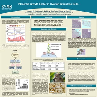
Four candidate antibodies from various suppliers were selected that could potentially
detect placental growth factor:
Supplier Type of Antibody Optimal Dilution Antigen Retrieval
Abbiotec Anti-rabbit 1:200 Basic
GeneTex Inc. Anti-rabbit 1:100 Basic
Sigma-Aldrich Anti-mouse 1:100 Basic
Thermo Scientific Anti-rabbit 1:100 Basic
Tissues and cells harvested from female
macaques were utilized to model human
ovarian follicles because this nonhuman
primate species experiences a true
menstrual cycle similar to humans. The
macaques were administered shots of
human chorionic gonadotropic (hCG) to
mimic the luteinizing hormone surge.
Paraffin ovary sections were collected via
oophorectomy (removal of the ovaries) from
the macaques after 0, 12, 24, and 36 hours
after the luteinizing hormone surge. The
ovary sections harvested after 36 hours
were believed to have the highest
expression of placental growth factor
protein. The candidate antibodies were
utilized in immunohistochemistry,
immunofluorescence, and antigen retrieval
procedures. Hematoxylin, a nuclear,
histologic stain, was used as the
counterstain and DAB substrate was used
as a tag because it has a brown reaction
product.2
Multiple trials of immunohistochemistry and immunofluorescence were
conducted on macaque ovarian sections testing each of the candidate
antibodies. Adjustments of concentration, hours after luteinizing hormone
surge, and antigen retrieval conditions were observed. Ultimately, none of
the four antibodies exhibited specific staining of placental growth factor
in the granulosa cells. Both the control sections, which received no antibody
exposure, the sections that were exposed to the antibody did not reveal
specific staining in the granulosa cell layer. Specific staining would have been
marked by a dark brown color from the DAB, but this color was only visible
nonspecifically in blood vessels.
example of ovary section anatomy
example of nonspecific staining