
Recommended
More Related Content
What's hot
What's hot (20)
Similar to Trigger Point Therapy Workshop 09.11.19
Similar to Trigger Point Therapy Workshop 09.11.19 (20)
More from Katie Emmett 🌐 Myofascial Decompression Therapy
More from Katie Emmett 🌐 Myofascial Decompression Therapy (9)
Recently uploaded
Recently uploaded (20)
Trigger Point Therapy Workshop 09.11.19
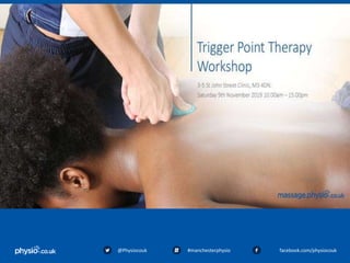
- 2. * 2 Trigger Point Therapy workshop With Katie Emmett @Physiocouk #manchesterphysio facebook.com/physiocouk Welcome
- 3. 3 @Physiocouk #manchesterphysio facebook.com/physiocouk Who are we? Katie’s LinkedIn: www.linkedin.com/katieemmett Twitter: @KatiePhysiocouk Website: www.massage.physio.co.uk Twitter: @physiocouk Facebook: www.facebook.com/physiocouk
- 5. @Physiocouk #manchesterphysio facebook.com/physiocouk This course does not qualify you / cover you to perform massage to clients.
- 6. @Physiocouk #manchesterphysio facebook.com/physiocouk ✓ Learn the theory of a trigger point ✓ Learn the theory of trigger point therapy technique ✓ Practice the trigger point technique to various muscle groups Aims of today
- 7. @Physiocouk #manchesterphysio facebook.com/physiocouk 10.00 - 10.30 - Induction / Arrival 10.30 - 10.50 - Quiz – What do you know about trigger point therapy 10.50 -11.30 - Theory: Trigger point therapy 11.30 -12.00 - Indications/ outcome measures 12.00 - 12.30 - Lunch 12.30 - 13.00 - Theory: Trigger pointing technique 13.00 - 14.00 - Practical: Muscle groups 14.00 - 14.30 – Practical: STR techniques 14.30 - 15.00 - Case Studies/Quiz answers Itinerary
- 8. @Physiocouk #manchesterphysio facebook.com/physiocouk Quiz…What do you know about trigger point therapy?
- 9. @Physiocouk #manchesterphysio facebook.com/physiocouk What is a trigger point? Question 1
- 10. @Physiocouk #manchesterphysio facebook.com/physiocouk How would patients describe Trigger Point pain? Question 2
- 11. @Physiocouk #manchesterphysio facebook.com/physiocouk Name some indications for Trigger Point Therapy? Question 3
- 12. @Physiocouk #manchesterphysio facebook.com/physiocouk Name 3 benefits of Trigger Point Therapy Question 4
- 13. @Physiocouk #manchesterphysio facebook.com/physiocouk Name some contraindications for Trigger Point Therapy Question 5
- 14. @Physiocouk #manchesterphysio facebook.com/physiocouk Where is the Trapezius muscle found? Question 6
- 15. @Physiocouk #manchesterphysio facebook.com/physiocouk Where is the Sternocleidomastoid muscle found? Question 7
- 16. @Physiocouk #manchesterphysio facebook.com/physiocouk What muscle group does Vastus Lateralis belong to? Question 8
- 17. @Physiocouk #manchesterphysio facebook.com/physiocouk Theory: Trigger Points
- 18. @Physiocouk #manchesterphysio facebook.com/physiocouk • Trigger points are hyperirritable areas of contracted muscle fibres that form a palpable nodule • On a microscopic level, the contracted muscle fibres accumulate into a small thickened area causing the rest of the fibre to stretch • The areas of contracted muscle restrict blood flow within the tissue causing an accumulation of waste products and reduced levels of nutrients available. What are Trigger Points?
- 19. @Physiocouk #manchesterphysio facebook.com/physiocouk • 1930s -Dr Hans used sclerometer to prove that tender areas in muscles are 50% harder than surrounding areas. • 1940s- Janet Travell developed trigger point injection therapy and termed the “tender areas” described by Dr Hans “Trigger points”. • Travell's therapy called for the injection of saline (a salt solution) and procaine (also known as Novocaine, an anesthetic) into the trigger point. • Travell mapped what she termed the body's trigger points and the manner in which pain radiates to the rest of the body. • Travell's work came to national attention when she treated President John F. Kennedy for his back pain. • Travell co-authored several books with David Simons which are considered the definitive reference for trigger point therapy. • Travell & Simons' Myofascial Pain and Dysfunction: Upper half of body • Travell & Simons' Myofascial Pain and Dysfunction: The Trigger Point Manual • Myofascial Pain and Dysfunction: The Trigger Point Manual, Volume 2 Brief History
- 20. @Physiocouk #manchesterphysio facebook.com/physiocouk • 1976- Bonnie Prudden, a physical fitness and exercise therapist developed Travells trigger point therapy. She found that applying sustained pressure to a trigger point using thumbs, knuckles and elbows produced superior results to those treated with injections when followed by corrective movements and stretching. Prudden later went on to author two books: • Myotherapy: Bonnie Prudden’s Complete Guide to Pain Free Living • Pain Erasure the Bonnie Prudden Way Brief History
- 21. @Physiocouk #manchesterphysio facebook.com/physiocouk • Trigger points are described according to location, tenderness and chronicity. • The main types of trigger points are: • Central/ primary trigger points • Satellite/ secondary trigger points • Active trigger points • Latent/inactive trigger points Different types of Trigger Points
- 22. @Physiocouk #manchesterphysio facebook.com/physiocouk • These are the most well-established and painful points • Pain is felt by the individual when they are active, and are usually what people refer to when they talk about trigger points • Central trigger points exist at a neuromuscular point, which is the meeting place of a nerve and muscle Central/primary
- 23. @Physiocouk #manchesterphysio facebook.com/physiocouk • These trigger points are “created” as a response to the central trigger point in neighbouring muscles that lie within the referred pain zone. • The primary trigger point is still the key to trigger pointing intervention: the satellite trigger points often resolve once the primary point has been effectively rendered inactive. • Satellite points may also prove unresponsive to treatment until the primary central focus is weakened. This is often the case in the paraspinal and/or abdominal muscles. Satellite/secondary
- 24. @Physiocouk #manchesterphysio facebook.com/physiocouk • This can apply to central and satellite trigger points. • A variety of stimulants, such as forcing muscular activity through pain, can activate an inactive trigger point. • This situation is common when activity is increased after trauma i.e a road traffic accident, where multiple and diffuse trigger points may have developed. • This trigger point is both tender to palpation and elicits a referred pain pattern. • Pain can limit range of movement. Active trigger points
- 25. @Physiocouk #manchesterphysio facebook.com/physiocouk • This applies to lumps and nodules that feel like trigger points. These can develop anywhere in the body and are often secondary. • These trigger points are not painful, and do not elicit a referred pain pathway. • The presence of inactive trigger points within muscles may lead to increased muscular stiffness and tension. They can build up for years. • It has been suggested that these points are more common in those who live a sedentary lifestyle (Starlanyl & Copeland 2001) • These points are “potential” trigger points and may reactivate if the central or primary trigger point is (re)stimulated • Reactivation may occur following trauma and injury Latent/inactive
- 26. @Physiocouk #manchesterphysio facebook.com/physiocouk Active trigger point referral symptoms •Dull ache •Deep •Pressing pain •“Stabbing” •Burning •Referred pain •Common reports of headaches, dizziness and pins and needles Symptoms of trigger points
- 27. @Physiocouk #manchesterphysio facebook.com/physiocouk Referral Pain Guide Sternocleomastoid and Masseter
- 28. @Physiocouk #manchesterphysio facebook.com/physiocouk Referral Pain Guide Trapezius
- 29. @Physiocouk #manchesterphysio facebook.com/physiocouk Referral Pain Guide Pectorals
- 30. @Physiocouk #manchesterphysio facebook.com/physiocouk Referral Pain Guide Quadratus lumborum
- 31. @Physiocouk #manchesterphysio facebook.com/physiocouk Referral Pain Guide Piriformis
- 32. @Physiocouk #manchesterphysio facebook.com/physiocouk Referral Pain Guide Glute maximus medius and minimus
- 33. @Physiocouk #manchesterphysio facebook.com/physiocouk Referral Pain Guide TFL
- 34. @Physiocouk #manchesterphysio facebook.com/physiocouk Referral Pain Guide Vastus lateralis
- 35. @Physiocouk #manchesterphysio facebook.com/physiocouk Referral Pain Guide Hamstrings
- 36. @Physiocouk #manchesterphysio facebook.com/physiocouk A sensation of: •Numbness •Fatigue •Weakness •Burning A loss of: •Flexibility •Range of movement •Muscular power and strength Other symptoms
- 37. @Physiocouk #manchesterphysio facebook.com/physiocouk • Repetitive overuse injuries (using the same body parts in the same way hundreds of times on a daily basis) from activities such as typing/mousing, handheld electronics, gardening, home improvement projects, work environments, etc. • Sustained loading e.g heavy lifting, carrying babies, briefcases, boxes or lifting bedridden patients. Why are they present?
- 38. @Physiocouk #manchesterphysio facebook.com/physiocouk •Poor posture due to our sedentary lifestyles, de-conditioning, poorly designed furniture and technology. •Muscle clenching and tensing due to mental/emotional stress. •Direct injury such as a strain, break, twist or tear e.g car accidents, sports injuries, falling down stairs. •Trigger points can even develop due to inactivity such as prolonged bed rest or sitting . Why are they present?
- 39. @Physiocouk #manchesterphysio facebook.com/physiocouk • Deep within muscles are spiral shaped nerve fibres called muscle spindles. • When muscles are excessively stretched muscle spindles activate and send signals to the brain to promote a protective muscular contraction- stretch reflex arc. Formation of a Trigger Point
- 40. @Physiocouk #manchesterphysio facebook.com/physiocouk • The problem occurs when the muscle spindle becomes sensitised. • Injury or overuse can over stimulate muscle spindles which can cause contraction within the muscle and subsequently forming localised muscular spasm…. A Trigger point. Formation of a Trigger Point
- 41. @Physiocouk #manchesterphysio facebook.com/physiocouk • Prolonged muscular contractions restrict blood flow through the area. • This causes a build-up of waste products and toxins within the area and a reduction in fresh, nutritious blood flowing through. • If the muscle spindle is active for prolonged periods of time the length of the muscle can shorten. • Subsequently patients may experience a reduction in ROM. Formation of a Trigger Point
- 42. @Physiocouk #manchesterphysio facebook.com/physiocouk • Trigger points are found all over the body. . • Trigger points are located within each sarcomere often where the nerve enters the muscle. • The motor end plate. Where are they formed?
- 43. @Physiocouk #manchesterphysio facebook.com/physiocouk • Chemoreceptors and mechanoreceptors are stimulated to send messages to the brain which results in the sensation of pain. • The brain stimulates decreased movement into these muscles which further tightens the structure. Trigger Point pain
- 44. @Physiocouk #manchesterphysio facebook.com/physiocouk Indications Outcome measures Pain NRS scale & subjective symptoms Reduced AROM Active range of movement Muscle tightness Palpation Muscle weakness Oxford rating scale Indications and outcomes
- 45. 45@Physiocouk #manchesterphysio facebook.com/physiocouk • Simple and easy • Before, during and after massage • Record change • Use with patient to see reduction in pain over the progression of treatments Outcome measure: NRS scale
- 46. 46@Physiocouk #manchesterphysio facebook.com/physiocouk • Pre and post measurements • Goniometer or visual • Standardise to produce reliable results • Review each session • Used to distinguish areas to treat and techniques types • Valuable in the success of treatment Outcome measure: ROM
- 47. 47@Physiocouk #manchesterphysio facebook.com/physiocouk • Measure nerve conduction and muscle recruitment. • Compare both sides. • If strengthening exercises are used alongside massage treatment patients will be able to feel a progression here. Outcome measure: muscle testing
- 48. 48@Physiocouk #manchesterphysio facebook.com/physiocouk Use palpation as a measure using “the four T’s” • Temperature Is the tissue hot? This could indicate presence of inflammation. ● Texture Swelling (acute-hard, chronic – “boggy”, congested) healthy tissues should have an even texture Adhesions feel like tissues are “stuck” and less mobile “audible crunching”. Outcome measure: palpation
- 49. 49@Physiocouk #manchesterphysio facebook.com/physiocouk Outcome measure: Palpation ● Tenderness Pain can be indicated through response. NRS can be used here. ● Tone Tissues may be tense, always compare to other side to see what is normal for the patient.
- 52. @Physiocouk #manchesterphysio facebook.com/physiocouk Theory: Trigger Point Therapy
- 53. @Physiocouk #manchesterphysio facebook.com/physiocouk Assessment •Find the most painful TP using patient response and Numeric Rating Scale. •Treat the highest rated point and radiate out from this point •Once the points are found – a good amount of pressure is applied (perform with precaution - keep communication with patient) •Initial pain is stimulated and you hold the pressure until the pain has eased completely or in some cases reduced slightly •Re-apply pressure onto the same point until the pain eases off quicker or it isn’t felt anymore (roughly 3 times) •Thumbs/elbows or tools can be used How to treat a Trigger Point
- 54. @Physiocouk #manchesterphysio facebook.com/physiocouk Guidelines Application of direct pressure onto the trigger points for around 30 seconds or until the patient’s pain has decreased to at least 3/10 NRS score. The applied pressure help to break-up the adhesive fibre connections within the trigger points and push out blood containing waste products and toxins. After 30 seconds, the pressure is released allowing a rush of fresh blood containing nutrients to circulate the trigger point. Repeat 3 times in conjunction with deep massage strokes. This can vary on the severity of pain/ how deep or superficial the TP is – subjective and variable to each patient How to treat a Trigger Point
- 55. @Physiocouk #manchesterphysio facebook.com/physiocouk • Reduced pain • Increased range of motion • Decreased muscle stiffness and tension • Reduction in headaches • Improved flexibility • Improved circulation • Fewer muscle spasms The benefits
- 56. @Physiocouk #manchesterphysio facebook.com/physiocouk • High pain scales • Patient Anxiety • Acute/ Inflammatory stage of healing • Hypersensitivity • Pregnancy • Epilepsy • Asthma • Hypertension • Prescribed medication Precautions
- 57. @Physiocouk #manchesterphysio facebook.com/physiocouk General Local Acute conditions requiring medical attention Acute flare-up of inflammatory arthritides Acute pneumonia Aneurysms deemed life-threatening (may be general contraindication depending on location) Advanced kidney, respiratory or liver failure Local contagious condition Diabetes with complications such as gangrene, advanced heart or kidney disease or very unstable or high blood pressure Local irritable skin condition Hemorrhage Malignancy Severe atherosclerosis Open wound or sore Severe and unstable hypertension Recent burn Shock Undiagnosed lump Systemic contagious or infectious condition Contraindications
- 58. @Physiocouk #manchesterphysio facebook.com/physiocouk • Posture – Bed height – Stance – Patient position • Use different parts of your hands/ arms to apply pressure • Keep arms straight to utilise body weight when applying pressure/resistance. • Move from the hips and knees as much as possible • Oil (or cream)- only needs to be a little bit, if any. Look after yourself before you look after the patient! Manual handling and posture
- 59. @Physiocouk #manchesterphysio facebook.com/physiocouk Very common for people to experience irritation for up to 72 hours after treatment. Side effects can include: • Bruising • Redness • Tenderness/Increased Sensitivity • Increased symptoms • Aching similar to DOMS Post treatment irritation
- 60. @Physiocouk #manchesterphysio facebook.com/physiocouk Causes • The release of toxins/waste products from muscular tissue • Neurological sensitisation • Increased blood flow and micro trauma can lead to bruising and redness Advice •Reassure the patient it's a normal response to be sore after soft tissue treatment •Recommend they drink water to keep hydrated Post treatment irritation
- 61. @Physiocouk #manchesterphysio facebook.com/physiocouk • Trapezius • Sternocleomastoid • Rhomboids • QL • TFL • Vastus Lateralis • Gastrocnemius Practical
- 62. @Physiocouk #manchesterphysio facebook.com/physiocouk The trapezius can be separated into three muscles: •The upper trapezius •The middle trapezius •The lower trapezius Anatomy: All three trapezius muscles originate along the spine to T12 and extend laterally to attach to the shoulder girdle. Function: Each muscle has a different direction of pull. Movements facilitated include scapula elevation, depression, retraction, upwards and downward rotation. Trapezius
- 63. @Physiocouk #manchesterphysio facebook.com/physiocouk The whole trapezius muscle creates various movements of the shoulder blade, neck, and head. To move your arm above your head you need muscular contraction pulling in opposite directions. Muscular contraction in both lower and upper fibre traps to upwardly rotate the scapula. This type of complexity makes it easy for trigger point activity to spread quickly through the muscle group as a whole. Trapezius
- 64. @Physiocouk #manchesterphysio facebook.com/physiocouk Four primary trigger points in the trapezius muscle group; two trigger points in the upper fibers, and one each in the middle and lower fibers. • The anterior trapezius trigger point • The upper trapezius trigger point • The middle trapezius trigger point • The lower trapezius trigger point Trapezius Trigger Points
- 65. @Physiocouk #manchesterphysio facebook.com/physiocouk Causes ● Poor posture- shoulders, neck and back ● Stress ● Carrying heavy handbags/ laptop bags on one side ● Dysfunction/ pathology within the shoulder complex Symptoms ● Ache and tightness in shoulders and neck ● Tension headaches ● Upper cross syndrome ● Struggle to look over shoulder Trapezius Pain
- 66. @Physiocouk #manchesterphysio facebook.com/physiocouk • Tip: squeezing UFT between finger and thumb can be very effective with upper and anterior trigger points. Rx – Trapezius
- 67. @Physiocouk #manchesterphysio facebook.com/physiocouk Anatomy: ● Originates from the mastoid process. ● The sternal division runs diagonally downward to attach to the sternum. ● The clavicular division attaches right behind it on the medial clavicle. Function: ● Turn head towards opposite side and bilaterally side flex the neck. ● Control and monitor the head’s position in space. Proprioceptive feedback from the SCM is essential to being able to maintain one’s balance. Sternocleidomastoid
- 68. @Physiocouk #manchesterphysio facebook.com/physiocouk • The sternal division typically has 3-4 trigger points spaced out along its length, while the clavicular division has 2-3 trigger points. • Trigger points are usually present in both left and right SCM muscles as they work together to control the head. Sternocleidomastoid Trigger points
- 69. @Physiocouk #manchesterphysio facebook.com/physiocouk Each SCM division has a separate and distinct referred pain pattern: • The sternal division’s referred pain is felt deep in the eye socket (behind the eye), above the eye, in the cheek region, in the back of the head, and on the top of the head. • The clavicular division’s referred pain is felt in the forehead, deep in the ear, behind the ear, and in the molar teeth on the same side. Causes/ symptoms: •Sore Neck •Tension Headaches •“Heavy head” •Poor head posture •Poor exercise technique (sit ups) Sternocleidomastoid Pain
- 70. @Physiocouk #manchesterphysio facebook.com/physiocouk • Locating and releasing these trigger points can be complicated due to their proximity to many blood vessels and nerves in the neck region. • Caution: do not massage somewhere you can feel a pulse. • Tip: Rotate head to side to find muscle but rotate back to neutral to treat. Rx: Sternocleidomastoid
- 71. @Physiocouk #manchesterphysio facebook.com/physiocouk Anatomy: ● The rhomboid muscle group originates from the spinous process of C6-T4 and inserts onto the medial border of the scapula. ● It is separated into rhomboid major and rhomboid minor muscles. Function: ● scapula retraction and slight elevation Rhomboids
- 72. @Physiocouk #manchesterphysio facebook.com/physiocouk 3 primary trigger points • The rhomboid minor trigger point lies just medial to the inside edge of the scapula, level with the scapular spine. • The rhomboid major trigger points lie one above the other, along the lower part of the scapular border. •Referred Pain: The pain concentrates in the region between the spine and the shoulder blade. Rhomboid Trigger Points
- 73. @Physiocouk #manchesterphysio facebook.com/physiocouk Causes •Poor posture •Rhomboid weakness •Scapular instability •Winging scapula Symptoms: •Pain Between the Shoulder Blades •Pain is usually felt at rest and not typically affected my movement. •Patients may hear snapping or grinding noises from the region around the shoulder blade during movements of the arm. Rhomboid pain
- 74. @Physiocouk #manchesterphysio facebook.com/physiocouk ● Make sure that you have released any trapezius trigger points first otherwise they may block you from reaching rhomboid trigger points. ● Try in both prone lying and side lying position. Tips: • Placing hand behind back can help to lift scapula out of the way. • Side-lying position to allow more forward movement of the scapula. • Prone to allow more pressure to be applied. Rx: Rhomboids
- 76. @Physiocouk #manchesterphysio facebook.com/physiocouk Anatomy: • Originates from the iliac crest and runs upwards and medially to attach onto the 12th rib and transverse process of L1-L4. Function: • Stabilise movement of spine and pelvis. • Control a upright posture. • Produce extension and side flexion of the lumbar spine. QL
- 77. @Physiocouk #manchesterphysio facebook.com/physiocouk • If one muscle develops trigger point activity, the muscle on the other side will become overloaded and develop trigger points as well. • From a clinical perspective, you should always address the trigger points in both the left and right QL muscles, even if the pain is limited only to one side. QL Trigger Points
- 78. @Physiocouk #manchesterphysio facebook.com/physiocouk There are four potential trigger points in the QL muscle: • The upper QL trigger point is found just lateral to where the lumbar paraspinal muscles and the twelfth rib meet. •The middle or deep QL trigger points lie closer to the spine next to the third and fourth lumbar vertebrae. •The lower QL trigger point lies deep in the region where the paraspinal muscles meet the iliac crest. QL Trigger Points
- 79. @Physiocouk #manchesterphysio facebook.com/physiocouk Causes ● Carrying children on hip ● Sitting with poor posture for prolonged periods of time ● Poor manual handling technique ● Poor workstation ergonomics Symptoms: • Usually described as an intense, deep ache • Occasionally can produce a sharp, knifelike symptoms particularly during movement. • Ache pain into small of back. • Pain when bending down. QL Pain
- 80. @Physiocouk #manchesterphysio facebook.com/physiocouk • The first step in the effective treatment of the QL trigger points is being able to accurately locate and contact the trigger points. • Try in both Prone and a extended side- lying position. • Tips: Angle inwards towards spine rather than directly posterior. Rx: QL
- 81. @Physiocouk #manchesterphysio facebook.com/physiocouk Anatomy: •Originates from outer aspect of the Iliac Crest and Anterior Superior Iliac Spine (A.S.I.S) it runs through illiotibial band which inserts onto lateral epicondyle of tibia. Function: • Its function is primarily to control movement of the leg during the stance phase of walking. •Assist with hip abduction, flexion and internal rotation on the hip. TFL – tensor fascia latae
- 82. @Physiocouk #manchesterphysio facebook.com/physiocouk • There is only one trigger point found in the TFL and it is located in the upper region of the muscle just below where it attaches to the A.S.I.S. • The referred pain pattern covers the entire hip joint and extends down the outside aspect of the thigh, sometimes nearly to the knee joint. TFL
- 83. @Physiocouk #manchesterphysio facebook.com/physiocouk Causes •Over foot pronation •Valgus knee position •Weakness in gluteus/ trendelenburg sign •Poor squatting/ lunging techniques •Poor landing biomechanics Symptoms: • Pain in the hip joint (greater trochanter) and down the outside thigh during movement of the hip. • Pain when sitting in low chair or flex their hip more than 90°. • Unable to lie on the affected hip during sleep and unable to lie on the unaffected side during sleep without a pillow between their knees. • Pain and limited ROM in hip adduction. TFL
- 84. @Physiocouk #manchesterphysio facebook.com/physiocouk Tips: Find ASIS drop fingers down and laterally. • If struggling can ask pt to flex and medially rotate hip joint to feel contraction. Rx: TFL
- 85. @Physiocouk #manchesterphysio facebook.com/physiocouk Location: The quadriceps femoris muscle group form the thigh musculature found on the front of the upper leg. The group is comprised of four muscles: • The Vastus Lateralis • The Rectus Femoris • The Vastus Medialis • The Vastus Intermedius Vastus Lateralis
- 86. @Physiocouk #manchesterphysio facebook.com/physiocouk Anatomy: •Vastus lateralis originates lateral aspect of the superior femur bone and runs down the outside of the thigh to attach to the lateral aspect of the patella. Function: •The vastus lateralis is the largest muscle in the group. •Contraction of this muscle produces extension of the lower leg at the knee. •Helps to stabilise the patella in patellofemoral groove. Vastus Lateralis
- 87. @Physiocouk #manchesterphysio facebook.com/physiocouk There are two sets of trigger points in the vastus lateralis muscle: • The upper vastus lateralis trigger points are located in mid-thigh region on the outside aspect of the leg. They refer pain all along the outside of the thigh and knee. • The lower vastus lateralis trigger points are found just above and to the outside of the knee joint. They refer pain around the outside aspect of the knee joint and below it. Vastus Lateralis Trigger Points
- 88. @Physiocouk #manchesterphysio facebook.com/physiocouk Causes ● Weakness in gluteus ● Over pronated feet ● Valgus knee position ● Overload from gym routine ● Skiing activities Symptoms: ● Pain on outside of thigh ● Pain into and behind the knee ● Pain on resisted knee extension ● Anterior knee pain ● Stuck patella ● Crepitus ● functional limitations. Vastus Lateralis Pain
- 89. @Physiocouk #manchesterphysio facebook.com/physiocouk Tips: try sliding thumbs up the outside of thigh until you feel the resistance of the trigger points. Rx : Vastus Lateralis
- 90. @Physiocouk #manchesterphysio facebook.com/physiocouk Anatomy: ● Largest muscle in the calf ● Originates from the achilles tendon and splits into two heads to attach onto the medial and lateral condyles of the femur. Function: ● plantarflexion of the foot and assists with knee flexion. Gastrocnemius
- 91. @Physiocouk #manchesterphysio facebook.com/physiocouk Gastrocnemius may contain up to four trigger points. •Two medial trigger points found in the medial head. One just below the knee crease and the other an inch down. •Two lateral trigger points mirror the medial trigger points except they are slightly more distal. Gastrocnemius Trigger Point
- 92. @Physiocouk #manchesterphysio facebook.com/physiocouk Causes •Prolonged wearing of high heels can leave gastroc in a shortened position •Achilles tendinopathy •foot pronation •Sudden increases in training programmes •Prolonged immobilisation e.g. cast •Sleeping on front for prolonged periods Symptoms •Pain in calf •Pain behind the knee •Pain when standing on top toes •Pain going upstairs •Suffer from calf cramp regularly Gastrocnemius Pain
- 93. @Physiocouk #manchesterphysio facebook.com/physiocouk • Tip: try using your elbow to trigger point. Rx : Gastrocnemius
- 96. Effectiveness of Myofascial Trigger Point Manual Therapy Combined With a Self- Stretching Protocol for the Management of Plantar Heel Pain: A Randomized Controlled Trial @Physiocouk #manchesterphysio facebook.com/physiocouk Renan-Ordine et al, (2011) •Aim: to assess the effect of trigger point therapy and stretching or stretching alone in the treatment for plantar heel pain. •Method: 60 patients with plantar heel pain were divided into 2 groups a)self-stretching b) self- stretching and trigger point therapy. •Outcome measures: assessed at baseline and at a 1-month follow up. – Physical function and pain assessed using a quality of life questionnaire. – pressure pain thresholds were assessed over affected gastroc, soleus muscles and over the calcaneus using a mechanical pressure algometer. •Results: trigger point therapy and self-stretching is superior to stretching alone in the treatment of patients with plantar heel pain. •Link: http://www.jospt.org/doi/full/10.2519/jospt.2011.3504
- 97. Comparative study on effects of manipulation treatment and transcutaneous electrical nerve stimulation on patients with cervicogenic headache @Physiocouk #manchesterphysio facebook.com/physiocouk Li et al, (2007) •Aim: To compare the effects of trigger pointing and transcutaneous electrical nerve stimulation (TENS) on patients with cervicogenic headache. •Method: 70 patients with cervicoigenic headaches were randomly allocated to receive trigger pointing or TENS every other day for 40 days. •Outcome measures: Taken 2 weeks pre-treatment and 4 weeks post-treatment. – headache degree, frequency and lasting time using a numeric rating scale – ROM of cervical spine. •Results: Trigger pointing was superior to TENS in headache frequency, lasting time and ROM scores. Response rate of trigger pointing treatment was 94.5%, significantly higher than 64.5% of TENS treatment. •Link: http://europepmc.org/abstract/med/17631795
- 98. Immediate effect of activator trigger point therapy and myofascial band therapy on non-specific neck pain in patients with upper trapezius trigger points compared to sham ultrasound: A randomised controlled trial @Physiocouk #manchesterphysio facebook.com/physiocouk Blikstad and Gemmell, (2007) •Aim: To determine the immediate effect of activator trigger point therapy and myofascial band therapy compared to sham ultrasound on non-specific neck pain •Method: 45 patients with non-specific neck pain of at least 4 on an 11-point numerical rating scale and upper trap trigger points, decreased cervical lateral flexion away from the active trigger points participated. Participants were assigned to one of three treatment groups; trigger point therapy, myofascial band therapy or sham ultrasound. •Outcome measures: assessed before and 5 min after treatment – pain levels assessed using numerical scale – cervical ROM using goniometer – pain perceived thresholds using pain pressure algometer. •Results: For the primary outcome measure of pain reduction the odds of a patient improving with activator trigger point therapy was 7 times higher than a patient treated with myofascial band therapy or sham ultrasound. •Link: http://www.sciencedirect.com/science/article/pii/S1479235407001083
- 99. Effect of myofascial trigger point therapy with an inflatable ball in elderly with chronic non-specific low back pain. Oh S, Kim M, Lee M, Kim T, Leed D, Yoon B Journal of Musculoskeletal Rehab (2018) 6;31(1):119- 126 Aim: To investigate the effects of myofascial trigger point with an inflatable ball for elderly individuals with chronic lower back pain. Measure: 50 elderly patients with CNSLBP were evaluated for pain, pressure sensitivity and physical function at baseline, week 1, week 3 and week 6. Outcome measure: Visual Analogue scale (VAS) and pressure pain threshold (PPT) were used to measure pain intensity and severity. Straight leg raise test and range of movement in the back was also used to assess physical function. Result: Significant differences were observed between the 3- and 6-week VAS scores baseline and 1- week (7%), 1- and 3-week (14%), and 3- and 6-week PPTs (18%); 3- and 6-week BROMs (Flexion, 7.1%; Extension, 41%); baseline and 1-week (6.9%), 1- and 3-week (3%) 3- and 6-week active SLR test scores (7%); and baseline and 1-week (2.6%), 1- and 3-week (8.34%), and 3- and 6-week passive SLR test scores (5.3%). CONCLUSION:Myofascial trigger point therapy with an inflatable ball relieved pain and improved physical function in the elderly with chronic non-specific lower back pain.
- 101. @Physiocouk #manchesterphysio facebook.com/physiocouk PC/HPC -21 year old female with an gradual onset of ache pain in shoulders over past 1/12 rating 4/10 on VAS scale. The pain is aggravated by sitting at a desk for long hours and eased with the application of heat. SH- final year art student with a sudden increase in workload as final project is due in 2/12. Carry heavy art portfolio to and from university. Attends a LBP class at the gym 1 x a month. PMH- nil to note DH- paracetamol when needed Case study – shoulder pain
- 102. @Physiocouk #manchesterphysio facebook.com/physiocouk Objective signs • Increased UFT tone • Reduced cervical lateral flexion due to UFT tightness • TOP of L and R UFT and Rhomboids • Active Trigger points in R and L Rhomboids • No neurological symptoms Case study – shoulder pain
- 103. @Physiocouk #manchesterphysio facebook.com/physiocouk Case study – shoulder pain
- 104. @Physiocouk #manchesterphysio facebook.com/physiocouk PC/HPC – 39 year old male 8/10 sharp pain in R lower back. Pain began suddenly when after lifting heavy box up which sent shooting pains down R leg. Aggravated by bending down and putting shoes on and eased by lying down flat. SH- full time receptionist, doesn’t perform regular exercise. PMH- history of lower back pain DH- analgesics Case study – lower back pain
- 105. @Physiocouk #manchesterphysio facebook.com/physiocouk Objective signs •Limited Lumber range of movement •Increase in pain during flexion and L lateral flexion •Pain eased during extension. •PALP – pain on palp of QL and L3 spinous process Case study – lower back pain
- 106. @Physiocouk #manchesterphysio facebook.com/physiocouk Case study – lower back pain
- 107. @Physiocouk #manchesterphysio facebook.com/physiocouk PC/HPC – 35 year old male runner. Felt a 6/10 sharp pain in R calf towards the end of a 5K run 2/52 ago. Had to stop running. No swelling or bruising was present. Pain reduced since 3/10 ache pain, tried running again but still feels painful. SH- work in a warehouse, on feet all day up and down ladders. PMH- prev R lateral ankle sprain 12/12 ago DH-nil to note Case study – Calf pain
- 108. @Physiocouk #manchesterphysio facebook.com/physiocouk Objective signs •Increased calf bulk L side •Thickening of R Achilles tendon •Reduced dorsiflexion of R ankle •Reduce muscular strength in R resisted plantarflexion •Reduced R calf length •PALP- pain on palp of medial gastroc •-ve Thomas test Case study – Calf pain
- 109. @Physiocouk #manchesterphysio facebook.com/physiocouk Case study – Calf pain
- 110. @Physiocouk #manchesterphysio facebook.com/physiocouk PC/HPC- 25 year old male 5/10 pain in L buttock. 1/12 ago increased pain following legs gym session, gradually worsening since. Aggravated by climbing multiple flights of stairs at work. Eased by resting. SH- Started going to the gym 1/12 ago after a 5 year break. Doesn’t do any stretching because he doesn’t know how to. Works on the 8th floor of a office building. PMH- over pronate both feet, especially bad in L side. DH- nil to note Case study – buttock pain
- 111. @Physiocouk #manchesterphysio facebook.com/physiocouk Objective signs •Over pronation in L > R foot •Valgus position of knees •Poor hamstring flexibility on 90/90 test in L>R legs •No neurological symptoms during SLR •PALP: tension L>R hamstring, glutes and piriformis •Very tender on PALP of piriformis Case study – buttock pain
- 112. @Physiocouk #manchesterphysio facebook.com/physiocouk Diagnosis? How would treat this? Case study – buttock pain
- 114. @Physiocouk #manchesterphysio facebook.com/physiocouk What is a trigger point? • Trigger points are hyperirritable areas of contracted muscle fibres that form a palatable nodule • On a microscopic level, the contracted muscle fibres accumulate into a small thickened area causing the rest of the fibre to stretch • The areas of contracted muscle restrict blood flow within the tissue causing an accumulation of waste products and reduced levels of nutrients available. Question 1
- 115. @Physiocouk #manchesterphysio facebook.com/physiocouk How would a patient describe trigger point pain? • Dull ache • Deep • Sharp • Pressing pain • Stabbing • Burning • Travelling pain • Head pain Question 2
- 116. @Physiocouk #manchesterphysio facebook.com/physiocouk Name some indications for Trigger point therapy • Pain • Reduced AROM • High muscle tension or tone • Muscle tightness Question 3
- 117. @Physiocouk #manchesterphysio facebook.com/physiocouk Name some benefits of Trigger point therapy •Reduced pain • Increased range of motion • Decreased muscle stiffness and tension • Reduction in headaches • Improved flexibility • Improved circulation • Fewer muscle spasms Question 4
- 118. @Physiocouk #manchesterphysio facebook.com/physiocouk Name some contraindications for Trigger point therapy General Local Acute conditions requiring medical attention Acute flare-up of inflammatory arthritides Acute pneumonia Aneurysms deemed life-threatening (may be general contraindication depending on location) Advanced kidney, respiratory or liver failure Local contagious condition Diabetes with complications such as gangrene, advanced heart or kidney disease or very unstable or high blood pressure Local irritable skin condition Hemorrhage Malignancy Severe atherosclerosis Open wound or sore Severe and unstable hypertension Recent burn Shock Undiagnosed lump Systemic contagious or infectious condition Question 5
- 119. @Physiocouk #manchesterphysio facebook.com/physiocouk Where is the Trapezius muscle found? The trapezius can be separated into three muscles: •The upper trapezius •The middle trapezius •The lower trapezius Anatomy: All three trapezius muscles originate along the spine to T12 and extend laterally to attach to the shoulder girdle. Question 6
- 120. @Physiocouk #manchesterphysio facebook.com/physiocouk Where is the Sternocleomastoid muscle found? Anatomy: ● Originates from the mastoid process. ● The sternal division runs diagonally downward to attach to the sternum. ● The clavicular division attaches right behind it on the medial clavicle. Question 7
- 121. @Physiocouk #manchesterphysio facebook.com/physiocouk What muscle group does Vastuslateralis belong to? The quadriceps muscular group. Anatomy: •Vastus lateralis originates lateral aspect of the superior femur bone and runs down the outside of the thigh to attach to the lateral aspect of the patella. Question 8
- 122. 122 Thanks for coming! Don’t forget to follow us on Twitter: @physiocouk @Physiocouk #manchesterphysio facebook.com/physiocouk