
Recommended
More Related Content
What's hot
What's hot (20)
Similar to Principles Of Amputation
Similar to Principles Of Amputation (20)
More from Joe Antony
More from Joe Antony (20)
Recently uploaded
Recently uploaded (20)
Principles Of Amputation
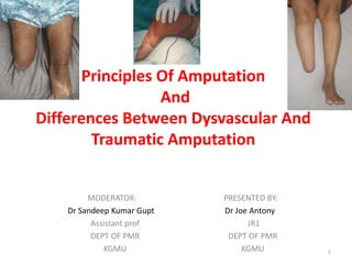
- 1. Principles Of Amputation And Differences Between Dysvascular And Traumatic Amputation MODERATOR: Dr Sandeep Kumar Gupt Assistant prof DEPT OF PMR KGMU PRESENTED BY: Dr Joe Antony JR1 DEPT OF PMR KGMU 1
- 2. Contents 1. Nomenclature and history 2. Definition of amputation 3. Epidemiology 4. Goals of amputation surgery 5. General principles of amputation 6. Diffrences between dysvascular and traumatic amputation 2
- 3. History and nomenclature • Derived from the Latin amputare. – "to cut away", from ambi- ("about", "around") and putare ("to prune").. • The English word "amputation" was first applied to surgery in the 17th century. • Most ancient of surgical procedure. • Historically were stimulated by the aftermath of war. • It was a crude procedure by which limb was rapidly severed from unanesthetized patient. • The open stump was then crushed or dipped in boiling oil to obtain hemostasis. • Hippocrates was the first to use ligature. • Ambroise Pare ( a France military surgeon) introduced artery forceps. He also designed prosthesis. Campbells operative orthopedics 13th edition 3
- 4. Definition • Amputation can be defined as “removal of diseased, protruding, functioning unit of body”. • Amputation surgical Removal or loss of limb through one or more bones. • Disarticulation—surgical Removal or loss of limb through a joint. Kulkarni textbook orthopedics and trauma 4
- 5. Epidemiology • Age- • Sex- approx. 86% male • Limb;- approx. 85% - lower limb, 15% -- upper limb Campbells operative orthopedics 13th edition Ghosh Das Pooja et al Prevalence and aetiology of amputation in India: A retrospective analysis 5
- 6. • 70 % of amputations accounts for trauma • 27 % of amputations account for peripheral vascular disease • Cancer accounts for 0.8% of the total amputations, and is the most common cause in above 80 year old Ghosh Das Pooja et al Prevalence and aetiology of amputation in India: A retrospective analysis 6
- 7. Amputation : Loss of a limb or part of a limb Causes • Congenital • Traumatic - Rail / Road accidents - Agriculture / domestic accidents - War / Industrial accidents • Vascular - Atherosclerosis / alcoholism - Burger’s disease /frost bite - Diabetes • Neoplastic - Malignant Tumors (Osteosarcoma) • Infection - Leprosy, syphilis, septic gangrene 7
- 8. Goals and objectives of amputation surgery • Preserving life • Preservation of function • Early return to function • Minimize energy expenditure • Preservation of Length • Painless residual limb • Prevention of symptomatic neuromas • Minimize phantom limb pain • Prevention of adjacent joint contractures • Early prosthetic fitting Campbells operative orthopedics 13th edition 8
- 9. Determination of level • Zone of Injury (trauma) • Adequate margins (tumor) • Adequate circulation (vascular disease) • Soft tissue envelope • Bone and joint condition • Control of infection • Nutritional status Campbells operative orthopedics 13th edition Essentials of prosthesis and orthotics, Dr AK Agarwal 9
- 10. General Amputation Principles • Skin • Muscle • Nerves • Hemostasis • Bone 10
- 11. Skin • Flaps should be kept thick • Unnecesary dissection should be avoided • Atypical flaps are preferable for proximal level • Tense sutures should be avoided • Painless and Non adherent scar • Removal of dog ear Campbells operative orthopedics 13th edition Grabs textbook of plastic and rehabilitative surgery 11
- 12. Muscle Myofascial closure Minimal muscle stabilization Myoplasty Opposing muscle groups Myodesis Attached to bone Tenodesis Tendon attached to bone 12
- 13. • Jaegers et al. showed that transected muscles atrophy 40% to 60% in 2 years if they are not securely fixed. • If possible, myodesis should be performed to provide a stronger insertion, help maximize strength, and minimize atrophy • Myodesed muscles continue to counterbalance their antagonists, preventing contractures and maximizing residual limb function. • Myodesis may be contraindicated, however, in severe ischemia because of the increased risk of wound breakdown Campbells operative orthopedics 13th edition 13
- 14. Non ischemic limb Fashioning of equal anterior and posterior skin flaps, each one half anteroposterior diameter of leg at level of bone section. Suture of myofascial flap to periosteum anteriorly. Fashioning of posterior myofascial flap. 14
- 15. Ischemic limb Fashioning of short anterior and long posterior skin flaps Separation and removal of distal leg Suture of flap to deep fascia and periosteum anteriorly. NO TORNIQUET 15
- 16. • Classically, surgeons evaluate for final closure based on the ‘‘4 C’s’’—colour, consistency, contraction, and circulation. • Traumatic amputations can be left open for serial debridement procedures, while primary amputations done— – those outside the zone of injury, infection, vascular compromise, or tumor— can often be closed at the time of surgery. 16
- 17. Hemostasis • Except in severely ischemic limbs, the use of a tourniquet is highly desirable and makes the amputation easier. • The limb may be exsanguinated by wrapping it with an Esmarch bandage before the tourniquet is inflated. • Contra indications- In amputations for infections or malignancy. – In such instances, inflation of the tourniquet should be preceded by elevation of the limb for 5 minutes. • Major blood vessels should be isolated and individually ligated. Arteries and veins should be ligated separately, and larger vessels should be doubly ligated. • The tourniquet should be deflated before closure, and meticulous hemostasis should be obtained. • A drain should be used in most cases for 48 to 72 hours. Campbells operative orthopedics 13th edition 17
- 18. Tourniquet Brunners ten rules of tourniquet • Venous tourniquet – More useless than not having tourniquet Sharma JP, Salhotra R et al. Tourniquets in orthopedic surgery. Indian J Orthop 18
- 19. Nerves • A neuroma formation is inevitable after transection since the axons are unable to locate the distal nerve stump. • A neuroma becomes painful if it forms in a position where it would be subjected to repeated trauma. • Normal physiologic stimuli such as – stretching, – pressure, – vascular pulsations – may be painful and thus limit prosthetic usage. 19
- 20. • Special techniques have been tried in the hopes of preventing the formation of painful neuromas. These include – end-loop anastomosis, – peri- neural closure, – Silastic capping, – sealing the epineurial tube with butyl cyanoacrylate, – ligation, – cauterization, – methods to bury the nerve ends in bone or muscle. 20
- 21. • Most surgeons currently agree that nerves should be – isolated, – gently pulled distally into the wound, – divided cleanly with a sharp knife so that the cut end retracts well proximal to the level of bone resection. • Strong tension on the nerve should be avoided during this maneuver; otherwise, the amputation stump may be painful even after the wound has healed. • Crushing also should be avoided. • Large nerves, such as the sciatic nerve, often contain relatively large arteries and should be ligated. Campbells operative orthopedics 13th edition 21
- 22. Bone Minimize sharp edges – by Beveling/filing Narrow metaphyseal flare/condyles To Minimize bleeding Minimize periosteal stripping 22
- 23. • A prominent distal fibula or fibular head can cause pressure related pain within the socket. • If fibula is left longer than the tibia, tenderness associated with bursitis, occur frequently. • GENU VALGUM- due to pull of biceps femoris muscle • Care should be taken to shorten the fibula approximately 1 inch more than tibia. 23
- 24. • In very short transtibial amputation (within 5 cm of the tibial tubercle) proximal shaft and head of fibula should be excised. 24
- 25. TYPES OF AMPUTATION GUILLOTINE MODIFIED GUILLOTINE REVISED PLANNED OPEN CLOSED 25
- 26. Braddom physical medicine and rehabilitatipn 26
- 27. Frantz and O'Rahilly classification Nomenclature for congenital skeletal limb deficiencies, a revision of the Frantz and O'Rahilly classification, Robert L.et al 27
- 28. Nomenclature for congenital skeletal limb deficiencies, a revision of the Frantz and O'Rahilly classification, 28
- 29. Levels of amputations • Around hip – Hemi-pelvectomy, Hip Disarticulation • Around knee –Transfemoral amputation, Knee disarticulation Transtibial amputation • Around ankle - Syme amputation • At foot - Pirgof , Chopart (Mid tarsal) Lisfranc (Tarsometatarsal), Boyd’s and Disarticulation of toes 29
- 31. Knee disarticulation Benefits • distal (or end) weight-bearing , with potentially greater comfort. • The intact femur provides a long mechanical lever powered by strong muscles for effective ambulation, better sitting balance and leverage. • Better proprioception from subchondral bone, gives better balance Demerits • The socket with distal padding, attachment brackets and knee mechanism results in a long “prosthetic thigh,” which locates the prosthetic knee axis lower to the ground than that of the sound knee. (Little evidence exists, however, that this knee level difference is in fact physiologically or functionally harmful.) 31
- 32. Knee disarticulation Benefits • Growth plates at both ends of the femur are preserved, a particular advantage for child patients. • bony overgrowth/spurs common in children with a transection is usually eliminated Demerits • Bulbous distal end of the residual limb typically requires a special socket design, sometimes including one or more cutout openings for donning 32
- 33. Knee disarticulation Benefits • By preserving the femoral condyles, a knee disarticulation provides a prominent base from which to suspend the prosthesis and help in controlling unwanted rotation. Demerits • With prosthesis applied, the residual limb may appear noticeably larger than the contralateral leg, presenting a self-image problem for some people. 33
- 34. Standard Levels of Upper-Limb Amputation 1. Transphalangeal 2. Transmetacarpal 3. Transcarpal 4. Wrist disarticulation 5. Transradial amputation 6. Elbow disarticulation 7. Transhumeral amputation—6.5 cm or more proximal to the elbow joint 8. Shoulder disarticulation 9. Forequarter amputation 34
- 35. Ideal stump • Length of the stump should be adequate. • Muscle power should good in the stump and proximal joint. • Full ROM in proximal joint • Healthy and Non adherent scar • Adequate muscle covering over distal end and around the stump • Normal skin sensation • No Neuroma 35
- 36. Bad stump • Small and inadequate size. • Flabby musculature around the stump • Bony stump • Restricted ROM at proximal joint. • Painful stump scar. • Presence of neuroma. 36
- 37. Postoperative goals 1. Adequate wound healing and Pain control 2. Preparation of residual limb for prosthetic fitting 3. Maintaining and optimizing ROM, especially in the remaining proximal joints of the amputated extremity 4. Independent mobility 5. Independence in self-care and activities of daily living 6. Education about prosthetic fitting and care 7. Support for adaptations to the changes resulting from the 37
- 38. Diffrences between dysvascular and traumatic amputation PRE OP Traumatic amputation • Usually acute presentation • Trauma zone and non trauma zone will be easy to identify • Level of amputation can be decided pre operatively in most cases • Vascular surgery reference not necessary Dysvascular amputation • Chronic presentation • Vascular zone and dysvascular zone are a complicated intra op decision, and surgeon and patient should be prepared for intra op review of level of amputation • Vascular surgery reference is necessary 38
- 39. INTRA OP Traumatic amputation • Usually patient will have hemodynamic instability, requiring surgeons to proceed quickly • May need to trade off rehabilitative benefits for immediate life saving Dysvascular amputation • Usually , can be managed conservativily till patient is optimized for surgery, and surgeon can take best surgical decisions for rehabilitative benefits 39
- 40. INTRA OP Traumatic amputation • Tourniquet must be applied • Equal posterior and anterior skin flaps • Long posterior myofascial flap • Myodesis and myoplasty can be done. • Reconstructive flaps are an option Dysvascular amputation • Tourniquet may harm the surgical decisions in severly ischemic limbs • Long posterior myocutaneous flap • Myodesis and myoplasty should be avoided in severely ischemic. • Reconstructive flaps are usually impractical due to non availability of good recepeint vessels 40
- 41. IMMEDIATE POST OP Traumatic amputation • Limb elevation • Compressive dressing to control edema • No need for anticoagulants if otherwise not indicated Dysvascular amputation • Limb elevation is controversial • Compressive dressing not recommended • Anticoagulants and vasodilators are recommended in severely ischemic limbs 41
- 42. PROSTHETIC PHASE Traumatic amputation • Hard sockets can be accepted Dysvascular amputation • Soft inner lining must be given – Silicone lining is the ideal – Ethoflex rubber • Detailed cardiac evaluation is necessary before lower limb prosthetic training • Wearing schedule must be increased very slowly and gradually 42
- 43. Thank you • Refernces – Campbells operative orthopedics 13th edition – Kulkarni textbook of orthopedics and trauma – Grabs textbook of plastic and rehabilitative surgery – Ghosh Das Pooja et al ,Prevalence and aetiology of amputation in India: A retrospective analysis – Braddoms textbook of PMR – Sharma JP, Salhotra R et al. Tourniquets in orthopedic surgery. Indian J Orthopedics – Nomenclature for congenital skeletal limb deficiencies, a revision of the Frantz and O'Rahilly classification, Robert L.et al – Essentials of prosthesis and orthotics, Dr AK Agarwal 43