1. Characterizing Novel Human Monoclonal Antibodies to PfCelTOS
Jacob Smith, Ms. Katherine Mallory, Dr. Evelina Angov
1Malaria Program, Walter Reed Army Institute of Research, 503 Robert Grant Avenue, Silver Spring, MD 20910, USA21
DISCLAIMER: The views of the authors do not purport or reflect the position of the Department of the Army or the Department of Defense (para 4-3, AR 360-5).
CelTOS Subunits & Peptides
Abstract
With malaria fatality rates close to one million deaths per year, the urgency for
discovering a vaccine candidate is imminent. The Plasmodium protein Cell-Traversal
protein for Ookinetes and Sporozoites (CelTOS) is a major player in the pre-erythrocytic
stages of malaria parasite infection, facilitating parasite cell-traversal necessary for
hepatocyte invasion. In addition, this protein is highly conserved among the Plasmodium
species, providing scientists with an opportunity to develop species transcending vaccine
approaches. Thus, we believe that targeting the immune response to this protein may
interfere with the parasite’s ability to elicit an infection and result in protection. This study
was focused on the most deadly species in humans, Plasmodium falciparum, and
specifically, the PfCelTOS antigen, to characterize 10 unknown (novel) human monoclonal
antibodies (Mabs). These Mabs were prepared via Kymouse technology, delivering high
quality human antibodies by complementing human immune system genes with
transgenic mice. Each Mab was initially tested via western and dot blot to the full length
PfCelTOS as well as its C/N termini for a qualitative assessment of their reactivity to the
protein. Next, enzyme-linked immunosorbent assay (ELISAs) were performed for more
sensitive, quantitative results. Subsequent ELISAs to the PfCelTOS C/N termini and their
respective long peptides determined fine specificity mapping of the Mabs to the protein.
Cross reactivity of the MAbs between alternate species antigens from Plasmodium berghei
(mouse), Plasmodium knowlesi (human) and Plasmodium vivax (human) were also
determined via ELISA and western blot. Finally, immunofluorescent assays (IFAs) were
performed to assess the ability of the Mabs to recognize the native protein on the surface
of a sporozoite. The results show that a few of the unknown MAbs are positive, with
strong binding to epitopes in both the C and N terminus regions of the PfCelTOS protein.
The immunoreactivity and immunogenicity of these promising Mabs will be examined in
more detail as the project develops.
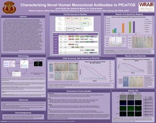
Results: IFA
907 (++)
BF
DAPI
FITC
DAPI/FITC
PfCelTOS
Rabbit (+++)
Pre-immune
Rabbit (-)
Mab
Level of
Detection
907 ++
909 -
916 ++
920 +
957 +/-
914 -
922 -
937 -
960 -
961 -
+ Control +++
- Control -
Methodology
Coomassie Stain / Western Blot
ELISAs
Dot Blot
Immunofluorescent Assay
(IFA)
Western blot: a qualitative assessment. Proteins are denatured via SDS, run through a gel and then transferred onto
a protein membrane. These membranes are probed with Mabs of interest and developed to reveal interaction with
proteins of interest.
Coomassie Blue Total Protein Stain: a replicate of the western blot experiment is stained with coomassie blue rather
than transferred onto a protein membrane. This gel is used as a reference to the western blot, separating proteins of
interest by molecular weight and visualizing a comparison of band intensity.
Enzyme-linked immunosorbent assay (ELISA): a quantitative assessment. Proteins are bound to high binding plastic
plates in several random orientations. Reactivity of the Mabs are measured by their optical density at different
dilutions, creating a titer concentration at an OD value of 1.
Dot blot: a qualitative assessment. Proteins are stacked heavily on the membranes like the western blot but are non-
denatured.
Immunofluorescence assay (IFA): a native protein assessment. Antibodies targeting protein of interest are identified
on the surface of a sporozoite via fluorescent microscopy. Sporozoites are probed on methanol fixed slides.
*Methodology requires corroboration
between assays!
Results: Fine Specificity Mapping
909 916
PfCelTOS
C-Term
Peptide 2
N-Term
Peptide 1
Peptide 5
Peptide 4
Peptide 1/2
MSP
0.00E+00
2.00E+05
4.00E+05
6.00E+05
8.00E+05
1.00E+06
1.20E+06
907 909
Using O.D. 1.0 Titers of MAbs Against PfCelTOS Antigens to Map Fine Specificity
PfCelTOS
C-Term PfCelTOS
N-Term PfCelTOS
Peptide 1
Peptide 2
Peptide 4
Peptide 5
PfCelTOS
C-Term
N-Term
MSP
907 916909SVP09 3D.11 4H9 3C3
Conclusions & Future Studies
Western Blot:
• Mabs 907, 909 & 916 are immunoreactive to the C-terminus (strongly) , N-terminus (strongly) & C-terminus (weakly) respectively
• Mabs 907, 909 & 916 are non-cross reactive to the Plasmodium species berghei, vivax & knowlesi
ELISA:
• Mab 907 can be mapped to PfCelTOS on the C-terminus at peptide 4 - likely to distal , coiled, or unstructured region
• Mab 909 can be mapped to PfCelTOS on the N-terminus but does not map to any of the N-terminal peptides
• Mabs 907, 909 & 916 are non-cross reactive to the Plasmodium species berghei, vivax & knowlesi
Dot Blot:
• Mab 909 can be mapped to PfCelTOS on the N-terminus on peptide 1 – likely to the coil region following histidine tag
• Mab 916 has weak, non-specific reactivity with the full length protein, PfCelTOS, the C-terminal & N-terminal fragments
IFA:
• Mabs 907, 916, 920 & 957 react on the Plasmodium falciparum 3D7 sporozoite using air-dried fixed sporozoites
• While Mab 909 tested positive using in the other detection methods, there was no recognition on native PfCelTOS protein by IFA
Future studies: Mabs that are immunoreactive by the various methods and recognize native CelTOS antigen on sporozoites by IFA can be further
analyzed using in vitro functional antibody assays such as inhibition of sporozoite gliding motility, traversal of host cells, invasion and development.
In addition, these positive Mabs can be passively transferred into mice and challenge using transgenic parasites to evaluate the specific role of
these antibodies in protection against infection.
I would like to thank Dr. Angov for giving me the opportunity to work at WRAIR this summer. I would also
like to thank all of the members in the lab for helping and teaching me through out my project and for
adding their unique personalities to the dynamic of the lab. You will all be missed.
Acknowledgements
References
1) Kariu, Tohru; Ishino, Tomoko; Yano, Kazuhiko; Chinzei, Yasuo; Yuda, Masao, (2006) Molecular Microbiology 59(5):
1369-1379
2) Bergmann-Leitner, Elke; Mease, Ryan; De La Vega, Patricia; Savranskaya, Tatyana; Polhemus, Mark; Ockenhouse,
Christian; Angov, Evelina, (2010) PLoS ONE 5(8)
3) Bergmann-Leitner, Elke; Chaudhury, Sidhartha; Steers, Nicholas J.; Sabato, Mark; Delvecchio, Vito; Wallqvist,
Anders S.; Ockenhouse, Christian F.; Angov, Evelina, (2013) PLoS ONE 8(8)
Results: Cross-Reactivity
0
0.5
1
1.5
2
2.5
3
3.5
4
200 400 800 1600 3200 6400 12800 25600
OD405nm
Dilution
Titration Curves of Mab Cross-reactivity to Plasmodium
Species 907 (PfCelTOS)
909 (PfCelTOS)
916 (PfCelTOS)
+ Control Avg
907 (PbCelTOS)
909 (PbCelTOS)
916 (PbCelTOS)
907 (PkCelTOS)
909 (PkCelTOS)
916 (PkCelTOS)
907 (PvCelTOS)
909 (PvCelTOS)
916 (PvCelTOS)
907 909 916
PfCelTOS
PbCelTOS
PkCelTOS
PvCelTOS
Gel loading:
1. PfCelTOS
2. PbCelTOS
3. PkCelTOS
4. PvCelTOS
Initial Screening: Mab Reactivity to PfCelTOS
0
0.5
1
1.5
2
2.5
3
3.5
200 400 800 1600 3200 6400 12800 25600
OD405nm
Dilution
Titration Curve of Mabs Against PfCelTOS:
907 & 909 = High Titer
907
909
914
916
920
922
937
957
960
961
+ Control
Avg.
SVP09 3D.11 4H9 3C3
957922 937 960 961
909907 916
920914
C-Terminus
PfCelTOS
N-Terminus
Gel loading:
1. PfCelTOS
2. C-Terminus
3. N-Terminus
Screening novel Mabs to the PfCelTOS full length peptide: Starting with western blot visualization, 3 of the 10 Mabs show reactivity to the protein at low
dilutions. Comparably, only 2 of the remaining 3 reactive Mabs had detectable optical density in the ELISA assay. At this point, the 7 non-reactive Mabs were
dropped from further assay exploration.
Fine Specificity Mapping : The 3 remaining Mabs were first probed to the full length PfCelTOS, C/N-Termini and their
corresponding long peptides by ELISA and then visualized in dot blots. Dot blot assays corroborated results that were
missing in the ELISA, showing that ELISAs can experience epitope blocking due to how the protein binds to the plastic.
Cross-reactivity:
Although their genomes
are highly conserved,
none of the Mabs show
cross-reactivity to
Plamsodium berghei,
knowlesi or vivax in
ELISA or western blot
analysis.
Immunofluorescence assay: detection was categorized by a +/- system shown in table to the right. Each
assignment is relative to the fluorescence of the positive and negative controls, respectively. 4 of 10 Mabs
showed positivity, 2 of which went undetectable in western, ELISA and dot blot analysis. Unfortunately, the
novel N-terminus Mab 909 did not show detection in the IFA.
![CRISPR Based Diagnosis For SARS-CoV-2[FELUDA]](data:image/gif;base64,R0lGODlhAQABAIAAAAAAAP///yH5BAEAAAAALAAAAAABAAEAAAIBRAA7)